Wednesday, December 28, 2005

Leg Ulcer in a Heart Transplant Patient

The patient is a 49 yo man with a one month history of a very painful leg ulcer.
He is 8 years s/p heart transplant.
His medications include: CsA, CellCept, Pred, Diltiazem, Ranitidine.
There was no history of trauma and no similar ulcers in the past. He works at a supermarket and is on his feet all day long at work.
This ulcer is periodically painful. He has had leg edema for over a year.

The examination shows an 8 mm in diameter ulcer with as slightly purulent surface and ragged edges.. The borders are grayish. The area surrounding the ulcer has 2+ pitting edema and is erythematous.


Bacterial culture shows only staph species
Peripheral pulses are present but weak.

No response to support stocking used for the past three weeks.

This is probably a venous ulcer, but it's a bit unusual for a 49 yo man. Diltiazem can cause edema.
The pain is out of proportion to what one would expect. I have known the patient for six years and he has not had pain like this before.

Please help with diagnostic and therapeutic suggestions.

Sunday, December 18, 2005

Retroauricular Dermatitis

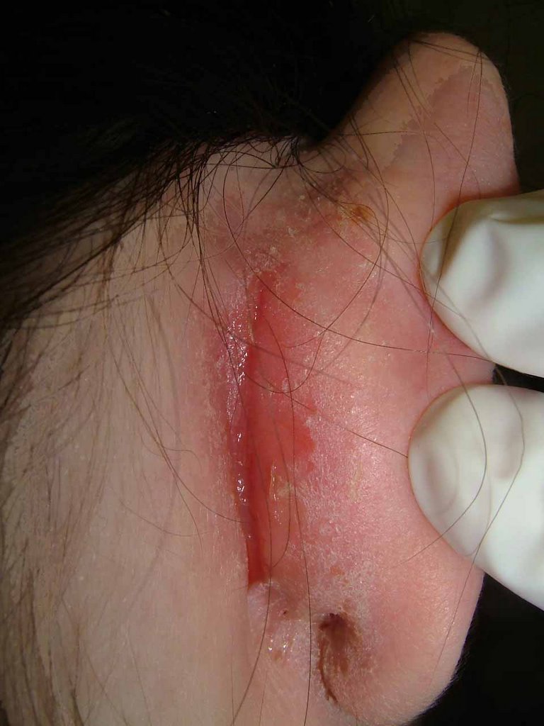
The patient is a 25 year old registered nurse with a few month history of a painful and pruritic process in the right retroauricular area. She also has mild scalp pruritus and some increased scaling of the scalp.

The exam shows a superficial erosion in the right retroauricular sulcus.


Bacterial Culture: Positive for Many Staph aureus - resistant to Pen and Erythro

Pt. Started on mupirocin cream

Note: She is an obstetrical nurse and has contact with newborns. Her staph infection is of potential import here.

Reference:
Marks MB, Gluck JC, Lavi E, Halem-Sinclair E.
J Am Acad Dermatol. 1981 May;4(5):519-22
An unsuspected sign of cutaneous allergy.
An eczematous eruption in the superior retroauricular areas of the scalp and often on the posterior aspects of the pinnas may be seen in about 30% of allergic children. The eruption is not generally noticed because the overhanging hair covers the affected areas. The dermatitis is seen mainly in those children afflicted with bronchial asthma, perennial allergic rhinitis, or both. A previous history of atopic or seborrheic dermatitis is, as a rule, not elicited.

Wednesday, December 07, 2005

Periocular Dermatitis

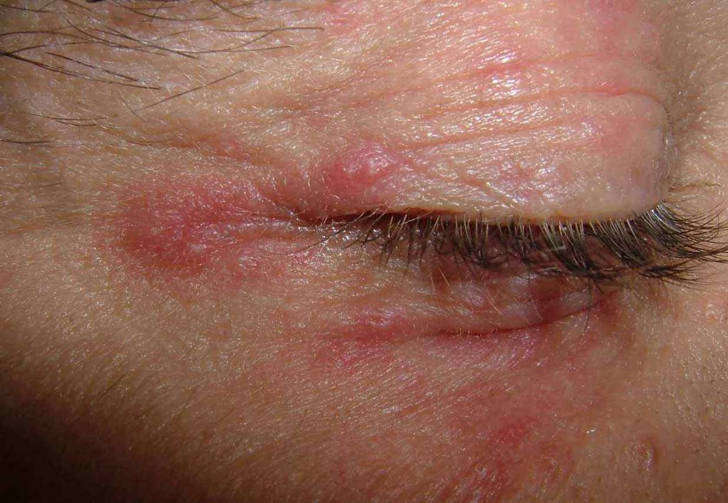
The patient is a 35 year old man who has had a right-sided eyelid dermatitis for around eight months. He was given a moderate strength topical steroid for this by his primary care physician and has been using it off and on since. When the process flares, he applies it again.

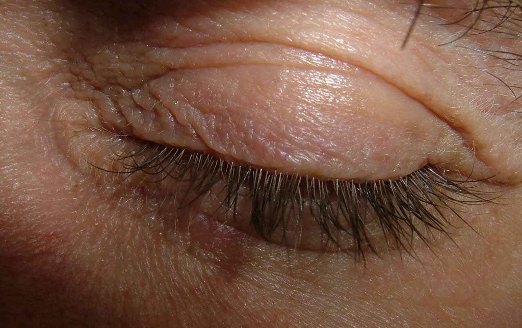
There are discrete erythematous papules around the upper and lower lids on the right side. The left eyelids are clear.

The clinical picture suggests a steroid-induced periocular acne.
Treatment: cold tap water compresses, doxycycline 100 mg. bid. Is there a role for tacrolimus ointment?

Affected Right Eye


Normal Left Eye


References:
1.
J Am Acad Dermatol. 1999 Sep;41(3 Pt 1):435-42.
Eyelid dermatitis to red face syndrome to cure: clinical experience in 100
cases.
Rapaport MJ, Rapaport V.

A retrospective review of all eyelid dermatitis patients seen over an 18-year
period revealed a large subgroup of patients who had, as the basis for their
ongoing problem, an addiction to the use of topical or systemic corticosteroids.
This group of 100 patients often sought many consultations with various
physicians. Unrelenting eyelid or facial dermatitis often resulted in the use of
increasing amounts of corticosteroids for longer periods of time. Soon the skin
became addicted. Once the work-up ruled out other causes, the remedy for the
problem was absolute total cessation of corticosteroid usage. This article
describes the typical history of the problem, the evaluation of these patients,
and the distinctive pattern of flaring erythema that ensued when the
corticosteroids were ceased. We stress the absolute necessity of total cessation
of corticosteroid use as the only treatment for corticosteroid addiction. We
also demonstrate that no additional therapy or further consultations were
necessary once remission was obtained after topical corticosteroid abuse was
stopped.

2.
West J Med. 2001 Jun;174(6):383-4.
"Tortured tube" sign.
Fowler KP, Elpern DJ.
Medical University of South Carolina Charleston, SC. Williamstown, MA, USA.
(This is available as full text - go to http://www.pubmed.gov find this article and you can see the full text version. This was a patient Dr. Fowler saw in my office 5 years ago.)

A Hunter in Deer Season

The Hunter as Hunted

[Your comments are welcomed]

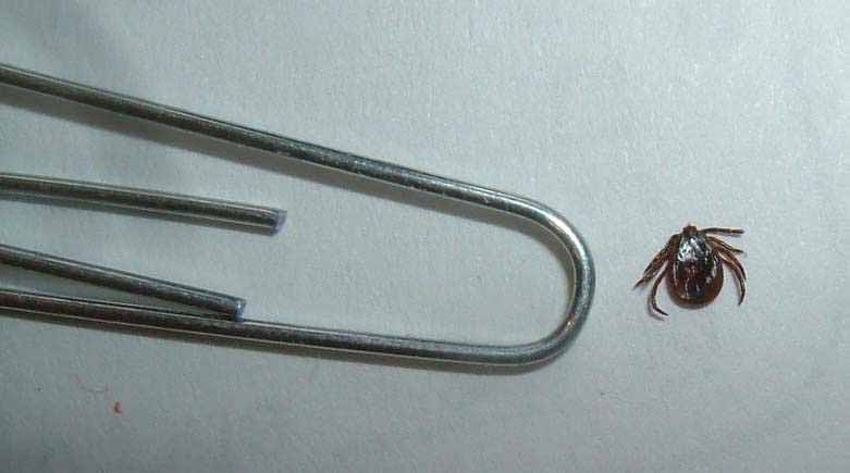
This 72 year old man was deer hunting on December 2, 2006. He remembers sitting on a log for 45 minutes and feeling as if he was being bitten. Later that day, his hunting companion removed three ticks from him. He presented on Monday, December 5 for an evaluation. The picture is taken from his left hip. The tick was gently removed and photographed with a paper clip.


Since the tick was attached for around three days, I elected to treat the patient with doxycycline 100 mg. bid for 10 days. The tick was sent to the lab for identification. I may get serologies in a few weeks. ADDENDUM: The tick was identified as a deer tick, Ioxides dammini.

Rationale for Treatment (from www.emedicine.com)
Ten days of doxycycline seem innocuous enough.
Although most patients do not require treatment, consider tick bite prophylaxis on a case-by-case basis. Base the decision on the species of the tick, duration of attachment (degree of engorgement of the tick is a surrogate marker), geography (percentage of ticks infected where the bite took place), method of tick removal, anxiety level of patient, and pregnancy (lower threshold to treat pregnant women).
After performing this exercise in clinical decision-making, one may decide to treat a given patient with prophylactic antibiotics. In the studies mentioned previously, no patient in the treatment group (which received 10 d of antibiotic treatment) had the disease. Historically, if one were to choose to treat, 10 days of oral amoxicillin, doxycycline, or cefuroxime axetil would seem prudent, depending on patient factors such as age, allergy, and pregnancy.
In one of the most recent studies (2001), a single 200-mg dose of doxycycline was used for prophylaxis with excellent results.

Wednesday, November 30, 2005

Difficult BCC


From A.R. Pito
Norfolk Island, S.P.

A friend from Australia sent me this photo with the following note.
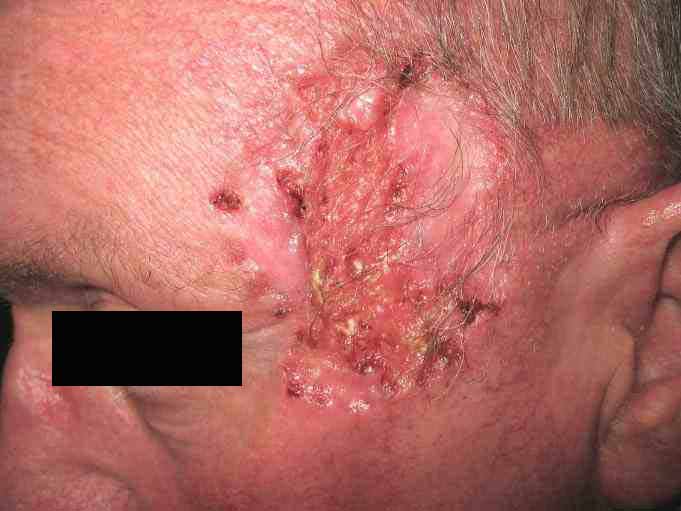
"A.R.: I just saw this 56 yo man with a two year history of a large ulcero-nodular plaque on his left temple. Biopsy is pending but this appears to be a morpheaform BCC. Assuming this is BCC, what would your therapeutic suggestions be.
Radiotherapy?
Mohs?
In our area, we do not have a specialized cutaneous radiation centre, but he could be sent to Sydney, I suspect."

Thank you for your suggestions. You can post here or send to oslerian@gmail.com. I will append the pathology when my colleague forwards it.

Sunday, November 27, 2005

5 year old girl with perioral rash

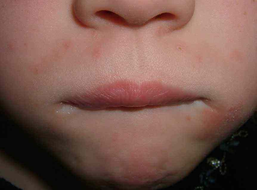
This 5 year-old girl presents with a 5 month history of a dermatitis at angle of mouth and upper lip. There are areas of patchy erythema with scale and discrete acneiform papules. She has been treated with mupirocin cream, ketoconazole cream, and 1% HC cream. None of these have helped. She is not atopic, has no family history of atopy and is not a lip licker.
Althoug subtle, I think this is perioral dermatitis of childhood. Tetracycline is contraindicated. I wonder if systemic erythromycin would be helpful. Based on the literature, I started her on metronidazole cream and told her mother that this may take weeks to months to get better.
I would appreciate your suggestions.


Wednesday, November 16, 2005

A Patient from Ghana - Please comment

Anak VGRD members have been asked to comment on a 16 year old Ghanaian girl from a small village without any specialized medical care. In otherwise good health and of normal intelligence, since age two she has a history of small hyperpigmented papules on her face. The larger lesions on her forehead appeared over the last two years. She patient states that she doesn't manipulate any of the lesions and that they are asymptomatic. There has been no treatment. HIV status and family history are unknown. This unfortunate young woman feels like an outcast because of these lesions.

We welcome you thoughts and advice. There are medical students in her community who could perhaps perform diagnostic testing if needed.

Tuesday, November 15, 2005

? Segmental Neurofibromatosis


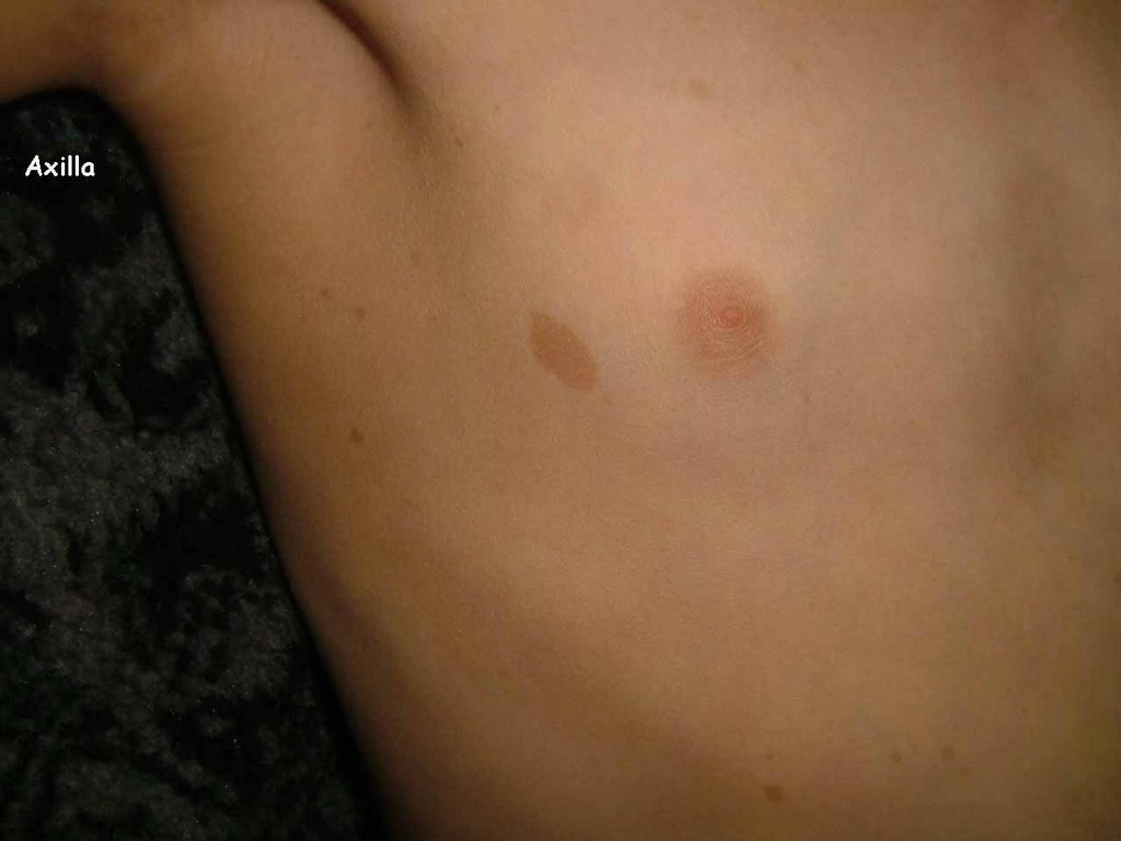
The patient is a three year-old girl with 2 cafe au lait spots on right arm and chest since she was a few months old and many small "freckles" on right chest and axilla (see photo). There are a few freckles on the right upper back as well. No pigmented lesions on any other area. Have not looked for Lisch nodules yet.

Athough it may be too early to tell, I suspect this is segmental neurofibromatosis. Most cases of segmental NF are diagnosed because of tumors. There are probably some that only manifest as CLS or freckling. Of course, we will have to wait years in this case to see what develops.

Does anyone have ideas as to wha to tell the parents?

Saturday, November 12, 2005

BCC of Eyelid

This is a 75 yo man who came in for an unrelated problem.
This tumor was obvioius, however.
Two years ago, and ENT physician told him he had a "blocked duct."
This 6 mm in diameter lesion looks like a BCC.
I propose to excise it but am worried about pulling down the lower lid.
I don't think C&E or Aldara would be appropriate. Mohs might be overkill.
Do you have any comments? I would, oif course, try to undermine the tissue.

84 yo Man with Atrophie Blanche

The patient is an 84 yo man with a ten year history of extremely painful ulcerations of his lower legs.
Other than whirlpool baths, I have found nothing to be of value.
His health is good and he has bounding dorsalis pedis and posterial tibial pulses.
I assume this is Atrophie Blanche.
Do you have any therapeutic suggestoins?


Tuesday, November 01, 2005

Localized Hyperkeratosis

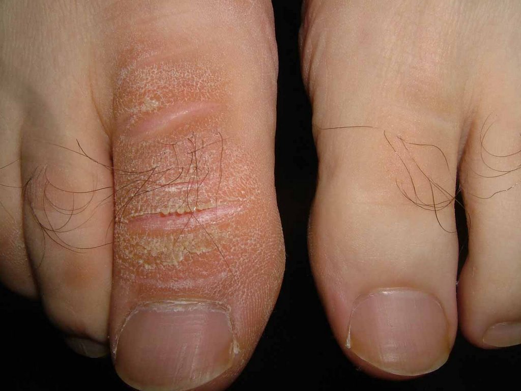
39 yo man with > 15 year hx of localized hyperkeratosis of right great toe. No unusual trauma to area. He's used mostly over the counter emollients without much help.

Are you aware of any localized forms of hyperkeratosis?

I have started him on Salex cream.

Pictured below are his affected right and normal left great toes.

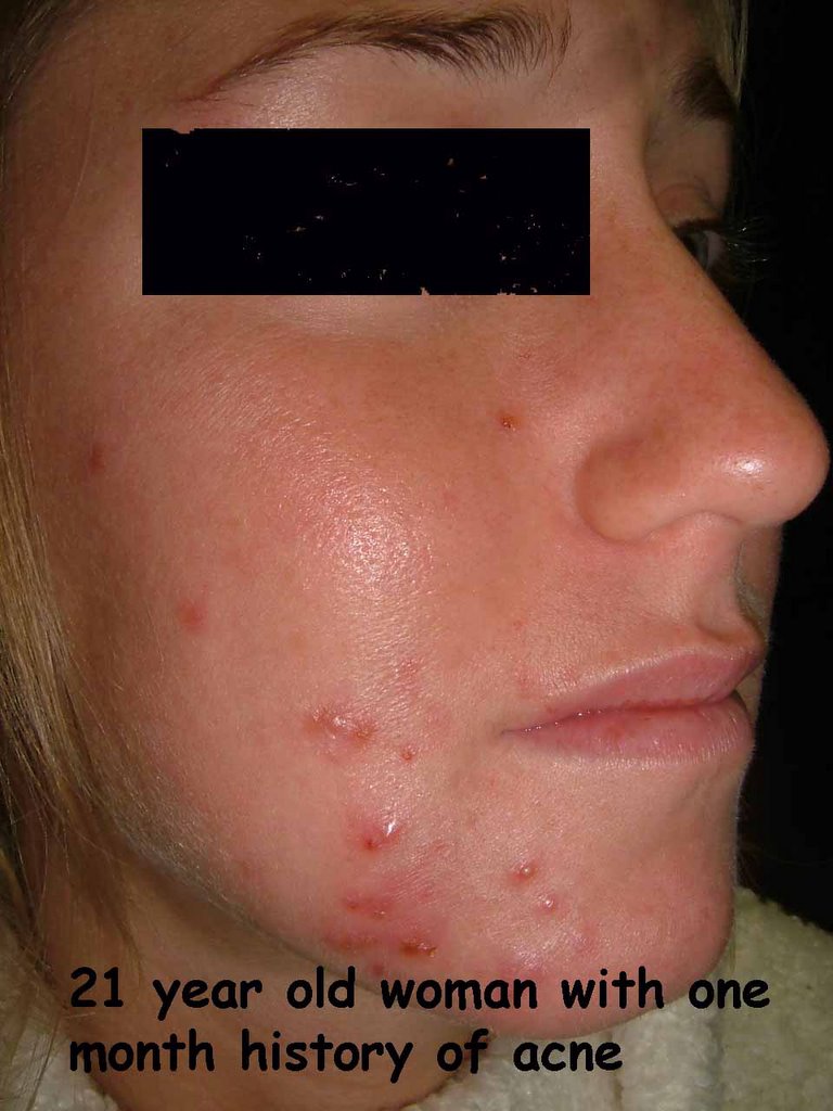
Recent Onset Acne in Young Woman


This 21 yo woman experienced the sudden onset of acne one month ago.
No new med. No unusual stresses.
She has never had acne like this before.

Do you think this is a mild case of pyoderma faciale rather than typical acne?

Monday, October 31, 2005

BCC Scalp


The patient is a 75 yo man with a three month hisory of a lesion at the vertex of the scalp.
Exam: Man with Type II - III skin. 2 x 1.6 cm diameter erythematous plaque scalp. There are some small crusted areas.
Biopsy shows superficial BCC.

Question: Is Aldara appropriate?
C + E will take ages to heal.
Excision could n ot be closed without a graft or a flap.

Wednesday, October 26, 2005

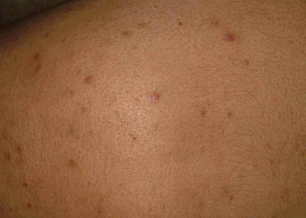
19 year old student with acneiform eruption


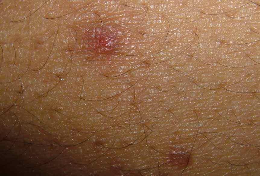
History: 19 year-old man with a 5 year history of acneiform eruption predominately on torso and proximal extremitiesl Some facial involvement, but less than on torso. He is in good health, no history of diabetes or steroid use. No antibiotics for months. In the past he has used benzoyl preoxide ceams and washes, topical retinoids, topical antibiotics and tetracycline and its derivatives. Mome has ever helped.

Exam: Healthy young man with Type IV skin. On torso and proximal extremities he has discrete erythematous papules and an occasional pustule. No cysts. Face largely clear.


Lab: nil

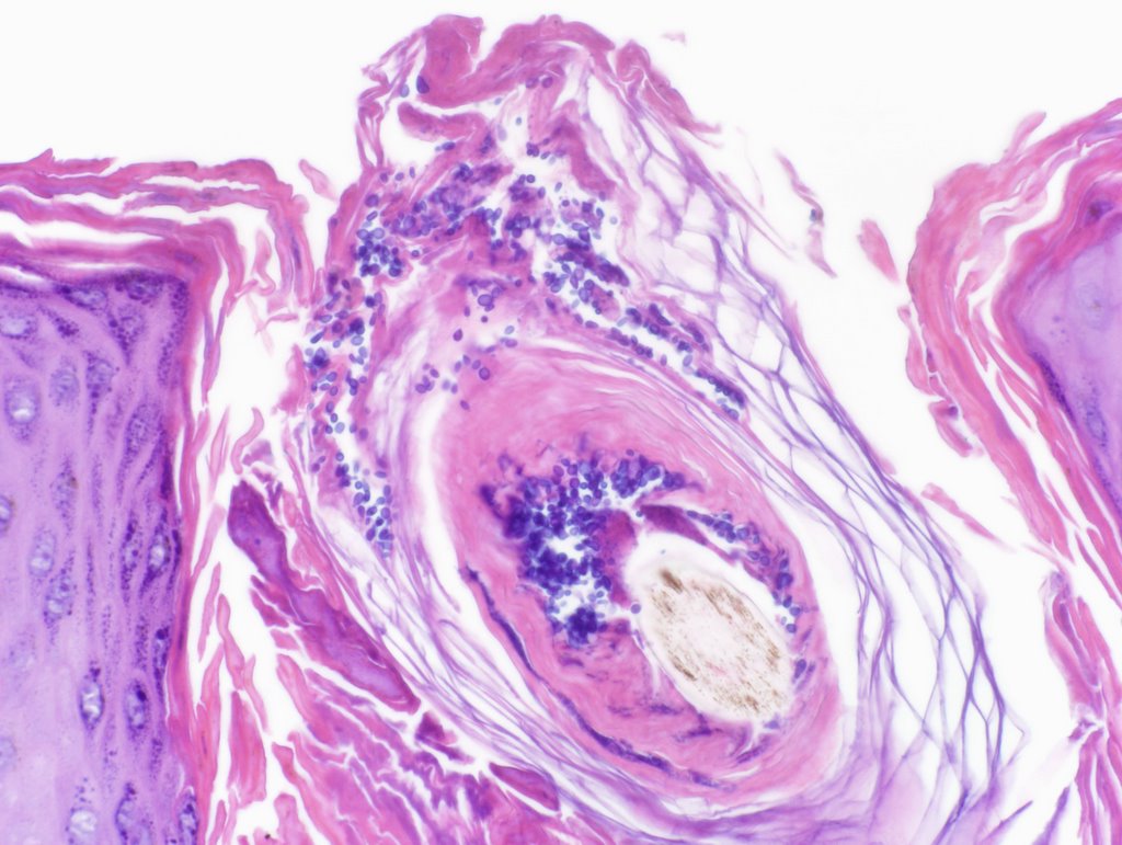
Pathology: Initial reading showed marked perifollicular lymphoneutrophilic infiltrate c/w acute folliculitis. PAS negative.


Diagnosis: Probable Pityrosporon folliculitis

Discussion: Are the yeasts incidental or indicative of pityrosporum folliculitis. I asked for more cuts and this showed numerous PAS (+)"fungal spores" in the follicular ostia.
I have started him on itraconazole 200 mg per day and Nizoral 2% shampoo to torso. The literature does not have good guidelins for how to treat this; but I suspect 4 - 6 weeks woth oral meds.
Please suggest diagnostic and therapeutic alternatives.

Thursday, October 20, 2005

Atypical Nail Pits


Case for Discussion:
This 40 year-old woman presented for evaluation of a nail dystrophy present for 2-3 months.
She has a history of Hashimoto's thyroiditis. About 9 months ago she developed vitiligo.
Her health is otherwise normal
Meds. Synthyroid and iron
Lab: Thyroid Perox AB 248 (Nl. 0 - 34 IU/ML)
ANA < 1:40

Physical Exam:
Vitiligenous patches left neck and left upper back
Around 4 finger nails show a distinctive pitting. The pits are fairly uniform and the affected nails are rough and lusterless. There are no cutansous lesions of psoriasis.

Discussion and Questikon:
The picture is atypical for psoriatic nail pits but that is not excluded.
I favor a relationship to the underlying autoimmunity that has caused the Hashimoto's and vitiligo.
The picture is similar to that seen with alopecia areata, but the patient has not has any alopecic patches.
Nail dystrophy has occasionally been described before the development of A. areata. And patients with Hashimoto's thyroiditis have a higher than expected incidence of alopecia areata.
We welcome your thoughts or suggestions.

Saturday, August 13, 2005

Hemangioma of Auricle


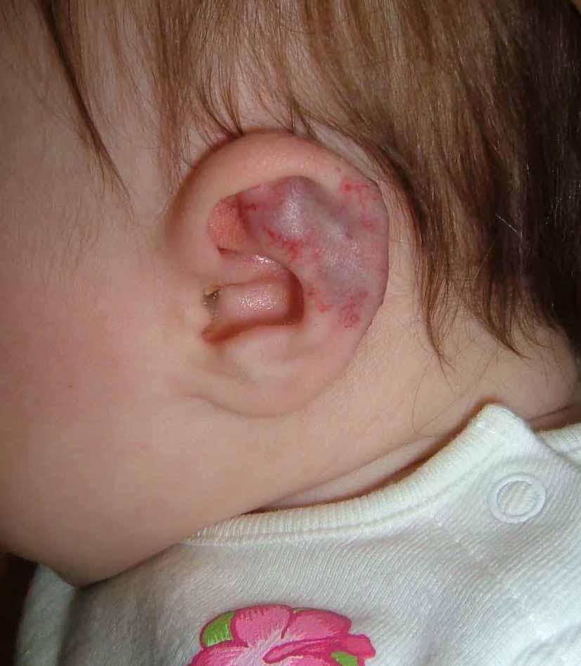
This 4 month infant girl was born with hemangiomas of the chin and left ear.
The lesion on the chin (not pictured) measures 1 cm in diameter and is typical of a congenital hemangioma.

The abnormality of the left ear involves the triangular fossa and the helix. I am concerned that as this involutes it could cause disfigurement. At the same time, I wonder if anyone has experience handling similar lesions in this site. A PubMed site found only a paucity of pertinent references.

Ref:
Cavernous hemangioma of the external ear canal.
Reeck JB, Yen TL, Szmit A, Cheung SW.
Laryngoscope. 2002 Oct;112(10):1750-2. Related Articles, Links
Division of Otology, Neurotology and Skull Base Surgery, Department of Otolaryngology-Head and Neck Surgery, Veterans Administration Medical Center, San Francisco, California, USA.

OBJECTIVE: To document the occurrence of a cavernous hemangioma of the external ear canal and to review the relevant literature.STUDY DESIGN Case report and literature review. METHODS: Review of a patient chart, imaging studies, operative report, and histologic findings. RESULTS: A cavernous hemangioma of the external ear canal not involving the tympanic membrane was surgically excised without complication. This is the third documented cavernous hemangioma of the external ear canal without tympanic membrane involvement in the English literature. Computed tomography scan is invaluable to narrow the differential diagnosis. Complete removal is curative. CONCLUSIONS: Cavernous hemangioma of the external ear canal with or without tympanic membrane involvement is a rare otologic entity amenable to surgical treatment. Temporal bone computed tomography scan imaging is an important preoperative diagnostic tool.

Wednesday, August 10, 2005

Nevus, Congenital


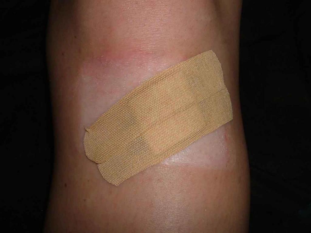
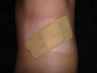
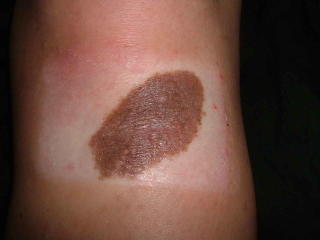
This 20 yo college student presented in consultation for removal of a congenital nevus.
The lesion is located just distal to her left knee. She's been embarrassed about this since childhood and for the past six or seven years won't let anyone see it. Indeed she covers it at all times with bandaids.
The lesion measures 5.2 cm in diameter.
I have referred her to a surgeon for removal since I think that's better than psychotherapy. To minimize the scar it may need staged excision.
The young woman appears normal in all other respects - she is very upset about the lesion and the questions she gets about it.
I wonder if this has been reported. I would hardly call this Body Dysmprphic Syndrome.
Your thoughts are appreciatged.

Not much written about this area -- here's a ref that may touch on it:


Congenital melanocytic nevi. Evaluation and management.
Marghoob AA., Dermatol Clin. 2002 Oct;20(4):607-16, viii.
Department of Medicine, Dermatology Division, Memorial Sloan-Kettering Cancer Center, 1275 York Avenue, New York, NY 10021, USA. marghooa@mskcc.org

This article discusses the care of patients with CMN, who often require
a multidisciplinary approach involving pediatricians, family physicians,
internists, dermatologists, psychologists, plastic surgeons,
neurologists, and radiologists. The cosmetic and psychosocial issues,
combined with the knowledge of the increased risk of developing
melanoma or NCM, is a huge burden that many of these patients
and their families have to carry. This article describes the importance
for physicians to help these patients and families come to terms
with these issues, as well as remind their patients and their family
members that although melanoma, NCM, or other complications can
develop, most affected individuals do not develop any complications.
The article mentions that there are many healthy, happy, functional
adults with large, small, and multiple CMN alive today.
Monday, August 08, 2005

Imiquimod Side-Effect?


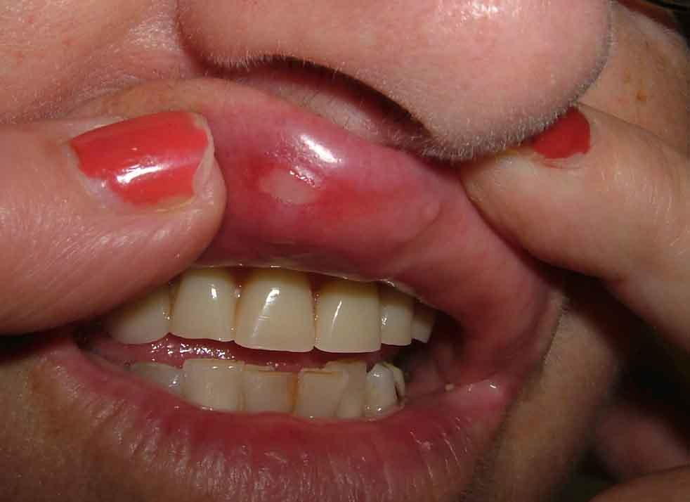
This 66 yo woman began topical imiquimod on July 27, 2005 for a biopsy proven superficial BCC of the right upper lip. One week later, she developed aphthous ulcers on the mucous membrane surface of the area just below the site treated. A literature search retrieved a reference to his occurence. This is another unexpected adverse effect. It is unclear how common this is.

Aphthous ulcers associated with imiquimod and the treatment of
actinic cheilitis.
Chakrabarty AK, Mraz S, Geisse JK, Anderson NJ.
J Am Acad Dermatol. 2005 Feb;52(2 Suppl 1):35-7.
Solano Clinical Research, Davis, California, USA. chakak3@yahoo.com

Our case series report is the first documented depiction of the
appearance of aphthous ulcers secondary to imiquimod
application. This case series presentation discusses the
underlying pathophysiology of aphthous ulcer development and
imiquimod therapy in terms of the stimulation of pro-
inflammatory cytokines, such as tumor necrosis factor alpha
(TNF-alpha). The literature review suggests more than just a
mere coincidence for the development of aphthous ulcers
subsequent to the treatment of actinic cheilitis with imiquimod
application.

Sunday, August 07, 2005

46 year old woman with indurated depressed lesion


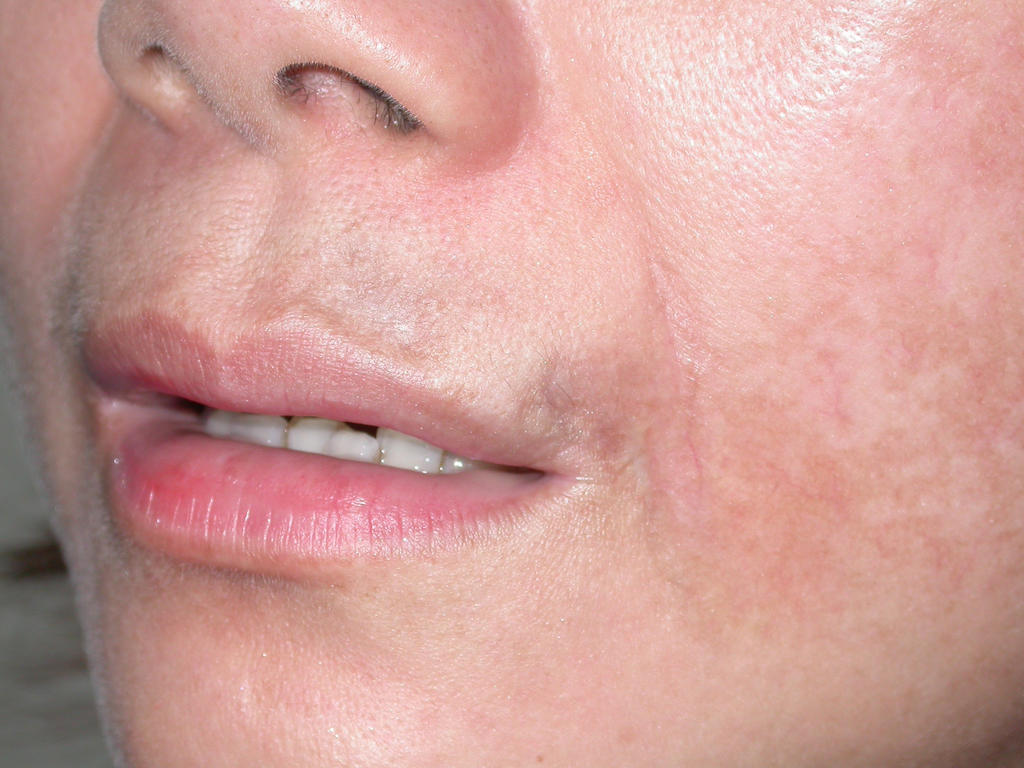
A 46-year-old woman presented with 7 years history of depressed lesion just above the left side of mouth. It was asymptomatic. She has seen few dermatologists and a plastic surgeon but none really helped her lesion. She did not have polyarthalgia or other constitutional symptoms. Drug history was nil significance.
Examination showed 2 areas of depression, almost tethered to the underlying dermis just above the left side of the mouth and another a bit above the angle of mouth. There is no induration on deep palpation. The inner buccal mucosa appeared normal.
Serology for lupus including ANA, anti Ro, Anti La, and other extractable nuclear antigens were negative. Possible diagnoses would include lupus profundus and morphoea. Biopsy with immunofluorescence studies should help. Anyone has any therapeutic pearls for this lady??

Basal Carcinoma - Deconstructed

August 9, 2006.

LATE BREAKING!! BIOPSY HERE SHOWED THE LESION TO BE AN INTRADERMAL NEVUS.
I should have paid more attention to the history.

MORE... The patient underwent an excision - and the final report was a Basal Cell + an intradermal nevus. Quite unusual. The initial clinical impression was more accurate that the incisional biopsy. This is sobering.


This 57 yo man presented with an 8 mm in diameter papule that has bled since he started to wear glasses a year or so ago. He has been aware of a slowly growing lesion in this area since age 19 (38 years ago).
The lesion has been biopsied and I await the results. It appears to be a BCC. The long history underscores the benign behavior of many of these lesions. While we have all seen case reports of aggressive BCCs that have caused loss of eyes, ears, nose - these are likely in the very small minority. It is the growth characteristics and behavior of these lesions which is likely key, not their appearance micorscopically. Our therapy for these indolent tumors may be too aggressive based on the slow growth and lack of metastatic potential.