Tuesday, January 31, 2006

Difficult BCC

On January 30, 2006, this 49 yo woman presented with a somewhat sclerotic plaque on the blub of the nose. By history, this has been present for 1.5 years. A 2 mm punch biopsy was taken at the site marked X.


If this turns out to be BCC, I will refer for micrographic surgery. Does anyone feel radiotherapy is appropriate for a patient of this age?

First Biopsy showed no evedence of BCC or malignancy. Repeat biopsy taken. Here is another clinical photo:

Thursday, January 26, 2006

Oral Hyperpigmentation

This 21 years student noticed hyperpigmentaiton of her buccal mucosae for one weeks.
She has gingival hyperpigmentation for years and has a family history of the latter.
Her health is good, and her only medication are albuterol and cromolyn sodium for asma.
No history of cancer in any first degree relation.


The examination show hyperpigmented buccal maculas measuring up to > 1 cm in diameter.
Gingival hyperpigmentation is present as well.


In the differential diagnosis I would consider pigmented fixed drug eruption, amalgam tattoo, melanoacanthoma. Biopsy may be offered. Melanoma is unlikely.

Presented by
Mordechai Pepe Grosbartt
Concepción, Chile

Tuesday, January 24, 2006

9 year old child with inflammatory alopecia

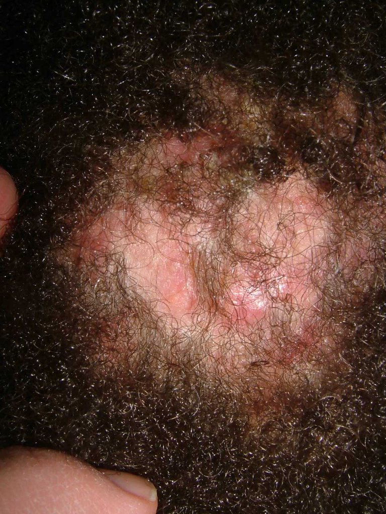
The patient is a 9 year-old girl who was in her usual state of health until September 5, 2005 when her mother applied a "hair relaxer" to her scalp. Within a few days, she developed inflammation. The process has persisted and over the past few months she has developed fluctuant areas over the scalp with alopecia and some scarring. There is moderate discomfort.

The examination shows a calm 9 year-old. Her scalp is involved globally with fluctuant nodules, some crusted. There are diffuse areas of alopecia. Some areas look scarred. Wood's light is negative. Posterior cervical lymph notes are enlarged and tender.


A bacterial and fungal culture were taken. Scalp biopsy will be performed and the patient will be started on prednisone 1 mg/kg/day and griseofulvin 25 mg/kg/day pending test results.

Working diagnosis is Inflammatory tinea capitis vs. dissecting cellulitis of the scalp.

Update February 9, 2006
The biopsies taken showed no fungfal elements, so the griseofulvin was discontinuted on Feb. 2, 2006 and patient was placed on cephalexin 50 mg/kg/day in divided doses. She did well.

On day 14, the fungal culture was positive and she was placed back on griseofulvin -- 25 mg/kg/day/ Her prednisone dose is 1 mg/kg/day.


Lesson: Scalp biopsy is not an absolute. Fungal culture takes ~ 2 weeks to be positive.
Dx: Inflammatory tinea capitis (the first diagnosis). Negative scalp bx threw me off.

Your comments are welcome.


Reference:
Tinea capitis mimicking dissecting cellulitis: a distinct variant.
Twersky JM, Sheth AP.
Int J Dermatol. 2005 May;44(5):412-4.
BACKGROUND: Tinea capitis is a common scalp dermatosis with several clinical
patterns. Only two patients with a presentation of tinea capitis mimicking
dissecting cellulitis have been described in the English literature.
OBSERVATION: We report a patient with tinea capitis mimicking dissecting
cellulitis who did not respond to griseofulvin therapy at 16 mg/kg/day but
eventually cleared after a protracted course of higher dose griseofulvin.
CONCLUSION: recognition of a dissecting cellulitis-like pattern of tinea capitis
will increase clinical suspicion and avoid inappropriate management of a
recalcitrant "dissecting cellulitis" in favor of prompt antifungal therapy of
appropriate dosage and duration for patients with this unusual variant of tinea
capitis.

Tuesday, January 10, 2006

Rapidly Growing Tumor

This 70 yo man presented on January 9, 2006 with a three to four month history of a lesion on his upper back. It was noticed by his wife. He has type III skin and a past history of renal cell carcinoma.


An excisional biopsy was performed.

Presumptive diagnosis is melanoma, probably nodular type.
Pigmented basal cell is another possibility, but rapid growth favors melanoma.
I do not think metastatic renal cell carcinoma would be pigmented.

January 25, 2006 -- Update
The biopsy showed a superficial spreading melanoma, 2.04 mm thick, Level IV.
The patient underwent a wide local excision with 2 cm margins. He elected not to have sentinal node biopsy.

Sunday, January 08, 2006

Suggestions for Wart Removal?

Dear Anak VGRD Members,

I am a G.P. trainee and have been having a hard time with a wart removal on a 3 year old's fifth toe. The difficulty is getting him to let anyone near it. I've seen him once, late in the day, and my preceptor asked that the child return for freezing with the help of sedation. We are now working on what to use. I prefer to avoid having to sedate the child, and have suggested duct tape in the interim (six days on, one day off with soaking). What suggestions or tricks would you have for treating a wart in this location in a three year-old? Thank you, Tammy Grimely, Hobart, Tasmania

Wednesday, January 04, 2006

Explosive Rosacea?

The patient is a 33 year-old executive V.P. who presents with a 6 month history of dramatic facial eruption. He had acne as a teenager but has been clear for many years. His health is good and he takes no medication by mouth. He has never used topical corticosteroids for his face.


The examination shows erythematous papules and small cysts on forehead, cheeks and chin. There are a few papules on the back, but the chest is clear.


Diagnosis: Florid Rosaca

Plan:
I will place on prednisone 20 mg bid for a week or so and isotretinoin 0.25 mg per kg for a cple of weeks and then increase to 0.5 mg per kg fir a month or two. Slowly incvrease isotretinolin to 1 mg per kg over 8 weeks and anticipate a six moth course. The prednisone will be tapered over a month to 6 weeks to avoid a flare.

I could have started with doxycycline; but isotretinoin may be more definitive; I welcom your thoughts.

Thank you,

David