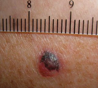
An excisional biopsy was performed.
Presumptive diagnosis is melanoma, probably nodular type.
Pigmented basal cell is another possibility, but rapid growth favors melanoma.
I do not think metastatic renal cell carcinoma would be pigmented.
January 25, 2006 -- Update
The biopsy showed a superficial spreading melanoma, 2.04 mm thick, Level IV.
The patient underwent a wide local excision with 2 cm margins. He elected not to have sentinal node biopsy.
My question: is the covering discoloration of the tht nodule a real pigmentary or just blackish crusting??If it is crusting,I would consider the possibility of keratoacanthoma.Here the management will be much easier.Biopsy will cofirm that.
ReplyDeletekhalifa sharquie
It smells something malignant and i vote for melanoma. Regarding the pigmentation (as you mentioned) a metastatic renal cell carcinoma is a less probability...besides, i think upper back is not a usual site for metastasis of renal origin. I beleive that dermatoscopy could be helpful in this situation. Thanks for sharing, Omid
ReplyDelete