Thursday, May 17, 2018

Painful Finger Tips Secondary to Opdivo

The patient is a 65-year-old woman with metastatic lung cancer who presents for evaluation of painful fingertips.  She has also had marked axillary pruritus which she handles with Lotrisone spray. 

Her medications include metformin, two types of insulin, metoprolol, Lipitor, Lexapro, and irbesartan.  Recently, she has been started on Opdivo (nivolumab) every two weeks.  Within a few months of starting Opdivo, she developed intense axillary pruritus, and a month or so later, intensively painful fissuring of the fingertips.  Each treatment of Opdivo re-exacerbates the fingertip problem, which is unbearable. Her cancer has gone into remission and she feels well otherwise.  She especially enjoys her grandkids.

Her oncologist put her on prednisone 20 mg q.i.d. for the fingertip eczema.  Because of the patient's diabetes, her fasting blood sugars while on prednisone have gone up over 500 mg%. 

EXAMINATION:  The examination shows erosions on a few of the fingertips without erythema.  Her feet and axillae are normal.  

Clinical Photos:


We did a literature search and there are a few early reports of dermatitis with Opdivo but they do noot not affect the hands.  It may be too early to say; but given the literature on painful hands and feet related to cancer therapies this is probably a significant reaction.  

PLAN:  I would try to keep this simple and avoid the prednisone.  Start with wet soak for 20 minutes in warm water, followed by clobetasol ointment, followed by gloves (a modified Soak and Smear protocol).  We will see how she does with this.  I have the patient's permission to post her case on this site where we may get ideas and/or be of help to others with a similar problem.   There is a reference to preventing Hand Foot reactions with chemotherapy.  It seems that Celebrex (celecoxib) may be the best option. Note: Celecoxib has been reported to be associated with an increased incidence of heart attack and stroke.  It's use should not be cavalier.

References:.

  1. Survey of cutaneous adverse reactions to targeted cancer therapies: value of dermatological advice. Damiani G OncoSkin working group. G Ital Dermatol Venereol. 2018 May 11. Abstract: From June to October 2012, 25 patients with cutaneous adverse reactions linked to targeted therapies were included in the study. The main prescribed drugs were cetuximab (52%) and erlotinib (20%) and the most common reactions were folliculitis/pustules (40%) and rash/erythema (40%). Hand-foot reaction syndrome was present in 8% of patients. A total of 30% of patients treated for a cutaneous reaction underwent a consultation by a dermatologist. In these patients the rate of oncologic therapy continuation without regimen modifications was higher (100%), while it was progressively lower in patients treated by oncologists (71%) or without any specific treatment (60%). Adverse reaction should be recognized by both dermatologists and oncologists and a multidisciplinary approach is mandatory.


2. Prevention strategies for chemotherapy-induced hand-foot syndrome: a systematic review and meta-analysis of prospective randomised trials.
Macedo LT, et.al. Support Care Cancer. 2014 Jun;22(6):1585-93.
Abstract
Amongst 295 studies identified, only ten met the inclusion criteria. Celecoxib prevented both moderate to severe (odds ratio [OR] 0.39, 95 % confidence interval [CI] 0.20-0.73, P=0.003) and all-grade HFS (OR 0.47, 95 % CI 0.29-0.78, P=0.003), whereas pyridoxine and topical urea/lactic acid formulations failed to prove efficacy. There were no proven benefits in mild HFS. The use of topical antiperspirant has not been shown to improve results, according to a single trial.


Friday, May 11, 2018

Generalized Pustular Eruption in a 27 yo woman


The patient is a 27 yo woman with a 2 week history of an evolving, wide-spread eruption.  The initial lesions were on the popliteal fossae.  These were described as erythematous areas studded with pustules.  Over a week or two these generalized.  She has a history of mild psoriasis (scalp and elbows) for over a decade.  About three months ago she was started on bupropion 75 mg a day for anxiety and restlessness.  This was increased to 150 mg per day ~ 2 week before the onset of the dermatitis.  The patient has moderate cognitive impairment and Type 2 diabetes.  Here other medications include thyroid supplementation, metformin and insulin.  In the days since her initial office visit, the eruption has become more extensive and is taking on an erythrodermic appearance.  She was admitted to hospital two days after her office appointment.               

O/E:  When seen on May 7, 2018, she had a widespread eruption on arms, legs and torso,  The lesions were large arcuate patches with pustules at the periphery.  She was experiencing considerable pain.

Clinical Images:


 Pathology: (courtesy of DR. Erin Tababa, Fellow in Dermatology, Boston University)  

The biopsy shows prominent, relatively large, subcorneal pustules that are filled with a dense exudate of acute inflammatory. The neutrophils extended into the underlying epidermis, which has evidence of mild spongiosis. The papillary dermis is slightly oedematous, and there is a moderately dense perivascular inflammatory infiltrate with a predominance of neutrophils, although no eosinophils are noted.


Lab:
WBC: 29,000
Differential:  Shift to left
Eosinophils: normal
G6PD Normal

Chemistries normal.

Wound Culture: Pending

Dx:  We are initially considering subcorneal pustulr dermatosis, but with more history, especially considering the recent prescription of bupropion a drug-induced annular pustular psoriasis evolving into pustular and exanthematous psoriasis seemed more accurate.  Histopathology supported that.  Similar reactions have been reported to bupropion.  Our patient had been on the bupropion for over a month when this began which is longer than the patients in the case report below.  Reference 2 is a similar patient with a long latent period between initiation of drug and development of GPP.

Follow-up in hospital.  The eruption continued to evolve. It became more exanthematous and desquamative. These pictures were sent us by her mother.


Plan: 
Cyclosporin 3 – 4 mg per kg per day in divided doses
Wet dressings followed by triamcinolone 0.1% ointment bid – tid
Adjunctive secukinumab has been reported to be effective.

Follow-up:
Patient is doing very well.  These photos were taken 5 days after starting cyclosporine 100 gm q.i.d.  Her dose was dropped to 100 mg t.i.d.  She also was treated with wet dressings and triamcinalone ointment 0.1% (although the hospital only gave her 15 mg tubes, so she could not cover most ot the lesions. 
References:
1. Generalized pustular and erythrodermic psoriasis associated with bupropion treatment. Cox NH, Gordon PM, Dodd H. Br J Dermatol. 2002 Jun;146(6):1061-3.
Abstract: Severe drug eruptions may cause diagnostic and therapeutic difficulty when they mimic or provoke endogenous patterns of dermatosis. We report three patients with known psoriasis in whom use of bupropion (Zyban), prescribed to assist with cessation of smoking, led to severe pustular or erythrodermic exacerbation of psoriasis within 3-5 weeks. All patients were systemically unwell and required hospitalization to control the disease flare.

2. A diagnostic challenge: acute generalized exanthematous
pustulosis or pustular psoriasis due to terbinafine
L. Duckworth et.al. Clin Exp Dermatol. 2012 Jan;37(1):24-7
Abstract:  A 72-year-old man developed a generalized erythematous pustular eruption 11 weeks after commencing terbinafine. Clinically and histologically, the appearance was that of acute generalized exanthematous pustulosis (AGEP), and the disease was managed with topical preparations. Initial improvement was marred by relapse of acute pustulosis, now more in keeping with terbinafine-induced pustular psoriasis (PP),which was successfully treated with acitretin. This case highlights the difficulty of differentiating between AGEP and PP.

3. Acute generalized exanthematous pustulosis mimicking toxic epidermal necrolysis in patients with psoriasis: a coincidence?
Worsnop F, et. a. Clin Exp Dermatol. 2015 Aug;40(6):688-9

.


Tuesday, May 08, 2018

An Infant Girl from Rwanda with a Congenital Dermatosis

presented by Caitlun Stiglmeier, M.D.

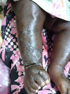
I was hoping to have some input on this case.The patient is an 11 month old female infant born via spontaneous vaginal delivery, to a G1P1 mom (young, but not sure of mothers age). No reported maternal history of perinatal infections. TORCH screening performed on the child during this admission is negative. The child has bilateral retinal detachment, but her hearing is retained. She has gross developmental delay, and the rash you see in the photos started after birth on the R forearm, and has migrated since to all areas of the body, including the chest, back, yo the upper and lower extremities, and scalp. Her mother states the rash begins as blisters (second photo), which then open (no drainage noted per mom) and leave areas of hypopigmentation (third photo) and finally hyper-pigmentation (first photo).

The child does not seem bothered by this rash, it is not pruritic, and she does not have any fevers. Despite negative TORCH screening, I'm still thinking along the lines of a congenital-type of disease. Your thoughts would be greatly appreciated!


Dr. Yoon Cohen wrote:
The clinical description and the images seems to go with incontinentia pigmenti in this her 11 month infant girl.
For management, the skin changes of IP usually do not requires any specific treatment other than wound care for blisters to prevent secondary skin infection. The baseline eye exam and close follow up by an ophthalmologist will be important, especially with her retinal detachment history. Neurological evaluation should be done for potential seizures, encephalopathy and ischemic stroke. Dental evaluation is recommended after teeth erupt for pegged or missing teeth. Other ectodermal abnormalities can be seen as alopecia and nail dystrophy.  In countries with more resources, this infant would benefit from being seen at a center where the pediatric dermatologists have more experienc with genodermatoses.

References:
1. I.P. NORD site.
2. I.P. GARG (NIH Genetic and Rare Diseases information site)
3. I.P. World Community  (some parts in French)