Presented by Will Shepard, M.D.
Gillette, WY
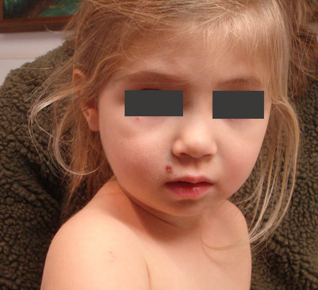
The patient is a two year-old girl with a two week history of
oral and skin lesions. She has been well
and healthy otherwise and all of her milestones have been normal. The present
illness began with two ulcers on her tongue.
A few days later she started to develop skin lesions, first on the
arms. The new lesions start with
erythematous macules and became crusted after 12 – 18 hrs. She has continued to develop new lesions on
the torso, face and extremities. Throughout
this period she has been healthy, no fevers, appetite normal and in no discomfort.
O/E: The tongue
lesions have disappeared. The skin
lesions are few in number and measure 0.5 to 1 cm in diameter. They are scaly annular macules on an
erythematous base.
 |
| Rough area from Bandaid |
New Lesion present since patient seen yesterday:
Impression: The onset of an acute problem with first oral and then skin lesions in an otherwise healthy toddler suggests a viral process.
References:
1
[Paraviral exanthems]. [Article in German]
Fölster-Holst R, Zawar V, Chuh A. Hautarzt. 2017
Mar;68(3):211-216.
Abstract: Paraviral exanthems are distinct skin diseases due
to infections with different viruses. Although no virus has been identified so
far in some exanthems, the main age of manifestation, the clinical course of
the exanthem, and the extracutaneous symptoms are suggestive for a viral
genesis. While many viral infections are a direct result of the infection,
paraviral exanthems reflect the response of the immune system to the infectious
pathogens. Viruses cannot be identified in the skin. Typical paraviral
exanthems include Gianotti-Crosti syndrome, pityriasis rosea, pityriasis
lichenoides, papular-purpuric gloves and sock syndrome, and asymmetrical
periflexural exanthema. Unilateral mediothoracic exanthem, eruptive
pseudoangiomatosis are rare and eruptive hypomelanosis has been described
recently.