Showing posts with label squamous cell carcinoma. Show all posts

Thursday, August 31, 2023

Dodging Scalpels

Presented by:
Dorinda Johnstone, M.D., Dermatologist
Scottsdale, Arizona

The patient is a vibrant, independent-living nonogenarian who saw a mid-level provider at a plastic surgery office for a skin screening. A lesion was noted on her right nasolabial fold and a shave biopsy was taken (expand image to see "x"). Also, a few actinic keratoses were also treated with liquid nitrogen.
The pathologist reported a superficial squamous cell carcinoma and the patient was scheduled for excision by the mid-level professional’s plastic surgeon employer.
The patient was anxious about the surgery and sought the opinion of a DJ, a dermatologist she had seen in the past.

"x" marks center of the lesion that was biopsied

DJ did not feel the lesion needed urgent treatment. She got a copy the path report and saw that it had been signed out by a general pathologist. She asked a dermatopathologist colleague of the general pathologist’s to take a look at the slide. The dermatopathologist felt the legion was an actinic keratosis.

The patient was called and the revised diagnosis who is related. She expressed great relief. She will make a follow up in three months to see the dermatologist and decide whether anything needs to be done.

Take a messages:
1. Some mid-level providers working for high-volume surgical and dermatology practices serve as feeders for big-ticket procedures to their employers.
2. These surgeons and dermatologists rarely question biopsy reports.
3. It can be important to have the pathology reviewed by a board certified dermatopathologist.
4. The dermatologist who saw this patient tries to apply a palliative approach to elderly patients to spare them unnecessary procedures.
5. As long as we have fee-for-service medical care this kind of comedy will continue to happen.

IT’S A JUNGLE OUT THERE.

Wednesday, June 07, 2017

Between a Rock and a Hard Place

This is the saga of a man whose care poses therapeutic dilemmas.

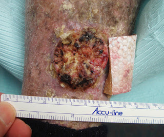
HPI:  The patient is a 69 yo man with a greater than 40 year history of severe psoriasis.  He was a research subject at a prestigious university hospital for many years.  Over that time, he has received PUVA, methotrexate and an investigational drug, Rapamycin.  He has been treated with NB UVB and acitretin with some improvement.  As a result of his therapies, he has developed numerous squamous cell carcinomas.  These pose therapeutic challenges They are painful, foul smelling and difficult for the patient to care for.  An additional problem is occasionally disabling trigeminal neuralgia.  He has had many well-differentiated SCCs excised over the past decade.  He has atrial fibrillation, Factor 5 deficiency and is on warfarin and a host of cardiac medication.

O/E:  Wide-spread erythema and scale covering entire integument except head and neck.  There is a 3 cm exophytic tumor of the left pre-tibial area and smaller similar tumors on right leg, back and chest.

Clinical Photos (6.6.17)
L. Pretibial
Diagnosis:  Generalized psoriasis, Squamous cell carcinomas

Questions:  Should these lesions be excised? Should we consider  one of the new targeted therapies?  Do any of our members have expertise with them?  This man has a number of co-morbidities which may make therapy more difficult.

References:

Oral therapy for nonmelanoma skin cancer in patients with advanced disease and large tumor burden: a review of the literature with focus on a new generation of targeted therapies.
Rudnick EW, Thareja S, Cherpelis B. Int J Dermatol. 2016 Mar;55(3):249-58
Author information
Abstract: This review of the literature aims to describe previous and current treatment options for oral therapy in locally advanced and metastatic NMSC otherwise unamenable to standard treatment. Oral Smoothened (Smo) inhibitors Vismodegib, Sonidegib, and Taladegib have shown to be effective in several trials. Oral tyrosine kinase inhibitors Erlotinib and Gefitinib, which target epidermal growth factor receptor (EGFR), have early supporting data and are currently undergoing large multicenter trials. Oral therapy in NMSC is useful in high risk patients with recurrent and aggressive disease who may not tolerate other systemic therapies.

Thursday, April 06, 2017

78 yo man with scalp lesions

Abstract:  78 yo man with 1 year history of scalp lesions.

HPI:
May 9,2016:  Cryotherapy 4 keratoses on vertex of scalp
August 11, 2016: Crusted lesions at site of cryotherapy.  Clinical diagnosos of erosive pustular dermatosis of scalp made.  Treated with mupirocin oint and clobeyasol ointment  Initially improved.
Sept 9 2016:  Resolved
10/24/2016: Continued to do well
April 4, 2017: Recurrent lesions on vertex of scalp.  Thick crusted lesions (see photo)  The crusts were brownish and dirty looking, but unfortunately I removed them before taking the photo of April, 2017.

O/E:  What was initially hypertrophic keratosit papules were transformed into ~ 1 cm crusted erosive lesions.

Clinical  Photos:
Note pustule
4.4.17 (after crusts removed)
s/p 1 week Chlorhexidine, 4 days tacrolimus 0.1%

Lab:  August 11, 2016: + Staph aureus from lesion Vx scalp - usual sensitivities

Diagnosis:
This is either erosive pustular dermatosis of the scalp or squamous cell ca.  The rapid worsening since cryotherapy suggests the former.  It appears that biopsy may be necessary.  Not all cases of EPD respond to clobetasol ointment.

Questions:  Is this EPD or are these lesions squamous cell carcinomas.  Patient is reluctant to have a biopsy done. This process appears to have been gtriggered by the trauma of liquid nitrogen and did respond initially to clobetasol.  10 - 20% of EPD cases appear to be non-responders ro clobetasol.

Follow-up.  Marked improvement following chlorhexidine wash daily and topical tacrolimus 0.1% ointment at the suggestion of a colleague.  He recurred after clobetasol ointment.

References:
1. Erosive pustular dermatosis of the scalp: Clinical, trichoscopic, and histopathologic features of 20 cases.  [Current and thorough review]
Starace M, et. al.: J Am Acad Dermatol. 2017 Feb 14.
BACKGROUND: Erosive pustular dermatosis of the scalp is a chronic eruption that leads to scarring alopecia.
OBJECTIVE: The clinical, dermoscopic, and histopathological features and the course of the disease in 20 patients were reviewed and compared with the reports in the literature.
RESULTS:The mean age was 59.4 years. Androgenetic alopecia was present in 12 patients, 6 of whom showed actinic damage. Trauma was reported in 9 patients. Four patients were affected by autoimmune disorders. The vertex was the most common location. In all 20 patients trichoscopy showed an absence of follicular ostia with skin atrophy. Histopathology revealed 3 different features, depending on the disease duration. A reduction of inflammatory signs was observed in 14 patients treated with topical steroids and in all 3 patients treated with topical tacrolimus 0.1%.
CONCLUSIONS: The relatively high number of patients allowed us to identify a better diagnostic approach, using trichoscopy, and a more effective therapeutic strategy, with high-potency steroids or tacrolimus, which should be considered as first-line treatment.

2.  Disseminated Erosive Pustular Dermatosis also Involving the Mucosa: Successful Treatment with Oral Dapsone (Free Full Text)
Jamison D. Feramisco.  Acta Derm Venereol. 2012 Jan; 92(1): 91–92.

3.  Erosive pustular dermatosis of the scalp: a review with a focus on dapsone therapy.
Broussard KC. J Am Acad Dermatol. 2012 Apr;66(4):680-6
Abstract
BACKGROUND: Erosive pustular dermatosis of the scalp (EPDS) is an inflammatory disorder of unknown origin characterized by pustules, erosions, and crusting in areas of alopecia that tend to be atrophic, actinically damaged, or both. The most common treatments reported include antibiotics and topical anti-inflammatories, which can be ineffective. In the search for effective treatment for EPDS, we share our experience with topical dapsone 5% gel.
OBSERVATIONS:We present 4 patients with EPDS, all with classic clinical presentations and histologic findings of EPDS, who had failed a variety of treatments including oral, intralesional, or topical steroids, tacrolimus, and antibiotics. All patients demonstrated rapid improvement or resolution with topical dapsone 5% gel.
CONCLUSION: Our observations demonstrate topical dapsone 5% gel to be a novel, safe, and efficacious therapeutic alternative for mild to moderate EPDS.

Tuesday, September 06, 2016

Postiive Band-Aid Sign

The patient is a 77 y.o. man who presented with a number of skin lesions.  He has a past history of non-melanoma skin cancer.

The lesion in question has been present for a few months.  It is an almost 5 cm in diameter exophytic tumor.

Diagnosis:  Probable Squamous Cell Carcinoma.

I anesthetized the lesion and shaved it off.  There was a fair amount of bleeding.  I curretted it and cautrized the base. It was not as soft as a typical SCC or BCC.  Specimen submitted and I'll attach a follow-up with the path.

Pathology:  Well-differentiated squamous cell carcinoma

This is a particularly good example of the "Positive-Band Aid" sign.  Most of us know this, but it has not been well-reported in the literature.  We presented this sign on the VGRD Blog in 2007.

Saturday, July 02, 2016

What is Right Care?


The patient is a 90 year-old man, homebound with a dementia.  His 87 year-old wife is a steadfast, loving and loyal caregiver.  His dermatologist has made house calls for the past four months. 

There are non-melanoma skin cancers on the left cheek  and mid upper lip.  The former lesion has increased from 1.3 to 1.5 mm in diameter and the the lip lesion has increased from 1.8 to 2.2 mm in diameter in the past two months.  Both are somewhat inflammatory and crusted. He picks on the lip lesion, but because of his dementia he can not articulate what it is that bothers him.
7.28/15
March 2016
6.28.2016

8.11.16
Thoughts: The question is what is the best treatment for this man.  The lesion on the left cheek could be curetted and desiccated in the office. The lesion on the lip is a more complicated problem. 

A trial of topical 5-FU plus imiquimod may be helpful, especially for the lesion on the lip, as a palliative procedure.  The lesion on the malar eminence which grew rapidly ~ 6 months ago and is either a squamous cell or a keratoacanthoma) could be curetted and dessicated.

The patient can not make a decision for himself and his wife wants to just watch these lesions.  She understands that treatment is not likely impact on his quality of life at this point and want’s to spare him the trauma or surgery.

As physicians, we feel compelled to “do something.”  Is this the right time to “not just do something, but to sit there.”

Reference:
1. Linos E. Treatment of nonfatal conditions at the end of life: nonmelanoma skin cancer.  JAMA Intern Med. 2013 Jun 10;173(11):1006-12
CONCLUSIONS AND RELEVANCE: Most NMSCs are treated surgically, regardless of the patient's life expectancy. Given the very low tumor recurrence rates and high mortality from causes unrelated to NMSC in patients with limited life expectancy, clinicians should consider whether these patients would prefer less invasive treatment strategies.  PubMed.  PMC Free Full Text.

2.  Knocking on Heaven’s Door by Katie Butler is an honest, sobering book that describes what awaits so many elderly people and their caregivers, who are often family members.  It is relevant to how one manages a patient such as the man described and discussed here.

Sunday, August 02, 2015

To Treat or Not to Treat: that is the question


Elani Linos and colleagues wrote a milestone paper on the treatment of nonmelanoma skin cancer (NMSC) that was published in JAMA – Internal Medicine in June 2013. In it, they stated:

“Nonmelanoma skin cancer (NMSC) is the most common cancer and predominantly affects older patients. Because NMSCs do not typically affect survival or short-term quality of life, the decision about whether and how to treat patients with limited life expectancy (LLE) is challenging, especially for asymptomatic tumors.

“The current standard of care in the United States is to treat NMSCs, and no guidelines exist about whether physicians should consider patient age or functional status in choosing treatments.  Treatment decisions for patients with NMSC with LLE require consideration that the benefits of treatment may not occur within the patient's
remaining life span, but any risks are immediate.”

We saw two such patients recently in our dermatology practice.  They are presented for your thoughts and discussion.

1. The patient is a 94 yo woman, status post CVA (12/24/13) with right hemiparesis.  She has a two year history of a rodent ulcer on the right nasolabial fold measuring 2.4 x 1.4 cm.  It itches and she picks it.  Biopsy shows “infiltrating basal cell carcinioma.”  She is a retired executive secretary, never married with no close relatives nearby.  Mentally, she is alert and oriented.  We discussed active surveillance, surgery and radiotherapy.  She is confined in a nursing home and was not keen on having XRT considering the number of treatments.


2.  This 89 yo man has a tumor of the mid upper lip for ~ 10 months.  The 1.4 cm in diameter lesion is firm with rolled borders.  Clinically, this is BCC, but it has not yet been biopsied.  His general health is good, but he has moderately advanced dementia and lives independently with his wife.  The couple have children who live at some remove.  We discussed active surveillance, XRT and surgery.  The latter would be fairly simple; but we recognize that the tumor may not ever significantly impact on his quality of life or longevity.


Discussion:  Both of these lesions could be treated or watched.  Lesion # 2 would be easy to excise and that may make management easier.  Excision of lesion #1 would entail a long trip for micrographic surgery which is difficult logistically.  In our opinion, how to proceed with these cases is a value judgement and input from the patient and/or the family is important.

Dr. Linos’ article (1) is helpful but each case presents unique management quandaries.  It has been said that “often it is more important to treat the patient with the disease, than it is to treat the disease the patient has.”  These two cases are examples of this conundrum.

An additional thought:   Topical imiquimod can be helpful in the management of superficial and nodular basal cell carcinomas.(2)  The marked inflammatory response is often difficult for patients to tolerate, but less frequent applications may allow for palliation and slowing of tumor progression.

You thoughts will be appreciated.

 Reference:
1. Treatment of nonfatal conditions at the end of life: nonmelanoma skin cancer.  Linos E, Parvataneni R, Stuart SE, Boscardin WJ, Landefeld CS, Chren MM.  JAMA Intern Med. 2013 Jun 10;173(11):1006-12.
Available Free Full Text.

2.  Surgical excision versus imiquimod 5% cream for nodular and superficial basal-cell carcinoma (SINS): a multicentre, non-inferiority, randomised controlled trial.
Bath-Hextall F, et. al.  Lancet Oncol. 2014 Jan;15(1):96-105.


Wednesday, February 12, 2014

Prurigo Nodularis with Squamous Cell Carcinioma


Abstract: 63 yo man with 10 month history of intense pruritus and excoriated papules and nodules

HPI:   This 63 yo retired radio announcer presents with a 10 month history of intense pruritus and excoriated papules and nodules. He is in reasonable health.  Medications include Welbutrin (bupropion) and occasional prednisone for his itching.  He's tried topical steroids and anti-histamines without relief.  Smokes ~ 5 cigarettes a day.

O/E: Skin thin from actinic damage.  There are excoriated papules and nodules on the torso and extremities.  There are two or three more exophytic lesions.

Clinical Photos:


Lab:  CBC, chemistries normal.  IgE 867 IU/Ml

Pathology:  Initial bx signed out as SCC.  Since he has scores of lesions repeat biopsies of an early and more developed lesion were taken.  Thanks to Dr. Lynne Goldberg (Boston University Skin Path) for the beautiful photomics.
Prurigo Nodularis


Well_differentiated Squamous Cell Carcinoma


Diagnosis: Prurigo Nodularis with Squamous Cell Carcinoma

Discussion and Questions:The association of SCC with Prurigo Nodularis has only been reported one time (ref 5).  Yet we do not feel this is a chance association.  There are also some articles about P.n. and KA in the literature.
Has anyone seen a similar case?  He will be treated with gabapentin and followed. A follow-up will be posted in a month or so.  The SCCs will not be re:excised at this time.
Thaldomide has been recommended for P.N. in the literature, however, it is now > $10,000 per month!

References:
1. Journal of the American Academy of Dermatology
Volume 69, Issue 3, Pages 426-430, September 2013
Keratoacanthomas arising in association with prurigo nodules in pruritic, actinically damaged skin
Timothy P. Wu, BA, Kristen Miller, MD, David E. Cohen, MD, Jennifer A. Stein, MD, PhD  jennifer.Stein@nyumc.org

2. J Clin Pharm Ther. 2013 Feb;38(1):16-8. doi: 10.1111/jcpt.12005. Epub 2012 Sep 26.
Treatment of prurigo nodularis with pregabalin.
Mazza M, Guerriero G, Marano G, Janiri L, Bria P, Mazza S.

3. Dermatol Ther. 2010 Mar-Apr;23(2):194-8. doi: 10.1111/j.1529-8019.2010.01314.x.
Therapeutic hotline: Treatment of prurigo nodularis and lichen simplex chronicus with gabapentin.
Gencoglan G, Inanir I, Gunduz K.  (no real data on patient background)

4. Eur J Dermatol. 2008 Jan-Feb;18(1):85-6. Epub 2007 Dec 18.
Gabapentin for the treatment of recalcitrant chronic prurigo nodularis.
Dereli T, Karaca N, Inanir I, Oztürk G.  Available Free Full Text.

5.  Saudi Med J. 2000 Mar;21(3):300-1.
Squamous cell carcinoma complicating prurigo nodularis.
Al-Waiz MM, Maluki AH.
Abstract:  Squamous cell carcinoma complicating ulcerative prurigo nodularis is described in 2 patients who were having prurigo nodularis on dorsum of the feet for duration of many years. Biopsy specimens from the ulcerating nodules showed features of squamous cell carcinoma. This finding has not been previously reported. Squamous cell carcinoma should be considered in the evaluation of long standing ulcerative lesion of prurigo nodularis especially when not responding to conventional therapy.

Wednesday, May 30, 2012

Skin Cancer in Renal Transplant Patient

The patient is a 64 yo man who received a renal transplant x years ago and is maintained on prednisone and Prograf.  He presented with a 4 cm biopsy proven superficial squamous cell carcinoma on the left parietal scalp.  This lesion would have necessitated a large micrographic surgical procedure with a graft.

An attempt was made to treat with topical chemotherapy.  Imiquimod was inititiated, but there was only minimal response after two weeks.  Five fluorouracil was then added and this achieved a moderate response.  The combination of imiquimod/5FU was continued for a total of six weeks, then stopped.  One month later there appears to be a clinical cure.  He will be followed closely.  

There is a possibility that this combination therapy can help selected transplant patients with low risk superficial nonmelanoma skin cancers.

Clinical Photos:
After Six Weeks Imiquimod/5FU


One month after Stopping Imiquimod/5FU


Comment:  This treatment made me a little nervous, but the surgical approach would have been major for a lesion that had only a small chance of of metastasizing.  The benefits and risks were discussed with the patient; however, his oncologist was unhappy about this approach.