Saturday, January 27, 2007

A Common Entity


How many times have you experienced this?
The waiting room is full and a patient comes in, settles down comfortably and says, "I won't take up much of your time today, I see how busy you are. So I wrote down a few questions." Thus spake a healthy 55 year old man as he sat down and pulled out this post-it from his pocket. "La Maladie du Petit Papier," I thought with resignation.

PubMed has only five references to this entity we all know. The first in Italian from 1967. * We each have our own way of handling this. Some docs grab the list from the patient and say, ""Let's have a look at this." Others say, we'll deal with only three today; you choose." I usually let them read all of them and then proceed.

How do you handle this event? A couple of years ago there was a piece in the BMJ comparing the malady of the petit papier with the newly emerging la maladie du grand printout. The latter may be more common in the major cities of North America than elsewhere.

* Iandolo C. [The "petit papier" sign] [Article in Italian]
Policlinico [Prat]. 1967 Mar 6;74(10):321-8.

Wednesday, January 24, 2007

Mystery from Micronesia

We received this note from a physician in Hawaii. Your thoughts will be appreciated.

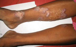
"Two years ago, I was shown photos of a severe skin condition which afflicted more than 80 persons on the tiny atoll of Satowan in Chuuk. Now, more than 100 persons on Satowan are affected and about 10 on the neighboring atoll of Lekinioch.

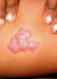
Affected persons develop thick, pink plaques, usually on the arms and legs – this appearance resulted in the nickname “Spam.” While the condition is not life-threatening, the people with the condition are sometimes ostracized and made to feel shame and embarrassment. Anecdotally, the plaques are more common in those who work or play outdoors (particularly taro farmers) and may sometimes occur following skin trauma.

The only treatment used on the plaques has been surgical debridement. Dr. Bosco Buliche who practices in Weno, the capital of Chuuk, has seen and photographed cases on Satowan. He has posted 5 cases on the Pacific Island Healthcare Project website (through TAMC), including one with biopsy results, which showed only nonspecific inflammation – we could transport only a fixed specimen, unfortunately. Opinions from various practitioners on the cause of the condition have run the gamut from “island psoriasis” to mycobacterial infection.

So, two years later, there is still no answer. It would be wonderful to find someone with the interest and resources to investigate the problem. Suggestions will be most welcome."

Clinical Photos:


Friday, January 19, 2007

Stranger than Fiction

Abstract:
50 yo man with unusual facial eruption

Presented by:
DJ Elpern

History:
This 50 yo man develops an eruption on his left malar eminence annually. This occurs after his late December deer hunting trip. He is exposed to cold air. Does not take any new meds at that time. The eruption has a burning sensation.

Exam:
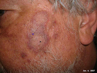
Annular plaques with central pallor and peripheral dusky red color left malar. No other similar lesions.

Clinical Photos:
"X"marks the biopsy sites


Lab and Path:

DIAGNOSIS: Tumid lupus erythematosus
NOTE : The specimen exhibits flattened epidermis , mild to moderate papillary dermal edema, dilated superficial blood vessels and a moderate superficial and deep perivascular and periappendageal lymphocytic infiltrate with numerous extravasated erythrocytes and marked superficial and deep interstitial mucin deposition. These findings support the histologic diagnosis . The differential diagnosis includes , in the appropriate clinical setting , lymphocytic infiltrate of Jessner. P.A.S. stain is negative for fungal hyphae and basement membrane zone thickening. Clinico-pathologic correlation is suggested.


Therapy and Course:
The patient was biopsied and seen a week later for suture removal. No treatment was rendered. Here is how he looked at day six post biopsy.


Questions and Teaching Points:
Histologically this is lupus. However, the patient is asymptomatic and the lesions cleared without therapy. The cold exposure is most likely key. He may have a form of L.E. There is an entity called chilblain lupus which could fit here. Most of those patients had SLE. Has anyone seen a similar case? I will suggest serologies to him. He does not need any therapy at this time.

References
This and other references can be found in PubMed if "lupus chilblain cold" is searched.

Franceschini F, et. al.
Chilblain lupus erythematosus is associated with antibodies to SSA/Ro.
Adv Exp Med Biol. 1999;455:167-71.

Clinical Immunology Unit, Spedali Civili, Brescia, Italy.

Chillblain Lupus Erythematosus (CL) of Hutchinson is a subtype of Lupus Erythematosus characterized by erythematous lesions symmetrically distributed on the face, nose, fingers and toes, knees and heels. The lesions are induced by cold, damp climates. A number of patients affected by CL eventually develop features of Systemic Lupus Erythematosus (SLE). We report here 7 patients, all but one affected by SLE, with chilblain cutaneous lesions on their hands, feet and face. The onset of CL preceded the diagnosis of SLE, from 1 to 10 years in 3 cases, it was concurrent in one case and was subsequent in the other 2 cases. Six out of the seven patients referred typical Raynaud's phenomenon and one had acrocyanosis. CL lesions developed and were aggravated by the cold during autumn and winter, they improved during summer. Skin biopsy performed in 5 patients from the lesions showed, on histology, a typical pattern of alterations with granular deposits at the dermo-epidermal junction on direct immunofluorescence. Laboratory findings showed: ANA and anti-SSA/Ro were detected in all the patients, anti-SSA/Ro were isolated in 4 patients and associated with anti-Sm in one case, anti-U1 RNP in one case and with anti-Sm and anti-RNP in a third case. Complement consumption was observed in 5 patients, anti-dsDNA in the six patients with SLE, hypergammaglobulinemia in 4 and rheumatoid factor in one. The fine specificity of anti-SSA/Ro as determined by immunoblotting using a human spleen extract as a substrate, showed: anti-60kD and anti-52 kD in two sera, anti-60kD isolated in 2 sera, anti-52kD isolated in one serum (from the patient without SLE) while 2 sera did not blotted. In conclusion, our study confirms the previous report of anti-SSA/Ro antibodies in association with CL. This clinical and serologic association widens the spectrum of cutaneous disease that is associated with antibodies to SSA/Ro to include conditions such as to SCLE, hypergammaglobulinemic purpura and neonatal lupus.

Wednesday, January 10, 2007

Sunday, January 07, 2007

Imiquimod + 5FU

Abstract 62 yo man with SCIN of finger. Failed Imiquimod and responded to combination of 5% 5FU + Imiquimod

Presented by: D.J. Elpern, Williamstown, Massachusetts, USA

History This 62 yo man had a 15 year history of a plaque on his left index finger. He had been told it was a wart in the past.

Exam
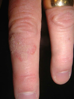
2.0 cm in diameter verrucous plaque.

Clinical Photos


Lab and Path
Because of long history a biopsy was done to r/o SCC. The path showed in situ squamous cell carcinoma.

Therapy:
We discussed micrographic surgery vs. topical chemotherapy and he elected the latter. After two weeks of nightly imiquimod there was no reaction. I then added 5% 5FU cream as described in a recent article. Within two weeks the area was ulcerated and painful. The treatment was stopped and he will have a repeat biopsy to test for cure in 2 - 3 months.

Questions and Teaching Points The combination of 5FU and imiquimod is interesting and will be a therapeutic advance, but like imiquimod, it will take some time to master the "Art." I was surprised at the intensity of this reaction. I suspect he may have done better if I used the 5FU three times a week. Your comments are welcome

References
J Am Acad Dermatol. 2006 Dec;55(6):1092-4.

Topical combination therapy for cutaneous squamous cell carcinoma in situ with5-fluorouracil cream and imiquimod cream in patients who have failed topical monotherapy.

Ondo AL, Mings SM, Pestak RM, Shanler SD.

Topical therapeutic options for cutaneous squamous cell carcinoma in situ include 5-fluorouracil cream and imiquimod cream. Such treatment may be preferable to surgical or destructive modalities in certain anatomic locations and in instances where patients are unwilling or poor surgical candidates. We present 4 such patients with cutaneous squamous cell carcinoma in situ involving a digit. Each patient failed treatment with imiquimod cream as monotherapy. In addition, two patients failed treatment with 5-fluorouracil cream as monotherapy. All 4 responded completely to 5-fluorouracil and imiquimod cream as combination therapy. In patients who have failed monotherapy with a topical agent for cutaneous squamous cell carcinoma in situ, combination treatment using both topical 5-fluorouracil cream and imiquimod cream may be considered as an alternative therapeutic strategy.

Thursday, January 04, 2007

Deadly Allopurinol Hypersensitivity Syndrome


The patient was a 59-year-old man who presented with 3-week history of generalised skin eruptions associated with high fever. He was well previously and was started on allopurinol for asymptomatic hyperuriacemia. About 3 weeks later, he started feeling unwell and giddy. Then he developed a generalised erythematous eruptions on the lower legs which then spread to the trunk and face. He was admitted at the general hospital but discharged five days later. He persisted feeling unwell despite discharged from the ward. The skin eruptions worsened.

On examination he was febrile. Temp: 38.5 degC He appeared jaundiced and sallow. There was marked pitting ankle edema. Generalised erythema all over the face, trunk and extremties were noted. Diffuse scaling was noted on te face, neck and upper limbs. There was no hepatosplenomegaly.

Clinical diagnosis: Allopurinol Hypersensitivity Syndrome

Blood urea was 27.6mmol/l and creatinine 9.3mmol/l

SGPT 242 mmol/l
SGOT 115 mmol/l
Alk Po4ase 567 mmol/l
Bilirubin 87.7 umol/l

Hb7.8gm%
TWBC 18 000

Immediately the allopurinol was stopped and he was dialysed and started on IV hydrocortisone 200mg 6hrly. IV ciprofloxacin 750mg bd was initiated was empirical treatment for possible septicemia. He responded well but when the IV hydrocortisone was tailed down, his condition deteriorated. His jaundice became deepened and he became more drowsy. We increased the dose of IV hydrocortisone and withhold other drugs as well - there may be some cross reaction. IV albumin was transfused. He improved and became more alert. CT Brain and ultrasound abdomen was unremarkable. We hope the supportive therapy can pull him through.

Drug hypersensitivity syndrome is a severe idiosyncratic reaction associated with taking drugs. Other names are "drug rash with eosinophilia and systemic symptoms" (DRESS) and "drug induced delayed multiorgan hypersensitivity syndrome" (DIDMOHS). The most common triggering agents are antiepileptic drugs (phenytoin, phenobarbital, and carbamazapine), sulphonamides, and allopurinol.

The exact mechanism for the development of allopurinol hypersensitivity syndrome is unknown; the pathological substrate is often a diffuse vasculitis induced by a type III hypersensitivity reaction, with formation of immune complexes that precipitate in vascular endothelium and promote an inflammatory reaction. Accumulation of oxypurinol (the principal metabolite of allopurinol) in renal insufficiency is considered a crucial factor for the development of allopurinol hypersensitivity syndrome and may lead to tissue damage by toxic or immunological mechanisms.

References:
Alfonso Gutiérrez-Macías, Eva Lizarralde-Palacios, Pedro Martínez-Odriozola, Felipe Miguel-De la Villa Clinical reviewLesson of the week. Fatal allopurinol hypersensitivity syndrome after treatment of asymptomatic hyperuricaemia BMJ 2005;331:623-624

Y C Chan, Y K Tay,S K Ng Allopurinol Hypersensitivity Syndrome and Acute Myocardial Infarction— Two Case Reports. Ann Acad Med Singapore 2002; 31:231-3

Wednesday, January 03, 2007

Unique Drug Eruption

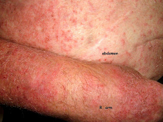
This 83 yo old man has chronic renal failure with a creatinine of 18 (!!) but does not need dialysis yet. His hemoglobin has been maintained with Procrit for the past four years. Because of a low transferrin saturation (30 %), his nephrologist added a new form of iron infusion around 2 months ago. (This was in another state and I don't have the name of the product) Within two weeks he started developing a generalized pruritic eruption. He stopped the iron infusions, but the rash persists.

There is only one report of a similar process (see below); but it is likely that more cases are extant as this therapy becomes more popular.
For the time being, he will be treated with triamcinalone 0.1% ointment after soaking in a tub. This is William James et. al's "Soak and Smear" technique. (see ref)

It's likely that the iron will stay in his system for some time. There are no treatment guidelines here; and as the patient can sleep at night, I prefer not to give him antihistamines at this time.



Reference

1. The safety and efficacy of ferumoxytol therapy in anemic chronic kidney disease patients.
Spinowitz BS, Schwenk MH,Jacobs PM et al
Kidney Int. 2005 Oct;68(4):1801-7.

BACKGROUND: Administration of safe and effective iron therapy in patients with chronic kidney disease is a time consuming process. This phase II clinical trial studied ferumoxytol, a semi-synthetic carbohydrate-coated iron oxide administered by rapid intravenous injection to anemic chronic kidney disease patients (predialysis or undergoing peritoneal dialysis). METHODS: Inclusion criteria included hemoglobin < or =12.5 g/dL and transferrin saturation < or =35%. Twenty-one adult patients were randomized to receive ferumoxytol in a regimen of 4 doses of 255 mg iron in 2 weeks or 2 doses of 510 mg iron in 1 to 2 weeks. Ferumoxytol was administered at a rate of up to 30 mg iron/sec. RESULTS: The maximum hemoglobin response following ferumoxytol administration occurred at 6 weeks, increasing from a baseline of 10.4 +/- 1.3 g/dL to 11.4 +/- 1.2 g/dL (P < 0.05). Ferritin increased from a baseline of 232 +/- 216 ng/mL to a maximum of 931 +/- 361 ng/mL at 2 weeks (P < 0.05), while the baseline transferrin saturation increased from 21 +/- 10% to 37 +/- 22% at 1 week (P < 0.05). Seven adverse events in 5 patients during this trial were deemed possibly related to ferumoxytol, none serious. These events included constipation, chills, tingling, a gastrointestinal viral syndrome, delayed pruritic erythematous rash, and transient pain at the injection site. CONCLUSION: Although larger studies are required, this small study demonstrates that ferumoxytol can be safe and effective in increasing iron stores, is associated with an increased hemoglobin response, and is well tolerated at a rapid infusion rate.

2. Soak and smear: a standard technique revisited. Gutman AB, Kligman AM, Sciacca J, Arch Dermatol. 2005 Dec;141(12):1556-9

BACKGROUND: Atopic dermatitis, nummular eczema, chronic hand dermatitis, palmar plantar psoriasis, and xerotic eczema are common inflammatory skin conditions. They may be refractory to conventional topical and even systemic treatment. Little evidence is available that demonstrates the benefits of aggressive topical treatment of patients with these disorders. OBJECTIVE: To describe a simple, inexpensive, effective topical treatment with an accompanying patient educational sheet. DESIGN: A retrospective study of 28 patients referred to a tertiary care center for refractory chronic pruritic eruptions. Intervention with a plain water 20-minute soak followed by smearing of mid-strength to high-strength corticosteroid ointment led to clearing or dramatic improvement. RESULTS: Objective and symptomatic improvement was obtained from aggressive topical treatment. It was well accepted in this group of referral patients. CONCLUSIONS: Hydration for 20 minutes before bedtime followed by ointment application to wet skin and alteration of cleansing habits is an effective method for caring for several common skin conditions. Prospective studies are needed to further validate these findings.

Monday, January 01, 2007

VGRD 2.0

"And gladly wolde he lerne and gladly teche" Chaucer

Welcome to VGRD 2.0

This updated version of VGRD will allow more interactive postings by dermatologists. Virtual Grand Rounds in Dermatology (VGRD) was formed in 2000 to serve as a place for dermatologists the world over to meet one another and share interesting and challenging patients. One may want to ask a question about diagnosis or therapy, present an interesting clinical photo or post a photo or photomicrograph. We are a group of clinical and academic dermatologists who believe that this form of web-based teledermatology can be both personally and professionally enriching. VGRD has hundreds of dermatologists on our mailing list and they cover all of the subspecialties in dermatology.

We would particularly like to invite dermatologists from both developing and developed countries to join us and share your experiences with our members. Together we will be able to provide better care for our patients.

We think you will find the new case presentation format to be clear and easy to follow. If you have suggestions on how we can improve it, please let us know.

We hope this form of electronic communication and teledermatology through shared knowledge will help to enhance our patient care. Your participation will help to make VGRD successful.


Yours sincerely,

David Elpern MD
The Skin Clinic
Williamstown, MA, USA
djelpern@gmail.com

Henry Foong FRCP
Foong Skin Specialist Clinic
Ipoh, Malaysia
bbfoong@gmail.com