Presented by Yoon Cohen MS IV, University of New England, Biddeford, Maine and David Elpern MD, Williamstown, Massachusetts
HPI: This healthy 68 yo woman with type II skin presented to the clinic with 4-6 months history of an atypical melanocytic lesion on the left knee. She had noticed an increase in size and change in color and was concerned about these changes in the lesion.
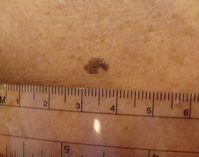
O/E: There was a 7 mm in a diameter asymmetrical brownish macule on the left knee. The lesion showed an irregular border with varied colors.
Clinical photographs:

Dermatopathology report:
The specimen exhibits a proliferation of moderate to severely atypical melanocytes distribubted in irregular nests, as well as singly at and above the dermal epidermal junction, with pagetoid spread to the granular layer and near confluence over at least three rete ridges. These findings support the histologic diagnosis of melanoma-in-situ.
Our appreciation to Dr. Deon Wolpowitz, MD from Boston Univeresity, Dermatopathology, for providing these photomicrographs for the case.
4X

10X
20X
20X
Diagnosis:
Malignant melanoma in situ
Discussion:
The BLINCK approach:
We followed the BLINCK checklist, introduced by Dr. Peter Bourne, a founder of the Skin Cancer College of Australia and New Zealand (SCCANZ). The score for the lesion was added up to 4 by criteria as following.
1. B. The lesion was not clearly benign at our first initial evaluation
2. L. The lesion appeared to be lonely without any other similar melanocytic lesion near by
3. I. The lesion appeared to be irregular outline and color on our dermoscopic exam
4. N & C. The patient was nervous about the change in color in past 4-6 months
5. K. The lesion exhibited known clues when viewed with a dermatoscope. See "Chaos and Clues" reference below.
BLINCK Score = 4
BLINCK Score = 4
According to the BLINCK approach, a lesion should be biopsied if the BLINCK score is 2 or more out of a possible 4. Therefore, the we excised this lesion and sent for a pathologic evaluation.
According to Dr. Bourne, the BLINCK approach is presented as a simple method to assist the clinician with the decision of whether to biopsy a skin lesion or not. The use of this algorithm will improve the pickup rate of potentially serious skin cancers as well as reduce the number of unnecessary benign lesion excisions. BLINCK may be especially helpful to clinicians who have only basic or intermediate dermoscopy skills but who are regularly called upon to assess skin lesions in their practices.
Questions:
1. Would you consider using the BLINCK approach at your practice?
2. If you already have adapted the BLINCK approach, how have your experiences been?
References:
1. McColl I. BLINCK. http://idsblinck.blogspot.com/2009/11/blinck.html. Updated November 19, 2009. Accessed September 7, 2010
2. Rosendahl C, Kittler H, Cameron A, et al. CHAOS & CLUES - The Algorithm. http://www.chaosandclues.blogspot.com. Updated November 19, 2009. Accessed September 7, 2010


