Thursday, October 17, 2019

21 year-old woman with solitary eschar


This 21 year-old college student presented with a 5 week history of an evolving lesion on the right leg.  She is in good health and takes no medications by mouth.  The lesion started with pruritus and pain and a solitary evolving bulla on her right leg.  She had walked through a wooded area the night before this developed. It has evolved into a dry eschar.  She has a history of a DVT on her right leg 2 years ago after tonsillectomy, bed rest and a long plane trip while on oral contraceptives.  To date, she has been treated with mupirocin ointment and a topical corticosteroid.

O/E: When seen there was a solitary 2 cm eschar on her right leg.  No erythema, no purulence.

Photos:
September 8, 2019 a.m.

September 8, 2019 p.m.


September 9, 2019


September 28, 2019


October 9, 2019

October 16, 2019 (Date of visit)


Labs: Pending

Diagnosis: Eschar.  Etiologic considerations:
Envenomation – Brown Recluse Spider Bite
Echthyma gangrenosum
Pyoderma gangrenosum (Antiphospholipid syndrome)

A lesion such as this in a young healthy immunocompetent woman suggests an antecedent insult such as a brown recluse spider bite, but we have no history to confirm that.  She is being worked up for underlying disorders that might predispose to echthyma.  However the antecedent DVT makes one consider an underlying problem such as the antiphospholipid syndrome.

Questions:
1.  What diagnoses do you entertain?
2.  At this time, what therapies do you recommend?

About Hydrocolloid Dressings.
1. Background.
2. Another useful resource on hydrogels.
3. Video Demonstration.


Reference:
The rash that leads to eschar formation. Dunn C, Rosen T.
Clin Dermatol. 2019 Mar - Apr;37(2):99-108. Author information
Abstract:  When confronted with an existent or evolving eschar, the history is often the most important factor used to put the lesion into proper context. Determining whether the patient has a past medical history of significance, such as renal failure or diabetes mellitus, exposure to dead or live wildlife, or underwent a recent surgical procedure, can help differentiate between many etiologies of eschars. Similarly, the patient's overall clinical condition and the presence or absence of fever can allow infectious processes to be differentiated from other causes. This contribution is intended to help dermatologists identify and manage these various dermatologic conditions, as well as provide an algorithm that can be utilized when approaching a patient presenting with an eschar.  Full Text.

Thursday, September 26, 2019

73 year-old woman with wide-spread plaques

The patient is a 73-year-old woman with a two month history of an eruption that began on the buttocks and thighs. It has spread to the arms. The clinical picture was not diagnostic, so biopsies have been done.

She is in her usual state of health.  There is no history of systemic illness. Her medications include: amlodopine, metooprolol, ASA, all for a number of years.  She had a tetanus booster a week before the onset of the rash.

I thought this would probably be indolent but she has has developed marked pruritus.  Because of her symptoms she was treated with fluocinolnide oinment. This had no effect. Doxycycline was not tolerated due to GI symptoms.
Laboratory studies were done. CBC and chemistries were within normal limits. Her Lyme tighter was negative.

Examination shows large plaques on the buttocks and thighs that they are now appearing on the arms. The remainder of the examination is unremarkable. 
Clinical Images:

Pathology:
Dermal interstitial proliferation of histiocytes with focally increased dermal mucin and increased dermal mucin.  Individual collagen fibers are circumferentially ringed with histiocytes.  The dermatopathologist  feels this is either interstitial granuloma annulare or interstitial granulomatous dermatitis.  Images courtesy of Lynne Goldberg, Boston University Skin Path.

 Diagnosis: Interstitial granuloma annulare versus interstitial granulomatous dermatitis.
There is one reference on PubMed to granuloma annulare following DT vaccination.

Reference:
1. A case of granuloma annulare in a child following tetanus and diphtheria toxoid vaccination.
Baskan EB, et. al. J Eur Acad Dermatol Venereol. 2005 Sep;19(5):639-40


What are your thoughts?

Hypopigmented Macules in a Child

The patient is a 4 1/2-year-old boy who is seen today for evaluation of hypopigmented macules on the arms and legs for less than a week. His parents first notice this four or five days ago. He has been in his usual state of health although two days ago he developed a fever to 104.7  and was seen by his pediatrician who felt it was a viral syndrome. Throat culture was negative for strep.

On examination: This is a healthy appearing child. He does have a raspy cough. He has 5 to 7 mm in diameter hyperpigmented macules scattered over the arms and legs. Some of these lesions have a so much angular outline.

The patient's mother is a neighbor who lives about a one minute walk from my house, so they walked over and I took a look.

My initial thoughts are that this may be the onset of vitiligo,  The lesions are larger than itiopathic guttate hypomelanosis; but if this occurs in children it must be very rare. 
Your thoughts will be appreciated.  Should any tests be done at this time?

Tuesday, September 03, 2019

Wart in a 9 y.o. girl

The patient is a healthy 9 year-old girl.  Her pediatrician referred her for a two-year history of a wart on the right middle toe after the child could not tolerate cryosurgery.
On questioning, the child states that the wart rarely bothers her.  She can walk and run without discomfort. 

My advice was to leave it alone as it will probably regress over time.  I discussed how this occurs with the girl and her grandmother.

Would you treat this?  And if so, how?

If the comment function of VGRD is too cumbersome, you can email me directly at DJE.

Friday, July 19, 2019

Gluteal Lupus Vulgaris

Case Presentation
by Dr. Henry Foong
Ipoh, Malaysia
A 50-yr-old man presented with painful fissures at the right perianal area for one month.  It started as a small lesion and subsequently increased in size.  it was occasionally painful. 

He had seen a general surgeon previously and was treated with recurrent courses of antibiotics but had not improved. There was no bleeding per rectal.  He had no history of contact with TB.

Examination showed an ulcerated indurated plaque 8 x 22 cm on the right perianal area extending to the right gluteal area.  A similar plaque 3 x 7 cm was noted on the left perianal area. The edge of the lesion was irregular, slightly raised and nodular.  His regional nodes were not enlarged. 


A skin biopsy was performed. Section shows a fragment of skin composed of epidermis and dermis. A granulomatous inflammation is seen in the dermis. The granulomas are composed of epithelioid cells, lymphocytes and plasma cells. Infiltrates of neutrophils and eosinophils are also seen. Multi-nucleated giant cells and Langhan's giant cells are seen. In one granuloma caseation necrosis is seen. The overlying epidermis is unremarkable. There is no evidence of malignancy. Ziehl-Neelsen stain for acid fast bacilli is negative.
Periodic acid Schiff stain for fungi is negative.

Diagnosis: Cutaneous tuberculosis (lupus vulgaris)

Discussion
Base on the clinical and histopathological examination features this patient most likely has cutaneous tuberculosis (lupus vulgaris).  The word "lupus" means wolf and indeed the appearance of the face chewed by a wolf. Apart from the face, it can also affect the buttocks and the legs. The plaque type as seen in this patient is the commonest, though there are several variants eg ulcerative, hypertrophic, vegetating and nodular type. 0.5 to 10% develop complications of malignancy eg SCC/BCC.The diagnosis of cutaneous TB is often delayed when the index of suspicion is low.  Hence, it is often missed when it should not be missed because of its sequelae.

We welcome comments.  If you have trouble uploading them, you can send them to DJ Elpern and he will upload them.  Thank you! 

Within an hour of uploading this, Professor Sharquie alerted us to a similar case he published this year.  See Reference 1.

Reference: 
1. Granulomatous Reaction at the Site of Positive Tuberculin Skin Test is a Marker of Active TB (Clinical and Histopathological Study) American Journal of Dermatology and Venereology 2019;  8(4): 55-60  Khalifa E. Sharquie, Adil A. Noaimi, Fatima A. Khalaf Department of Dermatology, College of Medicine, University of Baghdad, Iraq FREE FULL TEXT [Courtesy of Professor Sharquir)


 Mathur M, Pandey SN. Kathmandu Univ Med J (KUMJ). 2014 Oct-Dec;12(48):238-41.

Sunday, June 23, 2019

No Worst, There Is None


The patient is a 26 yo man with an eight-year history of Hidradenitis supprativa (HS).  This began in his axillae but has progressed to groin, perineum and scrotum.  He has been treated doxycycline, minocycline, resection for groin and buttocks sinus tracts three years ago, and lastly Humira for over the past year, utilizing the standard doses recommended in the literature.He has constant pain for which he takes oxycodone 20 mg six times a day.  He was seen this week because of continuing pain, drainage and low grade fever (38 C).   He is essentially home bound and is cared for by a devoted mother.

The most affected area at present is his genitalia.   He has massive involvement of his scrotum with inflammatory draining lesions. 

Clinical Image:

Impression:  Ongoing hidradenitis stage III mostly affecting the scrotum at this point.  The picture is similar to what some label as “Scrotal Elephantiasis.”

Discussion: I do not think the Humira is impacting on the local disease at this time and he may need a surgical approach.  This could be ongoing inflammatory disease; but may eventuate into chronic lymphedema.  Another possibility is that the resection of the groin lesions may have caused impaired drainage.  Against this is the lack of leg edema.

We are looking for therapeutic suggestions and whether any of our members have successfully treated similar patients.  There are a few articles on surgical approaches in the urology literature.

Follow-up June 16, 2020:  Since we last reported on this patient he has had scrotal surgery and has been back on Humira for three months at 40 mg a week.  He cannot urinate easily and is home bound.  He feels his scrotum is getting worse, but comparison with the photo of June 2019, today's photo is less impressive.  We'll see if anyone has helpful suggestions:
June 16, 2020
 

References:
1.  Scholl L1, et. al. [Surgical treatment options for hidradenitis suppurativa/acne inversa].
[Article in German] Hautarzt. 2018 Feb;69(2):149-161.
Abstract:  Hidradenitis suppurativa/acne inversa (HS/AI) is a chronic inflammatory skin disease. Therapy consists of conservative and surgical treatment options. In Hurley stages II and III, surgical intervention is regarded as the method of choice for areas with irreversible tissue destruction. Resection techniques with different grades of invasiveness are described in the literature. Nevertheless, there is no generally accepted concept regarding resection and reconstruction techniques or specific postoperative care. Due to lack of definitions of recurrence after surgery and poor study quality, recurrence rates are difficult to determine.

2. Kimball AB, et. al. Two Phase 3 Trials of Adalimumab for Hidradenitis Suppurativa.
N Engl J Med. 2016 Aug 4;375(5):422-34. Full Free Text.

3. Hormonal therapies for hidradenitis suppurativa: Review.
Clark AK, Quinonez RL, Saric S, Sivamani RK. Dermatol Online J. 2017 Nov 12;23(10)..
Abstract: Hidradenitis suppurativa is a recurrent inflammatory skin condition characterized by abscesses and boils, predominantly in the groin, armpit, and buttocks areas. HS is not a life-threatening condition, but severely impairs quality of life in those affected. Finding a successful treatment approach for HS has been challenging, in part because of the lack of a gold-standard treatment method, limited research-based information, and the nature of clinical variation in the disease. Treatment commonly consists of antibiotics, anti-inflammatory therapy, hormonal therapy, and more invasive clinical procedures. Treatment is chosen by the degree of severity by which the condition presents and is modified accordingly. This review describes the roles of hormones in the pathogenesis of hidradenitis suppurativa and describes the use of hormonal therapy such as, finasteride, dutasteride, spironolactone, and oral contraceptives. The outcomes of the use of these modalities in various clinical studies are summarized.


*  The discounted retail cost of Humira for HS (40 mg weekly) is $10,000 USD a month or $120,000 year year.
 

Wednesday, June 19, 2019

Congenital Nail Dystrophy


The patient is a nine month old fraternal twin with mild developmental delays. No other pertinent history

She has opaque toenails that grow at an upward angle from the nail bed since birth. The great toenails continue to have an increasingly severe upward slant so that they are almost at a 90 degree angle to the nail bed. The nail plates show no thickening. Her fingernails are all normal. The skin of the feet and the rest of the body appear normal. No peeling or scale. No rash. The foot anatomy is grossly otherwise normal. The twin does not have the same condition. The parents are unable to put shoes on her because she seems to be in pain from her toenails. She does not seem bothered by socks or soft slippers. She is not walking yet, but should be within 2-3 months. they cut the nails short, but it doesn't offer enough relief to use shoes.  

Clinical Image:


Has anyone seen a similar patient?  What are your thoughts?

Note:  The infant will be seen by a dermatologist in a few days and a KOH prep will hopefully be done.

Thursday, May 30, 2019

Infiltrating BCC of the Ala


The patient is a 58 yo man in fair health.  He suffers from anxiety and depression as well as diabetes and coronary heart disease and is status past CABG.  He was brought in by his female companion who noticed a lesion of the left ala.

O/E There is a nine mm indurated lesion with some surface erosion.

Image:


A 3 mm punch biopsy was difficult because of his severe agitation.  The pathology showed a deeply infiltrating basal cell.

Questions:
Given this patient’s pervasive anxieties, should one consider XRT over Mohs surgery?  The latter might also cause some deformity and may require a complicated reconstruction.  Of course, I will lay out the choices to the patient and his companion; but I thought this was a good question for our members to consider and weigh in on.  Some great unknown medical sage said, “Sometimes, it is may be more important to treat the patient who has the lesion, than it is to simply treat the lesion the patient has.”

Thursday, May 16, 2019

Extensive Pruritic Vulvar Plaque in an 84 year old Woman

An 84-yr-old woman presented with a pruritic vulvar plaque for more than a year.  It was increasing in size extending from the vulva to the surrounding areas. She was otherwise well and did not have any constitutional symptoms such as fever or weight loss. She had seen few doctors and a gynaecologist but did not improve. She was treated with topical fucidin cream, clotrimazole cream and moisturisers. Her past medical history was insignificant.  She is a housewife.

Examination showed an irregular asymmetrical extensive erythematous plaque on the vulva extending to the suprapubic, groins, perianal areas and anus.  Some of the lesions on the periphery appeared hyperpigmented and nodular.  Her regional nodes were not enlarged.

Diagnosis: Extramammary Paget Disease

Differentials needed to be considered included Bowen disease and malignant melanoma

A skin biopsy was done and pending results.  She may need assessment of other malignancy eg urological or gynaecological cancers.

Treatment could be challenging as the plaques are so extensive.  Surgical excision probably not advisable at her age  - may need multi disciplinary surgeons eg O & G, urologist, plastic and colorectal surgeon. Not sure about role of radiotherapy but am starting her on topical imiquimod cream every other day.


Thursday, May 09, 2019

A Young Girl with Ulcerated Lips

A 14-yr-old girl had severe blisters on the lower lip of 5 days duration.  It was painful and developed into superficial painful ulcerations of the lips.  Then she experienced eye discomfort with eye discharge esp in early morning.  There were no red eyes though.  She did not have any fever or any polyarthralgia.  No genital ulcerations. She is a secondary school student and stays with parents with no unusual habits. There was no family history of similar illness.  There was no recent drug history including OTC products, supplements and traditional chinese medicines. 

Examination was unremarkable except superficial ulcerations on the lower lip and to a certain extent on the upper lip too. The ulcerations was covered with yellowish slough and crusts.  Superficial erosions were noted on the inner buccal mucosa. No genital ulcerations.  No blisters elsewhere. 
Rest of exam unremarkable.

Diagnosis
Aphthous ulcerations - severe

Differentials considered were First episode orolabial HSV infection, drug eruptions, pemphigus vulgaris, erythema multiforme.

Blood counts and biochemistry was done as well as ANA serology.  HSV I and II serology was not done due to financial reasons. If she does not improve I think this patient may require a biopsy.

She was treated with oral prednisolone 20mg bd, topical triamcinolone gel bd and oral azithromycin 250mg daily. Your comments on this patient would be highly appreciated.


References:

1. Mucosal erosions as the presenting symptom in erythema multiforme: a case report. Spencer S, Buhary T, Coulson I, Gayed S. Br J Gen Pract. 2016 Mar;66(644):e222-4.  Free Full Text.

Follow up: Good response to treatment with oral prednisolone and azithromycin. Lesions were drying up and clearing.


-->

Saturday, March 02, 2019

Unusual Linear Dermatosis


Presented by Dr. Wagdy El Kifty
Giza Skin Specialist Centre

Pamukkale Baths
The patient is a 38 year-old businesswoman who presents with a six-month history of a progressive linear dermatitis that began on her left hand and has spread centripetally over the ensuing months.  It is mildly pruritic. She takes no medications by mouth.  The rash began shortly after she visited the Pamukkale baths in Southwestern Turkey with her family.  At a Turkish bath, attendants scrape and abrade one's skin for a vigorous exfoliation.

She saw another dermatologist in Amman, Jordan three months ago and a biopsy was performed.

O/E:  The patient has Type IV skin
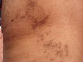
There is a linear dermatitis which follows Blaschko’s lines.  Acrally, it is erythematous, but more centrally it is hyperpigmented.  On her back the pattern is whorled.

Clinical Images:


Pathology:  We have not received the slide, but it was signed out as “Lichen planus.”

Diagnosis: Blaschkolinear Lichen Planus.

Discussion:   There are a number of rare and overlapping blaschkolinear dermatoses.  Histologically, some have features of lichen planus and some lichen striatus, with considerable overlap.  These are usually self-limited and resolve over time, but some can be followed by post-inflammatory hyperpigmentation in darker individuals.  Association with underlying pathology has not been found.

Questions: Do you have any specific comments?  It appears that potent topical corticosteroids can be helpful. Do you have any experience treating a similar patient?


References:
1. Adult blaschkolinear acquired inflammatory skin eruption (BLAISE) with simultaneous features of lichen striatus and blaschkitis.
Raposo I. et. al Dermatol Online J. 2018 Jan 15;24(1 Full Text.
Abstract

Blaschkitis and lichen striatus are generally distinguished in the literature by the age of onset, lesion distribution, and histopathology. However, there is currently no clear consensus among authors about whether to consider blaschkitis and lichen striatus different clinical entities or a spectrum ofthe same disease. We present a case of adult BLAISE with features of both lichen striatus and blaschkitis, which seems to support the theory that these clinical entities may in fact represent a spectrum of the same pathological process.
           
2. Lichen planus-like dermatosis with Blaschko line distribution: a case report. Stojanović S, Jovanović M, Vucković N. Acta Dermatovenerol Alp Pannonica Adriat. 2008 Sep;17(3):137-8. Full Text.
Abstract: The authors describe the case of a healthy 46-year-old woman with a unilateral linear papular band on the left side of the trunk that followed the lines of Blaschko from the lower back extending to the left anterior side of her abdomen. The lesions were flat-topped, slightly elevated, violaceous, agglomerated lichenoid papules. The biopsy specimen demonstrated the typical histology of lichen planus. A working diagnosis of linear lichen planus was confirmed. Because congenital and/or nevoid skin disorders in a blaschkolinear distribution may have a delayed onset after birth, these lesions must be differentiated from acquired dermatoses following the lines of Blaschko. This distinction should be made in cases with isolated lesions, such as the case presented here.

3. Linear lichen planus. Batra P.
Dermatol Online J. 2008 Oct 15;14(10):16. Full Text.
      

Thursday, February 28, 2019

Cupping Comes to the Boondocks


The patient is a 65 yo man who came in for a dermatitis of his scalp.  He requested a complete skin exam  This was remarkable for circular ecchymoses on his left back.  I asked about this.  He sees a massage therapist for back pain and she recommended cupping.  His wife was horrified to see these bruises until he explained how they were acquired.
Clinical Image: 

 With cupping, traditionally, a cotton ball soaked in alcohol is burned inside the glass cup and removed right before placement to create the vacuum. Bamboo and other materials can be used as alternatives to glass. The procedure breaks superficial blood vessels in the papillary dermis, creating ecchymoses, purpura, and petechiae, which is seen as evidence that the ailment is being drawn from the body.

Clearly, this traditional Asian remedy is entering the mainstream in Western Massachusetts. Our Asian readers probably see this all the time.  
For a few laughs, see Cupping Video.

Reference:
1. Dermatoses caused by cultural practices: Therapeutic cultural practices.
Vashi NA. J Am Acad Dermatol. 2018 Jul;79(1):1-16.
Abstract: With globalization and widespread immigration, physicians increasingly encounter patients from varying backgrounds and diverse customs. Although certain cultural practices are widely performed, there is limited medical literature describing their dermatologic and systemic effects and complications. Population diversity and sharing of traditions make it increasingly important for dermatologists to understand the role of cultural practices and recognize physiologic and pathologic sequelae. In addition, dermatologists are often adjured to assess skin findings that may be mistaken for abuse. Child abuse misdiagnosis can be traumatizing to all those involved, and immigrant families with limited English proficiency may have difficulty explaining their traditional practices. The first article of this 2-part continuing medical education series begins with a review of therapeutic cultural practices, including traditional Chinese medicine, Ayurveda, acupuncture, cupping, moxibustion, and coining, and the clinically relevant complications that may occur. Therapeutic practices can cause a range of complications, including contact dermatitis, heavy metal toxicity, and severe cutaneous adverse reactions.

Monday, February 18, 2019

2019 HOT SPOTS IN DERMATOLOGY CONFERENCE


SAVE THE DATES!

32nd  Annual Hot Spots in Dermatology
Turtle Bay Resort, Oahu, Hawaii
August 16 – 18, 2019
        
Please consider joining us for our 2019 meeting. Hot Spots addresses clinical dermatology, emerging technologies and humane aspects of medicine.  We keep the number of attendees small to assure meaningful informal sessions at which registrants and speakers have time to interact as colleagues.

Hot Spots specifically excludes speakers who are paid stipends from PhRMA or any special interest group.  Our registrants are not a captive audience to promote any products, either directly or subtly.

The Hot Spots web site, will answer most of your questions.

Nevus Lipomatosus Superficialis


There are some lesions that only a dermatologist could love.  Nevus Liopmatosus Superficialis (NLS) may be one.

Recently, a 63 yo woman presented with a pedunculated mass on her right costal margin.  It had been present for a few years and did not bother her; but her internist told her to see a dermatologist to have it removed.

Being compliant, she made an appointment.  It was a 3 cm, soft, fleshy, skin-colored nodule on a slender stalk.  At her request I removed it with a simple scissor snip and sent it for pathology.  The clinical diagnosis was “fibroepithelial polyp vs. nevus lipomatosus.”
Clinical Photo:
The pathology showed mature adipose tissue replacing large portions of the dermis.

Photomicrographs courtesy of Lynne Goldberg, dermatopathologist, Boston University Skin Path:

Diagnosis:  Solitary Nevus Lipomatosus Superficialis

Reference:  (PubMed has 30 citations on NLS)
1. Nevus lipomatosus superficialis: A rare cutaneous hamartoma.
Pujani M, et. al.  Indian Dermatol Online J. 2014 Jan;5(1):109-10. Free Full Text.

Sunday, February 03, 2019

Acute Lymphedema

Presented by Dr. Henry Foong
Ipoh, Malaysia

The patient is a 50-yr-old man with a history of swelling of the right leg for 2 months. He noted that the right foot was swollen initially and a few weeks later the swelling has progressed to the right leg.  Over the past week the swelling has progressed to the right thigh. The swelling was mildly painful but overall the past week the swelling has progressed a lot.  The swelling was so severe that he was unable to wear his normal shoe. He has a history of hypertension and a history of “stroke” where he lost his consciousness and was told by his doctor he suffered from a minor stroke.  He was treated as cellulitis by his doctor with penicillin but did not improve.  In fact he had developed a generalised maculopapular eruptions over the trunk a week after starting on IV penicillin.

O/E: Shows a severe unilateral swelling of the right leg extending from the foot to the thigh.  There was superficial erythema, desquamation with small blisters. The affected areas are well-demarcated especially on right thigh.  The leg swelling was indurated and mildly pitting.  The measured circumference of the right leg at mid-calf level was 51 cm compared with the corresponding left leg of 39 cm.  His right inguinal nodes were markedly enlarged. Rest of exam was unremarkable.

Images: 

Differential Diagnosis
1.  Erysipelas right leg
2.  Filariasis
3.  Deep vein thrombosis

Lab:
Hb 13.5 gm%, TWBC 17,900 (N87%, L10%, E0.1%, B1% M2%) ESR 79mm/hr Biochemistry unremarkable. Culture from the right leg grew Staphylococcal aureus.  Doppler ultrasound right leg did not show any evidence of deep vein thrombosis but enlarged right inguinal nodes.

He was advised to stop penicillin and started on IV moxifloxacin 400mg od, IV hydrocortisone 200mg qid,  and wet compress. Blood was sent for microfilaria (x3) which was negative.

Follow-up Photo after 5 days of IV moxifloxacin and IV hydrocortisone.

0


Marked improvement after 5 days of IV moxifloxacin and IV hydrocortisone.  There is a 5 cm reduction in the circumference of the right call. The patient would be scheduled for a MRI lower pelvis/right thigh soon, but he has refused since he is feeling so much better.
.

Your thoughts will be appreciated.

Friday, January 25, 2019

PRP and Kaposi’s: A Cautionary Tale


Our colleague, Professor Khalifa Sharquie from Baghdad, Iraq presents a 27-year old woman seen in his skin department with a dramatic facial rash and swelling of nine months duration.  Quite early on, the condition started as bruise-like discoloration and slight swelling around both eyes that slowly increased over a period of ~ three months “Figure. 1”.

The patient sought treatment at a  private clinic in Jordan.  A skin biopsy at that time showed "mucin deposition." She received peri-ocular plasma rich platelets (PRP) injections and afterwards, new lesions appeared more rapidly on her face; and then, after months, on other parts of her integument.  When seen at the Baghdad clinic she was severely ill with shortness of breath and difficulty in swallowing. “Figure. 2”.

Please see  the attached article.  and the click the link to download.
Your thoughts will be appreciated.

Figure 1

Figure 2


Reference:
Nathaniel Hawthorne:  The Birthmark
In which Beauty meets Science and is destroyed.  Science’s servant in this parable is called "Aminadab"!


Saturday, January 19, 2019

Retroauricular Dermatitis: An Orphan Disease


The patient is an 11 year-old girl with a one-year history of a dermatitis in the right retroauricular sulcus.  She has a history of  an eczematous  eruption on her thighs. She was prescribed mupirocin and betamethasone valerate ointmens.  They both caused burning and pruritus and she refused to use them.  There is a history of a vaginal dermatitis a year or so ago, which was successfully treated with topical corticosteroids.

O/E:  There is erythema, crusting and mild fissuring in the above-mentioned area.  The left retorauricular area is mildly affected.  Presently there are no findings of atopic dermatitis in the usual areas.

Clinical Image:


Lab:  Wound culture was positive for 3+ Staph aureus and 3+ Group B Strep.

Diagnosis:  Retroauricular dermatitis.

We prescribed cephalexin 250 mg q.i.d. and a small amount of Vaseline for the dryness and fissuring.  Patch testing will be recommended if there are further symptoms suggestive of contact dermatitis.

Comments:  Retroauricular dermatitis, also called "infra-auricular fissures," appears to be an under-reported entity.  Although it is common in atopic dermatitis, there have been few articles about it. I see a few cases a year and the vast majority yield coagulase positive Staph aureus and occasionally strep when cultured. Most respond quickly to mupirocin ointment and a  low to moderate strength topical steroid.  This patient’s symptoms make her an outlier.  In addition, she has no findings of atopic dermatitis at this time.
Do any of you have thoughts on this entity?  The take home message here is that, as with atopic dermatitis, these lesions are frequently colonized with Staph aureus.

References
1. Infra-auricular fissures in atopic dermatitis.
Tada J, et. al. Acta Derm Venereol. 1994 Mar;74(2):129-31.
Abstract: Retro-auricular or auricular dermatitis in atopic dermatitis (AD) is common and important for the diagnosis of AD in infancy and even in adulthood. Particularly, "infra-auricular fissures", acute eczematous changes like fissures at the adhesive junction of ear lobes, seem to be prominent features for the diagnosis of AD. Of 137 patients with AD, 81.8% showed present or past existence of infra-auricular fissures, but only one of the 30 controls. Of the 46 patients with severe AD, 98% had infra-auricular fissures, compared to 74% in those with moderate and mild AD. Our findings suggest that infra-auricular fissures are important for the diagnosis of AD and should be cited in a list of criteria for the diagnosis of AD.

2. Streptococcal perianal disease in children. Kokx NP, Comstock JA, Facklam RR.  Pediatrics. 1987 Nov;80(5):659-63.  PubMed Link.

3. Psoriasis inversa: A separate identity or a variant of psoriasis vulgaris?
Omland SH, Gniadecki R. Clin Dermatol. 2015 Jul-Aug;33(4):456-61.
Abstract: Psoriasis is a chronic skin disorder affecting approximately 2% of the European and American population. The most common form of psoriasis is the chronic plaque type. Inverse psoriasis, also named flexural or intertriginous psoriasis, is not considered a separate disease entity but rather a special site of involvement of plaque psoriasis, characterized by its localization to inverse/intertriginous/flexural body sites. We review current evidence and establish whether inverse psoriasis is a separate disease entity based on characteristics in terms of epidemiology, pathogenesis, clinical and histologic presentation, microbiology, and treatment.
.