Showing posts with label Psoriasis. Show all posts

Friday, May 11, 2018

Generalized Pustular Eruption in a 27 yo woman


The patient is a 27 yo woman with a 2 week history of an evolving, wide-spread eruption.  The initial lesions were on the popliteal fossae.  These were described as erythematous areas studded with pustules.  Over a week or two these generalized.  She has a history of mild psoriasis (scalp and elbows) for over a decade.  About three months ago she was started on bupropion 75 mg a day for anxiety and restlessness.  This was increased to 150 mg per day ~ 2 week before the onset of the dermatitis.  The patient has moderate cognitive impairment and Type 2 diabetes.  Here other medications include thyroid supplementation, metformin and insulin.  In the days since her initial office visit, the eruption has become more extensive and is taking on an erythrodermic appearance.  She was admitted to hospital two days after her office appointment.               

O/E:  When seen on May 7, 2018, she had a widespread eruption on arms, legs and torso,  The lesions were large arcuate patches with pustules at the periphery.  She was experiencing considerable pain.

Clinical Images:


 Pathology: (courtesy of DR. Erin Tababa, Fellow in Dermatology, Boston University)  

The biopsy shows prominent, relatively large, subcorneal pustules that are filled with a dense exudate of acute inflammatory. The neutrophils extended into the underlying epidermis, which has evidence of mild spongiosis. The papillary dermis is slightly oedematous, and there is a moderately dense perivascular inflammatory infiltrate with a predominance of neutrophils, although no eosinophils are noted.


Lab:
WBC: 29,000
Differential:  Shift to left
Eosinophils: normal
G6PD Normal

Chemistries normal.

Wound Culture: Pending

Dx:  We are initially considering subcorneal pustulr dermatosis, but with more history, especially considering the recent prescription of bupropion a drug-induced annular pustular psoriasis evolving into pustular and exanthematous psoriasis seemed more accurate.  Histopathology supported that.  Similar reactions have been reported to bupropion.  Our patient had been on the bupropion for over a month when this began which is longer than the patients in the case report below.  Reference 2 is a similar patient with a long latent period between initiation of drug and development of GPP.

Follow-up in hospital.  The eruption continued to evolve. It became more exanthematous and desquamative. These pictures were sent us by her mother.


Plan: 
Cyclosporin 3 – 4 mg per kg per day in divided doses
Wet dressings followed by triamcinolone 0.1% ointment bid – tid
Adjunctive secukinumab has been reported to be effective.

Follow-up:
Patient is doing very well.  These photos were taken 5 days after starting cyclosporine 100 gm q.i.d.  Her dose was dropped to 100 mg t.i.d.  She also was treated with wet dressings and triamcinalone ointment 0.1% (although the hospital only gave her 15 mg tubes, so she could not cover most ot the lesions. 
References:
1. Generalized pustular and erythrodermic psoriasis associated with bupropion treatment. Cox NH, Gordon PM, Dodd H. Br J Dermatol. 2002 Jun;146(6):1061-3.
Abstract: Severe drug eruptions may cause diagnostic and therapeutic difficulty when they mimic or provoke endogenous patterns of dermatosis. We report three patients with known psoriasis in whom use of bupropion (Zyban), prescribed to assist with cessation of smoking, led to severe pustular or erythrodermic exacerbation of psoriasis within 3-5 weeks. All patients were systemically unwell and required hospitalization to control the disease flare.

2. A diagnostic challenge: acute generalized exanthematous
pustulosis or pustular psoriasis due to terbinafine
L. Duckworth et.al. Clin Exp Dermatol. 2012 Jan;37(1):24-7
Abstract:  A 72-year-old man developed a generalized erythematous pustular eruption 11 weeks after commencing terbinafine. Clinically and histologically, the appearance was that of acute generalized exanthematous pustulosis (AGEP), and the disease was managed with topical preparations. Initial improvement was marred by relapse of acute pustulosis, now more in keeping with terbinafine-induced pustular psoriasis (PP),which was successfully treated with acitretin. This case highlights the difficulty of differentiating between AGEP and PP.

3. Acute generalized exanthematous pustulosis mimicking toxic epidermal necrolysis in patients with psoriasis: a coincidence?
Worsnop F, et. a. Clin Exp Dermatol. 2015 Aug;40(6):688-9

.


Wednesday, September 20, 2017

17 year-old girl with 8 year history of scalp dermatitis


The patient is a 17 year-old girl with an 9 year history of thick scales on her scalp.  She has used multiple medications without relief.  The patient has been bullied at school where she has been called “lice girl.”  Socially, this has been traumatic.

O/E:  She is a well-developed and well-nourished 17 yo with thick chestnut colored hair or normal intelligence.  There are no areas of alopecia. Thick, silvery adherent scales are present on the occipital, parietal and temporal scalp.  When these are removed, hair roots come out, too.  The remainder of the cutaneous examination is normal.  No nail dystrophy.
Clinical Images (July 2017)
Lab: Fungal culture negative.  Bacterial culture 3+ Staph aureus.

Failed Treatments (per mother):

"Every single otc dandruff shampoo
Every prescription medicated dandruff shampoo
Scalpicin
Prescribed scalp drops with and without coal tar
Every Tea Tree product you can find otc
Hot oil treatments
P & S Oil
Nutrogena T-gel and T-sal
Olive oil"
Terbinafine 250 mg p.o. x 1 month
Keflex 500 mg b.i.d. x 2 weeks

Scalp Biopsy read by Lynne Goldberg (Boston University Skin Path): was felt to be most compatible with psoriasis.  Seborrhea was in the differential diagnosis but less likely.

Diagnosis:  Working Dx:  Tinea amiantacea secondary to psoriasis.

Discussion: This 17 yo girl has suffered with what appears to be tinea aminatacea for almost a decade.  It appears unlikely that this is psoriasis. Tinea capitis has been ruled out by culture.  Her bacterial culture showed 3+ S. aureus but I suspect this is a secondary invader as she did not improve with cewplanexin.  Since the fungal  culture was negative and these approaches were not helpful, I may recommend isotretinoin.  The use of this has been reported for T. aminatacea only and in a Korean case report.

Dr. Goldberg's rotocol for Scalp Psoriasis, Tinea amiantacea and Related disorders:
1. Wet hair at night
2. Apply Dermasmoothe scalp oil liberally to scalp. Leave on overnight
3. Sleep with this overnight in a shower cap (to protect pillow)
4. Shampoo in the morning with T-Sal or other dandruff shampoo

Do this nightly at first if possible, but after a week or so she will be better and will not need to do it every night.


References:

1. Abdel-Hamid I et al. Pityriasis amiantacea: a clinical and etiopathologic study of 85 patients. Int J Dermatol. 2003 Apr;42(4):260-4.

2. Kwon JI.  Isotretinoin for Tinea amiantacea (A Case Report). Korean J Dermatol 2012;50(11):1002-1005 (In Korean)

3.  Mannino G, McCaughey C, Vanness E. A case of pityriasis amiantacea with rapid response to treatment WMJ. 2014 Jun;113(3):119-20.  Full Free Text.


4. Scalp psoriasis: European consensus on grading and treatment algorithm.  Ortonne J. J Eur Acad Dermatol Venereol. 2009 Dec;23(12):1435-44.

Thursday, July 20, 2017

Tinea amiantacea


Tinea amiantacea

Abstract:  17 yo girl with 10 year history of thick adherent scales over scalp

HPI:  The patient is a 17 yo girl who has suffered with wide-spread thick adherent scaly concretions over the scalp.  She has been bullied and teased at school for many years, often being called “lice girl” and similar epithets. She has tried many tar shampoos, ketoconazole shampoo, olive oil, and P&S liquid; all without effect.  There is no personal or family history of psoriasis or atopy.

O/E:  She is a well-developed and well-nourished 17 yo with thick chestnut colored hair or normal intelligence.  There are no areas of alopecia. Thick, silvery adherent scales are present on the occipital, parietal and temporal scalp.  When these are removed, hair roots come out, too.  The remainder of the cutaneous examination is normal.  No nail dystrophy.
Photo:
Laboratory: 
CBC, Chemistries normal.
Fungal Culture:Negative at 1 month
Bacterial Culture: 3+ Coag positive Staph aureus (sensitivities pending)

Scalp Biopsy read by Lynne Goldberg (Boston University Skin Path): was felt to be most compatible with psoriasis.  Seborrhea was in the differential diagnosis but less likely.

Diagnosis: Tinea amiantacea, aka Pityriasis amiantace.  In this case, the cause of T. aminatacea was most likley psoriasis.

Discussion:  This 17 yo girl has suffered with what appears to be tinea aminatacea for a decade.  It appears unlikely that this is psoriasis. Tinea capitis has not been ruled out.  I have found KOH preps from the scalp difficult, so did a fungal culture.  Her bacterial culture showed 3+ S. aureus but I suspect this is a secondary invader.  My plan at this time is to treat with two weeks of an antibiotic based on sensitivities, and start on terbinafine pending fungal culture.  If culture negative and if these approaches are not helpful, I may recommend isotretinoin.  The use of this has not been reported for T. aminatacea; but it makes some sense.  The other question I have is whether a scalp biopsy may be helpful.


Dr. Goldberg's rotocol for Scalp Psoriasis, Tinea amiantacea and Related disorders:
1. Wet hair at night
2. Apply Dermasmoothe scalp oil liberally to scalp. Leave on overnight
3. Sleep with this overnight in a shower cap (to protect pillow)
4. Shampoo in the morning with T-Sal or other dandruff shampoo

Do this nightly at first if possible, but after a week or so she will be better and will not need to do it every night.

References:
1. Pityriasis amiantacea: a clinical and etiopathologic study of 85 patients.
Abdel-Hamid IA. Int J Dermatol. 2003 Apr;42(4):260-4.
Abstract
RESULTS: A total of 85 PA patients were collected and studied. Pathological diagnosis of scalp psoriasis was confirmed in 35.3% of cases. Eczematous features suggesting a diagnosis of seborrheic and atopic dermatitis were detected in 34.2%. Diagnosis of tinea capitis, diagnosed by potassium hydroxide preparation, fungal culture, and periodic-acid Schiff staining, was detected in 12.9% of the PA patients. Staphylococcus isolates were detected in 96.5% of the PA patients compared with 15% in healthy persons as the control (P > 0.00001).
CONCLUSIONS: Pityriasis amiantacea represents a particular reaction pattern of the scalp to various inflammatory scalp diseases. The most frequent skin diseases associated with PA are psoriasis and seborrheic dermatitis. It is important to keep the diagnosis of tinea capitis in mind when evaluating PA patients. Staphylococci on the scalp could participate in the pathogenesis of PA.

2. Tinea capitis favosa misdiagnosed as tinea amiantacea.
Anane S, Chtourou O. Med Mycol Case Rep. 2012 Dec 28;2:29-3

Wednesday, June 07, 2017

Between a Rock and a Hard Place

This is the saga of a man whose care poses therapeutic dilemmas.

HPI:  The patient is a 69 yo man with a greater than 40 year history of severe psoriasis.  He was a research subject at a prestigious university hospital for many years.  Over that time, he has received PUVA, methotrexate and an investigational drug, Rapamycin.  He has been treated with NB UVB and acitretin with some improvement.  As a result of his therapies, he has developed numerous squamous cell carcinomas.  These pose therapeutic challenges They are painful, foul smelling and difficult for the patient to care for.  An additional problem is occasionally disabling trigeminal neuralgia.  He has had many well-differentiated SCCs excised over the past decade.  He has atrial fibrillation, Factor 5 deficiency and is on warfarin and a host of cardiac medication.

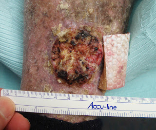
O/E:  Wide-spread erythema and scale covering entire integument except head and neck.  There is a 3 cm exophytic tumor of the left pre-tibial area and smaller similar tumors on right leg, back and chest.

Clinical Photos (6.6.17)
L. Pretibial
Diagnosis:  Generalized psoriasis, Squamous cell carcinomas

Questions:  Should these lesions be excised? Should we consider  one of the new targeted therapies?  Do any of our members have expertise with them?  This man has a number of co-morbidities which may make therapy more difficult.

References:

Oral therapy for nonmelanoma skin cancer in patients with advanced disease and large tumor burden: a review of the literature with focus on a new generation of targeted therapies.
Rudnick EW, Thareja S, Cherpelis B. Int J Dermatol. 2016 Mar;55(3):249-58
Author information
Abstract: This review of the literature aims to describe previous and current treatment options for oral therapy in locally advanced and metastatic NMSC otherwise unamenable to standard treatment. Oral Smoothened (Smo) inhibitors Vismodegib, Sonidegib, and Taladegib have shown to be effective in several trials. Oral tyrosine kinase inhibitors Erlotinib and Gefitinib, which target epidermal growth factor receptor (EGFR), have early supporting data and are currently undergoing large multicenter trials. Oral therapy in NMSC is useful in high risk patients with recurrent and aggressive disease who may not tolerate other systemic therapies.

Sunday, December 01, 2013

Arcuate and Circinate Facial Eruption


Presented by   Dr. Arnulfo Macadangdang
Cebu City, Philippines


Abstract:  18 year-old student with one week history of facial eruption

HPI:  The patient is an 18 yo man with a one-week history of an eruption on face and neck.  It is mostly asymptomatic.  He is an athlete, takes no medications and has no risky behaviors.  He had an upper respiratory infection around a month before onset. 

O/E:  There are arcuate and circinate lesions on cheeks, neck, and forehead.  Two similar lesions on u upper back. The scale is greasy.  No lymphadenopathy.


Clinical Photos:


Lab:  KOH negative


Diagnosis:  This has some features of seborrheic dermatitis, but it is not typical.  Sebosporiasis was another thought.  Evolving psoriasis?  Relationship to "URI" in October?

Questions:  Does this clinical picture suggest any specific diagnosis?

Follow-up:
5 Day F/U photos

 
The patient was seen 5 days after starting desonide 0.05% cream.  His skin lesions have esolved completely with mild post-inflammatory hypopigmentation.  The three lesions on back (not treated) have persisted. No new lesions.  Our tentative diagnosis is atypical seborrheic dermatitis.  Atypical Pityriasis rosea is also considered.  We do not feel serology for syphilis and HIV are indicated at this point, based on a discussion of risky behaviors.   Will taper desonide over the next two weeks and see patient back in a month as necessary.

Tuesday, December 25, 2012

Psoriasis vs. PRP: Your Thoughts

Presented by Dr. Henry Foong, Ipoh, Malaysia
Abstract: A 65 year old man presented with a history of erythroderma for 3 weeks.

History: A 65 year old man presented with pruritic scaly erythematous patches on the chest about 3 weeks ago.  Then he noticed the rashes spreading to the abdomen and back.  Now it has spread to the face and both upper and lower extremities. He had no fever or other constituional symptoms.  He denied taking any previous medication or health supplements.There was no past history of psoriasis or eczema.

Examination showed extensive multiple scaly erythematous patches on the abdominal wall, entire back and thighs.  His scalp was scaly too.  Over the lower back and abdominal wall, the erythema was confluent and generalised with multiple erythematous patches with islands of white in between. The lesions has an irregular margin. There is no follicular hyperkeratosis.  The nails were normal. Both palms and soles appeared normal and not hyperkeratotic.
Photos:


Pathology: 

The epithelium shows parakeratosis. The squamous epithelium does not show spongiosis or basal layer degeneration. The upper dermis shows mild perivascular and interstitial infiltrates of lymphocytes and eosinophils. The deep dermis is normal. Features do not suggest psoriasis. 

INTERPRETATION

Features are compatible with pityriasis rubra pilaris without follicular involvement.


Diagnosis: Erythroderma - Extensive psoriasis vs pityriasis rubra pilaris
Reason for posting: This is an interesting diagnostic problem as whether erythroderma is due to PRP or psoriasis.  Though there are features of psoriasiform changes over the extremioties and lower back, the presence of "islands of white" within patches of erythema is suggestive of PRP. As our experience with generalised PRP is limited, it is difficult to make a definitve diagnosis.  A biopsy was done to help to make a more definitive diagnosis.  In terms of treatment, however, oral retinoids such as acitretin would be useful in both conditions.

Saturday, February 21, 2009

Interesting Follow-up: Paronychia in a Child

In October 2007, we presented the case of an eight year old girl with chronic paronychial inflammation located on the left index finger (Paronychia in a Child). She had no other dermatoses. The patient is adopted so we have no family history. We assumed this was some kind of localized psoriasis or acrodermatitis continua. Clobetasol ointment was prescribed which she has used since. (Photo above from 10/2007)

The patient was seen in follow-up recently. The paronycial inflammation had subsided but the finger tip was still abnormal, especially on the palmar surface and there is now hypopigmentation and atrophy distal to the area of inflammation. This latter is likely secondary to the clobetasol. Her topical therapy was switched to calcipotriene cream (the ointment is no longer available in the US.)

Photos:


Questions:
1) What do you think the diagnosis is?
2) Side-effects on the fingers from super-potent topical corticosteroids are rarely reported. One suspects that they are not that unusual. When does the treatment get worse than the disease? (I should have been more diligent in follow-up)
3) Who thinks that these preparations can cause bone changes?
Your comments will be appreciated.

References:
1. Deffer TA, Goette DK.. Distal phalangeal atrophy secondary to topical steroid therapy. Arch Dermatol. 1987 May;123(5):571-2.

2. Tosti A, Fanti PA, Morelli R, Bardazzi F. Psoriasiform acral dermatitis. Report of three cases. Acta Derm Venereol. 1992;72(3):206-7.
Department of Dermatology, University of Bologna, Italy.
The authors report 3 patients affected by psoriasiform acral dermatitis, a distinctive clinical entity characterized by a chronic dermatitis of the terminal phalanges, associated with marked shortening of the nail beds of the affected fingers. The skin biopsy showed in all cases the pathological features of a subacute spongiotic dermatitis. X-ray examination of affected fingers showed no bone or soft tissue changes. Differential diagnosis of psoriasiform acral dermatitis included psoriasis, atopic or contact dermatitis and corticosteroid-induced distal phalangeal atrophy.

3. Brill TJ, Elshorst-Schmidt T, Valesky EM, Kaufmann R, Thaçi D. Successful treatment of acrodermatitis continua of Hallopeau with sequential combination of calcipotriol and tacrolimus ointments. Dermatology. 2005;211(4):351-5.
Department of Dermatology, J.W. Goethe University, Frankfurt, Germany.
Acrodermatitis continua of Hallopeau (ACH) is a rare type of pustular psoriasis affecting the digits. We report on a 43-year-old female patient who had been suffering from ACH for more than 20 years. Despite the fact that the disease was localized on one finger during the whole period, several topical and systemic treatments resulted in only temporary or partial improvement of the lesion. Although the monotherapies with calcipotriol and tacrolimus ointments gave no satisfying results in the long-term management of the disease, the combination of both agents led to a continuous improvement of the patient's skin condition. Copyright 2005 S. Karger AG, Basel.

Saturday, January 03, 2009

Magic Cure?

Abstract: 45 yo man with two year history of painful fingers
Posted by DJ Elpern
HPI: The patient is a 45 yo electrician and professional pianist who developed hyperkeratotic patches on his hands two years ago. Nothing new in exposures. After much questioning, he remembered that his mother-in-law moved in with them around that time. (not kidding). The fissures are very painful, especially when playing keyboard. He can use gloves doing electrical work. Patch testing has not been done but is planned.
O/E: Hyperkeratotic areas around thumb and middle finger tips bilaterally. Fissures are deep but clean. He has had similar areas on thenar and hypothenar eminences in past. Remainder of cutaneous exam is unremarkable.
Photos:


Diagnosis: Hyperkeratotic Hand Eczema, Psoriasis variant? Fristional Contact Dermatitis
Treatment: He has tried potent topical steroids with occlusion and with the Soak and Smear technique. Crazy glue for fissures. Intralesional triamcinalone 10 mg/cc helped the palmar keratoses. He has had one month of methotrexate 10 mg per week. Only the intralesional TAC has helped but he does not want finger tips injected at this time.
Questions:
1. What do you think the diagnosis is? The role of trauma may be key as he works with his hands as an electrician and his fingers are "traumatized" on the keyboard.
2. Do you have any magical therapeutic suggestions?
3. I have heard that X-ray treatment was used in the past. Any rational for Grenz?
4. Further work-up
Reason Presented: This man is at his wit's end with pain. He can't play the piano since every time he hits a key he has exquisite pain. I have had one or two similar patients -- they just got better over a few years seemingly not related to treatment.
Reference:
E. McMullen, D.J. Gawkrodger, Physical friction is under-recognized as an irritant that can cause or contribute to contact dermatitis. Br J Dermatol. 2006:154;154-156
Department of Dermatology, Royal Hallamshire Hospital, Sheffield U.K.
Full Text of Article
Background The role of physical friction as an irritant in the causation of contact dermatitis is under-recognized. Frictional dermatitis is defined as an eczematous process in which physical frictional trauma contributes to the induction of a dermatitis process.
Objectives To examine the clinical background of patients in whom friction was contributing to dermatitis.
Methods Over a 30-month period during which 2700 new patients were seen, frictional irritancy was identified as playing a role in the dermatosis in 31 cases: in 27 of these, case notes were evaluated for a range of parameters.
Results Physical friction was identified as causing or contributing to the dermatitis in 18 men and nine women, mean age at onset 42 years. The hands, usually the fingers of the dominant hand, were affected in all but two cases. Occupational frictional activities were found in 25 cases: commonly handling small metal components, paper, cardboard or fabric, and driving. Potential frictional activities in hobbies were noted in 12 cases. Wet work irritancy contributed in four cases (15%). Patch testing showed relevant contact allergies as cofactors in seven of 25 subjects tested (26%). Psoriasis was a cofactor in four (15%), and atopic dermatitis in 11. The study was selective, being based in a teaching hospital clinic with a special interest in contact dermatitis. Frictional irritancy is often one of several factors contributing to dermatitis.
Conclusions The contribution of friction to contact dermatitis is under-recognized probably because dermatologists do not think about the potential for physical forces to induce eczematous changes in the skin.