Tuesday, July 10, 2012

Nasal Tumor for Diagnosis

Presented by Henry Foong, Ipoh, Malaysia


Abstract: 45 yo man with one year history of a tumor on the nasal tip.

HPI: The patient is a 45 yr old man with a growth on the tip of nose for a year. It started as an asymptomatic papule last year and was shaved off by a GP.  He remained well till 3 months ago when it started to recur.  He had seen few doctors and the last doctor, a plastic surgeon decided to refer to a dermatologist. He lives in Teluk Intan about 100 km south from Ipoh. He is an excavator driver, does mainly outdoor work and has no family history of skin cancer.

O/E: Examination showed a firm non tender nodule 1.5 x 1.5 cm on the tip of the nose with a crateriform centre. Regional nodes were not enlarged.  

Clinical Photos:


Pathology: An incisional biopsy was done on the tumor. Biopsy done showed diffuse and nodular infiltration of lymphocytes in the reticular dermis and deeper layers, separated by variable amount of fibrocollagenous tissue. Admixed with the lymphocytes are few eosinophils and plasma cells. There is no epidermotropism. The epidermis is unremarkable. Mitotic figures are discernable.  

Immunohistochemical studies were performed on the tissues. The lymphoid population showed a polyclonal population of both T cell and B cell markers. A diffuse positivity of pan T markers CD 3 along with CD5 were noted. Patchy distribution of the B cell markers throughout is noticed for CD20, CD79a, PAX5 and MUM 1. Ki67 shows a proliferation index of 30-40% in the entire population.


Diagnosis (Tentative): Lymphocytoma Cutis

Questions:  Is the immunohistochemistry supportive of lymphocytoma cutis (cutaneous B-cell pseudolymphoma)  or cutaneous lymphoma? Will excision of the tumor help? If surgery is not contemplated, what would be recommmended treatment?


Sunday, July 01, 2012

Nail Dystrophy in a Nurse

A colleague requested our opinion regarding a woman who contacted him, but has not seen.  Here are her words and photos.
" I am a 61 yo palliative care nurse who has had a nail dystrophy for around 14 months. There is no pain or discomfort. I take no meds by mouth and am in good general health. Every 2 weeks I get a professional manicure and have never had acrylic, gel or silk.  I tried "formaldehyde free" products for a while, as well as Biotin 5000mcg daily and noted no improvement.  As part of my job, I visit patients in their homes and wash my hands before & after each visit. Rarely do I have the need to wear gloves.  Your suggestions will be welcome."


Questions at this time:
1) Are all nails involved (including toe-nails)?
2) Has a dermatologist been consulted and have KOH prep and/or fungal culture been done?


Saturday, June 23, 2012

Superficial Acral Fibromyxoma

The patient is a 61 yo woman with a 1 - 2 month history of a nodule on the dorsum of the left foot.

O/E: 1 cm nodule without any diagnostic features.

Clinical Photos:


Pathology: (Microscopic photos courtesy of Jag Bhawan, Chief of Dermtopathology, Boston University Department of Dermatology)
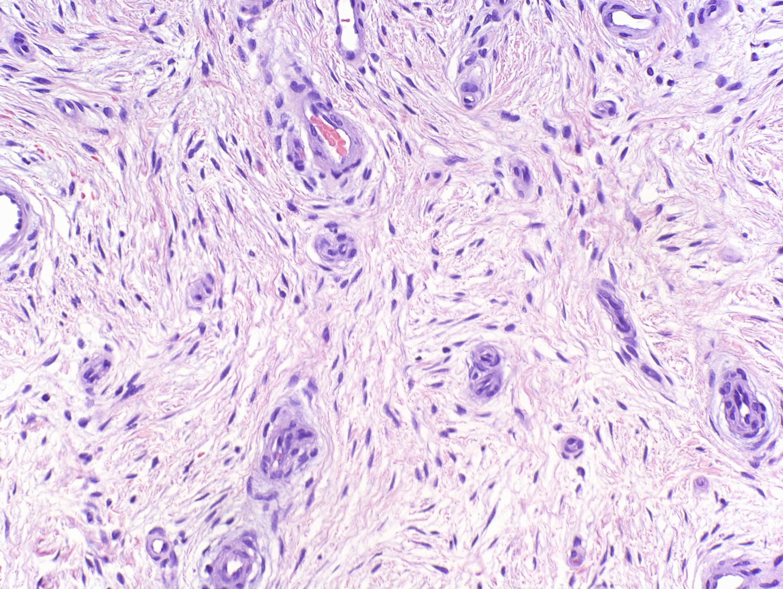
The lesion was shave-excised and the base cauterized.  Pathology showed a nodular dermal lesion comprised of fibromyxoid stroma with stellate to spindle-shaped fibroblasts and traversed by prominent vascular channels. (Photomicrographs 2x, 4x, 10x, 20x, 40x)


Diagnosis:  Superficial Acral Fivbromyxoma

Discussion:  This is a rare lesion.  I am not sure if the diagnosis could have been made prior to pathology.  Most of these lesions are periungual  This one was not.  This case is being presented for interest since little is known about this kind of lesion.

Reference:

Goo J, et. al. A case of recurrent superficial acral fibromyxoma. Ann Dermatol. 2010 Feb;22(1):110-3. Epub 2010 Feb 28
Department of Dermatology, Wonju College of Medicine, Yonsei University, Wonju, Korea.  Free FullText.

Wednesday, May 30, 2012

Skin Cancer in Renal Transplant Patient

The patient is a 64 yo man who received a renal transplant x years ago and is maintained on prednisone and Prograf.  He presented with a 4 cm biopsy proven superficial squamous cell carcinoma on the left parietal scalp.  This lesion would have necessitated a large micrographic surgical procedure with a graft.

An attempt was made to treat with topical chemotherapy.  Imiquimod was inititiated, but there was only minimal response after two weeks.  Five fluorouracil was then added and this achieved a moderate response.  The combination of imiquimod/5FU was continued for a total of six weeks, then stopped.  One month later there appears to be a clinical cure.  He will be followed closely.  

There is a possibility that this combination therapy can help selected transplant patients with low risk superficial nonmelanoma skin cancers.

Clinical Photos:
After Six Weeks Imiquimod/5FU


One month after Stopping Imiquimod/5FU


Comment:  This treatment made me a little nervous, but the surgical approach would have been major for a lesion that had only a small chance of of metastasizing.  The benefits and risks were discussed with the patient; however, his oncologist was unhappy about this approach.


Thursday, May 24, 2012

Interstitial Granulomatous Dermatitis in a Child

Abstract: 8 year-old girl with a one week history of a symmetrical eruption
This patient is presented with permission from her parent.

HPI:  The patient is an otherwise healthy 8 year old girl with a one week history of a mildly pruritic eruption;  No antecedent illnesses.  She has molluscum diagnosed in other areas a few months ago.

O/E:  On May 1, 2012, she had discrete and confluent erythematous papules on both thighs and to a lesser extent on both elbows and abdomen.  There was marked cervical lymphadenopathy (present, by history, for years).  By May 3rd, the lesions had enlarged and coalesced into symmetrical plaques on the thighs, buttocks,  and lower extremities.

Clinical Photos (May 3, 2012)

Labs:  CBC, UA normal

Pathology: There is a superficial and mid-perivascular and interstitial lymphocytic infiltrate with neutrophils and rare eosinophils. (Photomicrographs courtesy of Dr. Marjan Mirzabeigi, Dermatopathology, Boston University Department of Dermatology)
 
 
Diagnosis:  Clinically, I considered a viral exanthem or the urticarial phase of Henoch-Schoenlein Purpura.  Histologically, the suggestions were interstitial granulomatous dermatitis (IGD) or an interstitial variant of granuloma granuloma annulare.

Course:  Over the course of two weeks, the lesions completely resolved.  No therapy had been offered.  The child remains well at this time and is at full activity.

Questions:  Have you seen similar cases?  What are your thoughts about "interstitial granulomatous dermatitis."  It is only rarely seen.

Discussion: IGD is a relatively newly described disorder and not much is known about etiology or course.  There may be short-lived variants such as this that are reactions to viral illnesses, drugs or other causes.  Case reports such as this may help to define such rare disorders.

Reference:
Interstitial granulomatous dermatitis
Hillary Johnson MD PhD, Stephanie Mengden MD, Ronald R Brancaccio MD 
Dermatology Online Journal 14 (5): 18 Free Full Text.

Saturday, April 28, 2012

Cheilitis Oscura

Abstract:  59 yo woman with eight month history of cheilitis.

HPI: The patient is a 59 yo woman with an eight month history of a pruritic dermatitis which began on the upper lip. It spread to the lower lip and recently to some fingers. For dental hygiene, she has used Crest toothpaste, a white ceramic nasal irrigator, dental floss, a WaterPic and a Sonic Dental Care apparatus on a daily basis.  She has changed this protocol somewhat over the past few months.  Initially, she used Desonide ointment for three to four weeks with minimal help.  She also used clobetasol ointment 0.05% for 10 days with complete clearing. After a protocol of erythromycin gel, steroid ointment, protopic 0.03% the iritation largely disappeared. However, she continues to get flare ups with no apparent cause. Symptoms always include burning, itching and redness. She uses Vaseline on a regular basis to moisten and sooth her lips. A similar dermatitis has appeared around the nail of her right ring finger, and has lingered there for 3 months. In addition, her left ear has several similar spots, and other similar spots have come and gone around her eye lids, eye brow and forehead. Most recently, she has used tacrolimus 0.03% ointment with minimal help, and for the last few weeks only Vaseline, which seems to help the most.  Because of the concern for "steroid addiction" of the facial skin, she only used topical steroids for short periods and only under close supervision.  The process waxes and wanes unpredictably.

O/E:  The process is localized, erythematous and scaly.  The secondary lesions on the fingers and ears look similar.

Clinical Photos:
12/11/11


4/8/12

4/28/12


Laboratory:
Patch Test (T.R.U.E.) all 29 negative.  More focused patch testing is indicated.
Lip culture:  Staph epidermitis and alpha hemolytic strep (interpreted as normal skin organisms)
Biopsy:  Considered, but not done at this time.

Diagnosis and Discussion: Initially, I presumed this to be an allergic contact dermatitis.  I suspect that part of her regimen is the culprit. The sharp border is unusual and could suggest a koebnerized psoriasiform process from an oral hygiene devices that comes in contact with the area.  In children, lip licking would be considered (lick eczema) but the history here does not support that diagnosis.  If the process recurs, I would ask patient to let it develop and then biopsy the lesion.  If that was not helpful, then patch testing with a dental tray would settle the issue of allergic contact dermatitis. 

Questions: Have you encountered similar cases? What are your thoughts?

Reference:  
1.  This is a helpful reference which goes into the many causes of contact dermatitis around the mouth and lips.  Available Free Full Text.  Andrew Scheman, MD, et. al.  Part 3 of a 4-part series Lip and Common Dental Care Products: Trends and Alternatives.  Data from the American Contact Alternatives Group.  Clin Aesthet Dermatol. 2011 September; 4(9): 50–53. 

2.  Here is another useful reference on persistent cheilitis, also available Free Full Text. 

Friday, April 27, 2012

Plexiform Neurofibroma

The patient is a 33 yo man seen for evaluation of a large tumor involving the left thigh.  He lives in a rural area and his last medical visit was at age sixteen.  Sadly, he was without medical insurance until recently.  He lived with his father until recently when the latter died, and now he has been taken in by a family in his community.

The examination showed a pleasant man with a sunny disposition and a broad smile.  While mildly retarded he answers questions appropriately.  His care-giver accompanied him and it is clear that she and her family have welcomed him.

The patient has the stigmata of neurofibromatosis (NF1) with scores of cafe au lait spots and neurofibromas.  Most compelling is a large plexiform NF of the right thigh.  He has other plexiform NFs of the buttock and left chest, but these are smaller.

We will attempt to have him evaluated and treated at a center which specializes in the care of patients with NF.  This post will be updated periodically.

Photos:  Left thigh, anterior and posterior views

 Your comments will be appreciated.

References: 

1. Type 1 NF  eMedicine.  Full text online.

2, KT Power, et. al. Management of Massive Lower Limb Plexiform Neurofibromatosis – When to Intervene?  Ann R Coll Surg Engl. 2007 November; 89(8): 807  Free Full Text Online

Wednesday, April 25, 2012

Dermoscopy Rocks

Presented by Hamish Dunwoodie, MBBS
Locum Tenens Physician,
Dakota First Nation, Portage La Prairie, Manitoba

Overview:  The patient is an otherwise healthy artist from Moncton, New Brunswick who is studying native pottery production with a First Nation's band in Manitoba.  She presented to our clinic with a one week history of a black macule on the left thenar eminence.  The patient has a past history of nonmelanoma skin cancer and is worried that this may be a melanoma.

O/E:  There is a 1 mm black macule in the above-mentioned area.  Dermoscopic image suggests a vascular lesion.  Note reddish black globules.

The lesions was shaved off, a small amount of H2O2 was applied and the residual hemorrhagic area was digested. The lesion disappeared!

Photographs:  Clinical Image, Dermatoscopic Image before shaving. Dermatoscopic image after shave and H2O2.


Discussion:  Dermoscopy established that this is a vascular lesion, a "subcorneal haematoma."  Once the dermatoscopic image was appreciated, it was gently shaved off with a # 15 scalpel and it vanished.  End of story.  We needed no special tools in this isolated practice setting to put the patient's mind at ease.  I am sure that some of our dermatoscopy experts will have more to day.

Reference:
Zalaudak I, et. al.  Dermoscopy of subcorneal hematoma. Dermatol Surg  Dermatol Surg. 2004 Sep;30( 9):1229-32.
Abstract
BACKGROUND:
Subcorneal hematoma is a pigmented skin lesion usually occurring on palms or soles after a trauma or sport activity. Clinically, it may exhibit overlapping features with acral melanoma or acral melanocytic nevi, leading to unnecessary excision of this otherwise harmless skin lesion.
OBJECTIVE:
The objective was to describe the dermoscopic features in a series of subcorneal hematomas.
METHODS:
Dermoscopic images of 15 subcorneal hematomas were evaluated for the presence of different colors and dermoscopic structures.
RESULTS:
In our series, a red-black hue was the most frequent color seen by dermoscopy (40% of the lesions) and a homogeneous pattern of pigmentation was the most frequent dermoscopic structure (53.3%). Remarkably, 40% of the lesions exhibited a parallel-ridge pattern that is usually found in early melanoma of palms and soles. In 46.7% of the lesions, red-black globules were additionally seen at the periphery as satellites disconnected from the lesion's body. Only two lesions showed either parallel-furrow or fibrillar pattern. A scratch test performed in four lesions, allowed complete or partial removal of the pigmentation.
CONCLUSION:
Dermoscopic features of subcorneal hematomas may be similar to those observed in acral melanocytic lesions. Nevertheless, in most cases the correct diagnosis can be facilitated by the presence of a red-black homogeneous pigmentation, often combined with satellite globules. A positive scratch test may be considered as an additional diagnostic clue.


Friday, April 20, 2012

Imiquimod and Keloids

The patient is a 42 yo man who had a cyst I&D's on his mid-back four years ago. Postoperatively,  a keloidal scar developed.  It is very painful and pruritic.  He has had intralesional triamcinalone acetonide 40 mg/cc without much effect.

The lesion measures almost 4 cm in diameter, but, being sessile, the base is only ~ 2 cm wide.

Plan and Question:  We propose to shave this off and use imiquimod post-operatively as has been done with earlobe keloids.  Does anyone have any experience with this for keloids at sites other than earlobes?  Any other suggestions?  The literature on imiquimod use after keloid removal is all over the map.  One wonders whether employing imiquimod followed by judicial use of intralesional triamcinaloine might be appropriate.

5 Weeks p Shave excision: C&E, followed by imiquimod 5 days per week.  At this point we will stop the imiquimod and follow.  Patient does not live near to my office and can be seen only once a month or less frequently.

8 weeks p shave excision:  The patient stopped imiquimod 2 - 3 weeks ago and just applied Vaseline.  The wound is looking better.  There's a slightly raised area in the middle of the erythema.

6 months after surgery.  These is a subtle scar in the mid-portion of the excision.  The area is still quite pruritic.  Will try clobetasol ointment to area, Monday, Wednesday and Friday.  Scars are rich in mast cells and this likely explains the itching;
1 year follow-up shows small hypertrophic scar which is a considerable improvement over baseline.

 References:
1.  Treatment of keloid scars post-shave excision with imiquimod 5% cream: A prospective, double-blind, placebo-controlled pilot study. J Drugs Dermatol. 2009 May;8(5):455-8.  URL

2.  Successful treatment of earlobe keloids with imiquimod after tangential shave excision. Dermatol Surg. 2006 Mar;32(3):380-6.  URL

3.  Failure of imiquimod 5% cream to prevent recurrence of surgically excised trunk keloids.  Dermatol Surg. 2009 Apr;35(4):629-33.  URL

Thursday, April 05, 2012

54 yo man with necrotizing vasculitis

Abstract: A 54 year old man presents with long-standing rosacea and a few week history of mildly pruritic papules on his thighs.

HPI: The patient is otherwise well and has been treated with doxycycline for greater than five years for severe rosacea. Recently, it has not been effective. He presented for alternative therapy; and at the time of the visit he mentioned an a pruritic papular eruption of his thighs for two to three weeks. He has had recent onset hypertension and was started on HCTZ about a month ago.

O/E: Erythematous papules and small nodules on face. There are scattered three - four mm papules erythematous papules on the medial thighs. The remainder of the examination is unremarkable.

Clinical Photos:
Subtle Lesions on Thighs


Pathology: Necrotizing vasculitisof deep dermal artery. (Photomicrographs courtesy of Marjan Mirzabeiji, M.D., Boston University Department of Dermatology, Dermatopathology Section)


Lab: CBC normal, Chemistries normal, BUN/Cr normal, ANA 1:1280 Homogenous, ANCA panel negative

Diagnosis: Cutaneous Polyarteritis Nodosa (drug-induced) or microscopic polyangiitis. Doxycycline or HCTZ may be putative.

Discussion: This is an "interesting" case. A man walks in with rosacea and winds up with necrotizing vasculitis. He has some protein in his urine and a positive ANA. There's an old saying: It is often more important to treat the patient who has the disease than the disease the patient has. This may be a case in point.

Questions: What is your diagnosis and what more would you do?

Reference:
Rogalski C, Sticherling M. Panarteritis cutanea benigna--an entity limited to the skin or cutaneous presentation of a systemic necrotizing vasculitis? Report of seven cases and review of the literature. Int J Dermatol. 2007 Aug;46(8):817-21
Abstract: In 1931 Lindberg described a limited and benign subcutaneous form of panarteritis nodosa, which, in contrast to systemic panarteritis, only affects the skin. The terms panarteritis nodosa cutanea benigna, cutaneous polyarteritis nodosa, apoplexia cutanea Freund as well as livedo with nodules are used synonymously for this vasculitis which predominantly affects women in the fifth decade of life. Cutaneous lesions characteristically comprise painful subcutaneous nodules or vasculitis racemosa at the lower extremities. The cutaneous panarteritis may be regarded as its own entity or an isolated skin manifestation within systemic panarteritis nodosa. Full Abstract.


Wednesday, April 04, 2012

Cutting

Once or twice a month, I see patients with distinctive scars, mostly, but not always, confined to their arms. A recent patient prompted this post. The photo and text are presented with her approval as she feels her story needs to be told.


The patient is a 66 year old insulin dependent diabetic who presented with a dermatitis of the abdomen. It had non-specific features and was KOH negative. Most likely a xerotic eczema or an irritant contact dermatitis. Linear scars were noted on her arms and I questioned her about this. She had been the object of sexual abuse for many years as an adolescent and cutting was her outlet.

Cutting is a form of self-injury (aka nonsuicidal self-injury - NSSI). In my, albeit limited, experience, most cutters are or have been victims of abuse, often sexual abuse. These patients usually are receptive to discussion about their cutting and its causes. Viewing these lesions may be an opportunity to show compassion and understanding for these patients.

Cutting, almost always, is a sign of "Adverse Childhood Experiences." These, ACEs have negative impacts on one's health as an adult and are well described in a New Yorker article, "The Poverty Clinic."

For more on cutting:
Wikipedia
Why People Hurt Themselves
The Poverty Clinic by Paul Tough. There is a full text pdf online which I can not link here.

If you encounter a person with acute cutting, Brief Therapy Heals Trauma in Children, by Jane Brody, is worth reading.

Saturday, March 24, 2012

Recurrent BCC with Perineural Invasion

The patient is a 56 yo woman who had micrographic surgery for a BCC on the tip of the nose in August of 2008. The initial typing could not be done b/c the specimen was a superficial shave and deeper component could not be appreciated.

She presented in March 2012 with a subtle area of hypopigmentation at the site of the tumor. Because of the firmness of the nasal tip, induration could not be appreciated. The patient was worried that this might be a recurrence.

Clinical Photo:

A 3 mm punch biopsy showed "infiltrating BCC with perineural invasion (PNI)."

Photomicrographs courtesy of Dr. Jag Bhawan. Please click on Picasa for more images.
Teaching point: The initial shave bx was not adequate to type the lesion and this was also not commented on by Mohs surgeon. Complex BCCs of the nasal tip pose special problems. Dr. highlight some of these.

Questions to Mohs surgeons: How would you approach this woman who is concerned about cosmetic appearance of nose after second Mohs procedure? Is it likely that after almost four years of insidious growth this tumor may pose special problems for closure and necessitate plastic surgical reconstruction?

View Dr. Michael Albom's Comments on this patient.

References:
1. Leibovitch I, et. al,
Basal cell carcinoma treated with Mohs surgery in Australia III. Perineural invasion. J Am Acad Dermatol 2005 Sep;53(3):458-63.
Abstract Conclusion:
PNI is an uncommon feature of BCC. When present, PNI is associated with larger, more aggressive tumors, and the risk of 5-year recurrence is higher. This emphasizes the importance of tumor excision with margin control and long-term patient monitoring.

2. Geist DE et. al. Perineural invasion of cutaneous squamous cell carcinoma and basal cell carcinoma: raising awareness and optimizing management. Dermatol Surg: 2008 Dec;34(12):1642-51. Division of Dermatology, Department of Medicine, University of Massachusetts Medical School, Worcester, Massachusetts 01605, USA. david.geist@umassmemorial.org

ABSTRACT: BACKGROUND: Perineural invasion (PNI) by cutaneous squamous cell carcinoma (CSCC) and basal cell carcinoma (BCC) is an infrequent but not rare complication of traditionally low-morbidity skin cancers that can lead to catastrophic sequelae; 2.5% to 14% of CSCC and approximately 3% of BCC exhibit PNI. Tumors with PNI tend to be larger, have greater subclinical extension, have a higher rate of recurrence, and have a greater risk of metastases. Tumors with PNI may result in major neurologic deficits.

OBJECTIVE: To review current recommendations for the management of PNI and to evaluate a treatment strategy involving excision using Mohs micrographic surgery (MMS) followed by adjunctive radiotherapy.

MATERIALS AND METHODS:Cases of PNI treated with MMS and radiotherapy were reviewed for recurrence, disease-free follow-up, and adverse events.

RESULTS:Twelve patients with incidental PNI treated with MMS and adjunctive radiotherapy are presented. After 3 to 32 months of follow-up, there had been no recurrences. Adverse events from radiotherapy were minor and self-limited.

CONCLUSIONS: The use of adjunctive radiotherapy in these patients remains controversial. When managing superficial skin tumors with PNI, a multidisciplinary team including a cutaneous surgeon and a radiation oncologist familiar with PNI is recommended.

Wednesday, March 14, 2012

A 61 year-old disabled mason was seen for evaluation of bugs which had been burrowing into his skin for the past six months. He was an anxious-appearing man with fresh and resolving excoriations on his arms, legs and torso. His medications included lisinopril, oxycodone 60 mg three times a day, oxycodone 15 mg as needed for breakthrough pain, diazepam, montelukast, and various vitamins. Elaborately wrapped samples of the insects were presented for examination, at one point during the office visit, he noticed a dark spot on his right knee which “had just crawled out from under the skin.” A dermoscopic picture of that spot shows that this artifact is comprised of fibers from clothing. On viewing this image, the patient still felt this was insect parts, but he also alluded to his online readings about Morgellon’s disease and speculated that the fibers may have come from his skin.


Dermoscopy is a hitherto unreported aid for examining the artifacts that patients with delusions of parasitosis present to their physicians. A dermatoscope can be quickly attached to a digital camera and the nature of the specimens can be verified. Unfortunately, it is difficult to dissuade these patients of their delusions. Patients with delusions of parasitosis often doctor-shop when their clinicians do not accept their theories of infestation. Medications such as opioids and cocaine can occasionally precipitate formications (the feeling of bugs crawling on his skin) and this man’s high doses of oxycodone may well be related to his fixed ideas.

Reference:
Smith MJ, Thirthalli J, Abdallah AB, Murray RM, Cottler LB. Prevalence of psychotic symptoms in substance users: a comparison across substances. Compr Psychiatry. 2009 May-Jun;50(3):245-50. Epub 2008 Sep 23. Full Text.

Case 2 March 20, 2012
The patient is a 56 year old carpenter with a ten month history of matter being extruded from his groin and scrotum. He feels this is coming from his skin but does not specify the nature of the artefacts. He is very guarded and suspicious of me. He states he does not belong in a skin clinic, but that is where his internist sent him. For the past few months he has been treated with a variety of antifungal creams. He had a bovine aortic valve replacement done a year ago. He presented specimens on a paper towel in a zip-lock baggie. A dermoscopic picture was taken (see below) and when he was told that the material looked like it could have come from the fibers of his blue jeans, he got defensive and left the office a few moments later saying he'd get other opinions. There is one report of Delusions of Parasitosis after cardiac surgery in the literature. What is the risk/benefit relationship of the antipsychotics used to treat this disorder? He is, after all at this time, able to work. It's remarkable how similar this dermoscopic image is to the previous one. In another culture, shamanism might help these people.

See: W.B. Shelley,E.Dorinda Shelley Delusions of parasitosis associated with coronary bypass surgery. British Journal of Dermatology v. 118, p. 309-10, February 1988


Sunday, March 04, 2012

Central Centrifugal Cicatricial Alopecia (CCCA)

Abstract: 46 year old Ghanaian woman with scarring alopecia

HPI: The patient is a 46 yo woman from Ghana with 3 - 4 mo history of progressive alopecia. She has lived in the U.S. for ten years, takes no medications p.o. and has used hot combs only infrequently in the past.

The Examination shows patchy areas of complete hair loss on frontal, parietal and vertex areas of the scalp.
Clinical Photos:


Pathology: (Photomicrographs courtesy of Marjan Mirzabeigi, M.D. Department of Dermatopathology, Boston University School of Medicine.)


These show: Marked decrease in the number of follicular units which have been replaced with extensive fibrosis.

Diagnosis:
Central Centrifugal Cicatricial Alopecia (CCCA)

Discussion: Dr. Lynn Goldberg, Boston University Department of Dermatology: "The patchy alopecia in the vertex is consistent with CCCA. If the frontal loss is contiguous it could be CCCA, although these patients often have coexistent traction. My first line of therapy is a topical steroid. Most patients will experience stabilization. I reserve ILK and doxycycline for those patients with persistent symptoms and loss, or for those patients who also have pustules, which, in my experience, is infrequent. Some physicians will start with 6 months of topical and intralesional steroids and doxy. There are no controlled trials!

Reference:
Gathers RC, Lim HW. Central centrifugal cicatricial alopecia: past, present, and future. J Am Acad Dermatol. 2009 Apr;60(4):660-8.
Abstract: Clinical scarring alopecia in African American women has been recognized for years. The classification of this unique form of alopecia dates back to Lopresti, who first described the entity called "hot comb alopecia." More recently, the term "central centrifugal cicatricial alopecia" has been adopted to describe a progressive vertex-centered alopecia most common in women of African descent. While this form of hair loss is widely recognized, and may even be on the rise, the causes of central centrifugal cicatricial alopecia are a constant source of debate and remain to be elucidated. This review outlines the descriptive evolution of central centrifugal cicatricial alopecia and the historical controversies ascribed to its pathoetiology; it also examines African hair structure and discusses how hair structure along with common physical and chemical implements utilized by individuals with African hair type may play a causal role in the development of central centrifugal cicatricial alopecia.