This 23 yo woman has had facial erythema and acuminate micropapular lesions since childhood. She has extensive keratosis pilaris of her arms and thighs in addition. She has not gone out in public without thick make-up for 10 years. Her fiance has never seen her without make-up. She is desperate to haved this treated. I suspect this is a variant of KP rubra facei, possibly with ulerythema oopryogenes
Tuesday, May 24, 2005


Saturday, May 07, 2005
Seven Year Old Boy with Chronic Dermatitis
A.D. is a 7 year-old boy who was adopted from Siberia by a family in western Massachusetts at 21 months of age. Since adoption he has had recurrent dermatitis on torso and extremities. The lesions are mostly nummular by history. They have responded to systemic antibiotics on occasion. He was seen here yesterday for the first time for a second opinion.
The child appears normal otherwise. He has no evidence of atopy. This is the largest lesion. All are plaques, all covered with some crust, intensely pruritic. I applied some pressure to the large plaque with a cotton tipped applicator and a small amount of creamy pus was extruded.
My working diagnosis is nummular eczema driven by hypersensitivity to staph. A culture was taken and I'll wait for results before treating. This has been going on for 5 years. I may do a biopsy, but am not sure it will be helpful.
Any thoughts would be appreciated. The parents are at their wits end. He has also been treated with mupirocin cream in past with some success. Tacrolimus was not helpful.
The culture grew out coagulase positive staph sensitive to everything; even Penicillin G. This is unusual in the U.S. where most Saph is resistant to penicillin. One wonders if this is a strain he brought over from Russia when he was adopted. I started him on Pen VK 250 mg qid. I will add a topical corticosteroid and mupirocin - the latter for nares and crural folds. Will give follow-up after a couple of weeks. If he continues to have staph infections like this, I will look into his Ig status.
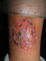
Right leg
The child appears normal otherwise. He has no evidence of atopy. This is the largest lesion. All are plaques, all covered with some crust, intensely pruritic. I applied some pressure to the large plaque with a cotton tipped applicator and a small amount of creamy pus was extruded.
My working diagnosis is nummular eczema driven by hypersensitivity to staph. A culture was taken and I'll wait for results before treating. This has been going on for 5 years. I may do a biopsy, but am not sure it will be helpful.
Any thoughts would be appreciated. The parents are at their wits end. He has also been treated with mupirocin cream in past with some success. Tacrolimus was not helpful.
The culture grew out coagulase positive staph sensitive to everything; even Penicillin G. This is unusual in the U.S. where most Saph is resistant to penicillin. One wonders if this is a strain he brought over from Russia when he was adopted. I started him on Pen VK 250 mg qid. I will add a topical corticosteroid and mupirocin - the latter for nares and crural folds. Will give follow-up after a couple of weeks. If he continues to have staph infections like this, I will look into his Ig status.

Right leg
Wednesday, April 27, 2005
A Change of Pace
The patient accompanied her "mother" to the office. She is a 12 yo chihuaha with autoimmune thyroid disease and extensive alopecia areata. I was aksed what could be done and injected large areas of her skin with intralesional triamcinalone. Unfortunately, there is no billing code for this - the dog lacked health insurance.
(Actually, I deferred treatment until I hear from my august colleagues)
Note tail alopecia in Fig. 3

Fig 1
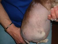
Fig 2
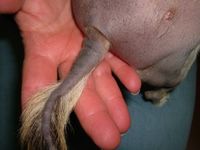
Fig. 3
Ref:
A natural canine homologue of alopecia areata in humans.
Tobin DJ, Gardner SH, Luther PB, Dunston SM, Lindsey NJ, Olivry T.
Department of Biomedical Sciences, University of Bradford, Bradford, UK.
Br J Dermatol. 2003 Nov;149(5):938-50.
BACKGROUND: Alopecia areata (AA) is suspected to be an autoimmune disease directed preferentially against hair follicles (HF) affecting both humans and various mammalian species. Recently, two rodent models of AA were described, namely the ageing C3H/HeJ mouse and the DEBR rat. Despite several case reports of canine AA in the literature, there has been no systematic assessment of the disease in these companion animals, and it is also not known whether dogs with AA could be useful as an outbred homologue of this disease in humans. OBJECTIVES: To evaluate the clinical, histopathological and immunopathological features of 25 dogs with AA and compare these data with those found in the human disease. PATIENTS/METHODS: Twenty-five client-owned dogs exhibiting macroscopic alopecia with peri- or intrabulbar lymphocytic infiltrates were selected for study. Biopsies and sera were obtained and assessed by histopathology, direct immunofluorescence of immunoreactant deposition, immunohistochemistry for lymphocyte markers, indirect immunofluorescence and immunoblotting analysis of circulating serum IgG, selective immunoprecipitation of HF proteins by serum IgG, and passive transfer of purified canine IgG into naive C57BL/10 mice. RESULTS: Clinical signs including alopecia, skin hyperpigmentation and leucotrichia usually developed during adulthood and were first seen on the face, followed by the forehead, ears and legs. Spontaneous remission of alopecia occurred in 60% of dogs and regrowing hair shafts were often non-pigmented. Histological examination of skin biopsy specimens revealed peri- and intrabulbar mononuclear cell infiltrates affecting almost exclusively anagen HF. Direct immunofluorescence analysis detected HF-specific IgG in 73% of dogs, while indirect immunofluorescence revealed circulating IgG autoantibodies to the HF inner and outer root sheaths, matrix and precortex. Immunoblotting analysis revealed IgG reactivity to proteins in the 45-60 kDa molecular weight range and with a 200-220 kDa doublet. The latter was identified as trichohyalin by selective immunoprecipitation. Purified HF-reactive IgG, pooled from AA-affected dogs, was injected intradermally to the anagen skin of naive mice where it was associated with the local retention of HFs in an extended telogen phase in AA-treated skin compared with that seen in controls. CONCLUSIONS: These findings are very similar to those reported for human AA patients; therefore, they support the consideration of dogs with AA as a useful homologue for the study of the pathogenesis of this common autoimmune disease of humans.
(Actually, I deferred treatment until I hear from my august colleagues)
Note tail alopecia in Fig. 3

Fig 1

Fig 2

Fig. 3
Ref:
A natural canine homologue of alopecia areata in humans.
Tobin DJ, Gardner SH, Luther PB, Dunston SM, Lindsey NJ, Olivry T.
Department of Biomedical Sciences, University of Bradford, Bradford, UK.
Br J Dermatol. 2003 Nov;149(5):938-50.
BACKGROUND: Alopecia areata (AA) is suspected to be an autoimmune disease directed preferentially against hair follicles (HF) affecting both humans and various mammalian species. Recently, two rodent models of AA were described, namely the ageing C3H/HeJ mouse and the DEBR rat. Despite several case reports of canine AA in the literature, there has been no systematic assessment of the disease in these companion animals, and it is also not known whether dogs with AA could be useful as an outbred homologue of this disease in humans. OBJECTIVES: To evaluate the clinical, histopathological and immunopathological features of 25 dogs with AA and compare these data with those found in the human disease. PATIENTS/METHODS: Twenty-five client-owned dogs exhibiting macroscopic alopecia with peri- or intrabulbar lymphocytic infiltrates were selected for study. Biopsies and sera were obtained and assessed by histopathology, direct immunofluorescence of immunoreactant deposition, immunohistochemistry for lymphocyte markers, indirect immunofluorescence and immunoblotting analysis of circulating serum IgG, selective immunoprecipitation of HF proteins by serum IgG, and passive transfer of purified canine IgG into naive C57BL/10 mice. RESULTS: Clinical signs including alopecia, skin hyperpigmentation and leucotrichia usually developed during adulthood and were first seen on the face, followed by the forehead, ears and legs. Spontaneous remission of alopecia occurred in 60% of dogs and regrowing hair shafts were often non-pigmented. Histological examination of skin biopsy specimens revealed peri- and intrabulbar mononuclear cell infiltrates affecting almost exclusively anagen HF. Direct immunofluorescence analysis detected HF-specific IgG in 73% of dogs, while indirect immunofluorescence revealed circulating IgG autoantibodies to the HF inner and outer root sheaths, matrix and precortex. Immunoblotting analysis revealed IgG reactivity to proteins in the 45-60 kDa molecular weight range and with a 200-220 kDa doublet. The latter was identified as trichohyalin by selective immunoprecipitation. Purified HF-reactive IgG, pooled from AA-affected dogs, was injected intradermally to the anagen skin of naive mice where it was associated with the local retention of HFs in an extended telogen phase in AA-treated skin compared with that seen in controls. CONCLUSIONS: These findings are very similar to those reported for human AA patients; therefore, they support the consideration of dogs with AA as a useful homologue for the study of the pathogenesis of this common autoimmune disease of humans.
Sunday, April 24, 2005
Asymptomatic Blanching Palmar Eruption
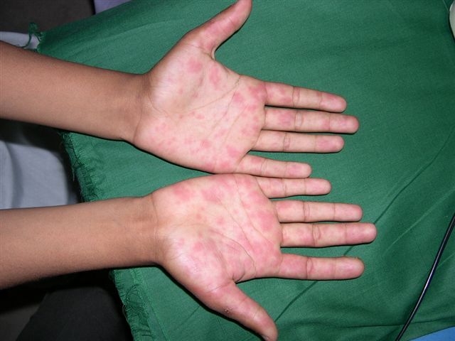
Asymptomatic non blanching eruption

A 14-year-old, otherwise healthy boy presented with an asymptomatic red blanching eruption on the palms that developed about one week earlier. Soles were not involved. No inciting factor including drugs or illness could be recognized. Rest of the physical examination and routine blood and urine tests were unremarkable. What conditions can give rise to this eruption and how should we work it up ?
Shahbaz A Janjua MD
Case for diagnosis (Alberta, Canada)
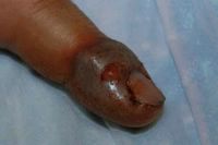
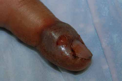
Day #1 this guy helped deliver a breech calf by c-section which was so traumatic that the calf was still-born and then the following day he helped butcher a cow. On Day #3 he noted swelling of his left index finger and by
Day #4 it blew up to look like these pictures. The more I read about orf the more I feel that this is likely the case. The risk factors are there and the time of year and setting is classic. Anyway, what do the viewers think?
(The break in the skin/expelled fluid in the pictures is my doing. There
were no vesicles or absesses and the tissue was tough and non-fluctuent.)
Duncan

Day #4 it blew up to look like these pictures. The more I read about orf the more I feel that this is likely the case. The risk factors are there and the time of year and setting is classic. Anyway, what do the viewers think?
(The break in the skin/expelled fluid in the pictures is my doing. There
were no vesicles or absesses and the tissue was tough and non-fluctuent.)
Duncan

Sunday, March 27, 2005
20 yo man with cutaneous larva migran

The patient is a 20-year-old man who noticed multiple intensely pruritic creeping lesions on his abdominal wall for more than a month. He is a body building enthusiast.
Examination showed multiple 2-3mm wide serpiginous raised erythematous tunnels on the left periumbilical and right upper quadrant of the abdominal wall.
Treatment was with freezing 1cm distal to the end of the trail with liquid nitrogen and adding oral mebendazole (zentel) 200mg bd for 3 days. It was successfully eradicated within a week of treatment.
Henry
Wednesday, March 23, 2005
10 year old girl with endogenous cheilitis
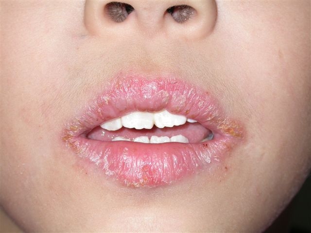
This patient presented with recurrent dry and fissured lips for more than 2 years. She has a strong family history of atopy. Her brothers and sisters have asthma and allergic rhinitis. Fortunately she didn't use any lipstick.
Examination showed fissures and dryness on both the upper and lower lips. There were crusts formations on the lips
Clinically she has endogenous (atopic) cheilitis
There are few factors that need to be considered here. is there a contact allergen element here? Lipstick would be the biggest culprit here. Is there a need to do a patch test?
Irritants can be an aggravating factor too. Lip smacking and hot spicy food can aggravate the eczema. Even toothpaste with strong mint flavour can be a factor too. I have seen several patients whose cheilitis was aggravated by our local "Darlie" toothpaste.
Moisturisers eg vaselin would be very helpful and they can be applied 4-5 times daily. Mild topical corticosteroids eg 1% hydrocortisone ointment or cutivate ointment helps. I prefer ointment to cream base in this situation.
Henry
34 year old man with multiple erythematous plaques
Recently I saw this interesting patient: a 34 yr old factory worker presented with redness over the thigh and lower abdomen for a week. Started as "burning discomfort" over the left knee and then spread to the right knee, thigh and lower abdomen. He has no fever. No history of diabetes. He denied any insect bite. There was no preceding drug history.
Examination showed diffuse and glistening redness on the thighs and lower abdomen. On the right thigh, there was a single non hemorrhagic blister. Regional nodes were not enlarged. The lesion was not particularly tender. He was afebrile.
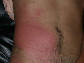
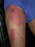
Impression: cellulitis - right thigh. Unusual site for cellulitis though. what is your take on this patient?
Henry
Examination showed diffuse and glistening redness on the thighs and lower abdomen. On the right thigh, there was a single non hemorrhagic blister. Regional nodes were not enlarged. The lesion was not particularly tender. He was afebrile.
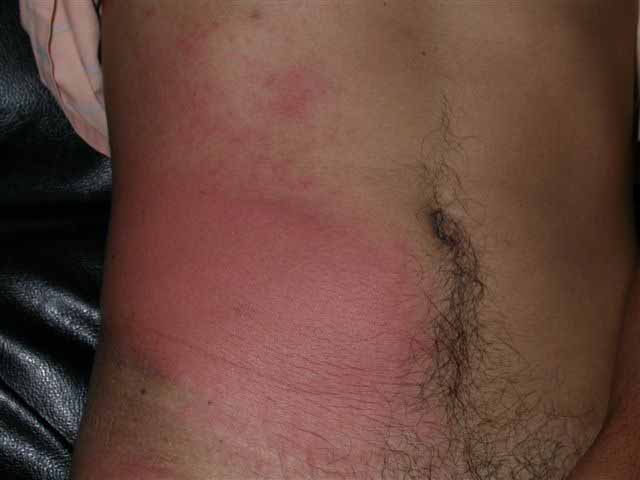
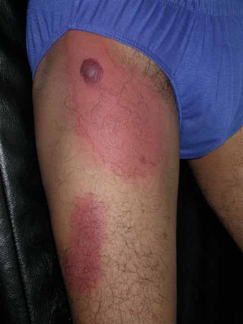
Impression: cellulitis - right thigh. Unusual site for cellulitis though. what is your take on this patient?
Henry
Tuesday, March 22, 2005
18 yo girl just returned from Costa Rica
This 18 year old girl just returned from a school trip to Costa Rica. Her group explored streams and swamps. She developed asymptomatic papules and pustules on the dorsae of her feet after being in stagnant muddy water and a running stream. I am wonderind if this is a gram negative organism or a parasite. Any thoughts?


36 yo man with lesion on back
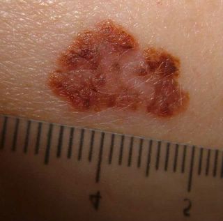
This 36 yo man presented with a 1.6 cm in diameter pigmented plaque on his back for approximately 2 years. Barely elevated. Has enlarged over past few months. Dx: Melanoma vs. Seborrheic keratosis. An excisional bx was performed today.
Path Report
DIAGNOSIS: Skin - Right Mid Back:
Malignant melanoma.
Type: Superficial spreading
Greatest thickness: 0.90 mm.
Anatomic level: II
Margins: Complete excised
Radial growth phase: Present
Vertical growth phase: Absent
Mitoses: None
Tumor infiltrating lymphocytes: Present, non-brisk
Ulceration: Absent
Regression: Present
Microsatellites: Absent
Vascular invasion: Absent
Precursor lesion: Not identified
NOTE: The lesion represents a severely atypical compound melanocytic neoplasm characterized by a predominantly intra-epidermal component with marked confluent lentiginous and nested melanocytic hyperplasia , pagetoid spread, extension into adnexae, and by a severely atypical dermal component with papillary dermal regression.
Path Report
DIAGNOSIS: Skin - Right Mid Back:
Malignant melanoma.
Type: Superficial spreading
Greatest thickness: 0.90 mm.
Anatomic level: II
Margins: Complete excised
Radial growth phase: Present
Vertical growth phase: Absent
Mitoses: None
Tumor infiltrating lymphocytes: Present, non-brisk
Ulceration: Absent
Regression: Present
Microsatellites: Absent
Vascular invasion: Absent
Precursor lesion: Not identified
NOTE: The lesion represents a severely atypical compound melanocytic neoplasm characterized by a predominantly intra-epidermal component with marked confluent lentiginous and nested melanocytic hyperplasia , pagetoid spread, extension into adnexae, and by a severely atypical dermal component with papillary dermal regression.
Sunday, March 20, 2005
2 yo girl with segmental hypopigmentation
This is a 2 year old girl who has a patch of hypopigmentation on her left shoulder since birth. It extends from back of neck to the left shoulder. It was asymptomatic. Examination showed a hypopigmented macule 5cm by 10cm on the back of left shoulder. It is segmental in distribution and has an irregular border. There was no central hypoaesthesia. She has no ophthalmic or CNS defects.
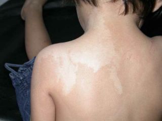
Clinically she has nevus depigmentosus or some form of pigmentary mosaicism. Biopsy of the lesion was not done.
This is usually a benign skin disorder and is caused by the functional defects of melanocytes and the morphologic abnormalities of melanosomes.
Clinically she has nevus depigmentosus or some form of pigmentary mosaicism. Biopsy of the lesion was not done.
This is usually a benign skin disorder and is caused by the functional defects of melanocytes and the morphologic abnormalities of melanosomes.
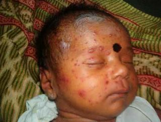
Newborn with Varicella
Posted for Dr. Jayakar Thomas of Royapuram, Chennai, India. Please comment here or reply directly to Dr. Thomas at thomas_j@vsnl.com
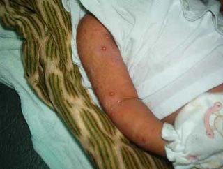
The patient is a 21 day old female infant who presented with multiple generalized vesicles of two days duration. Her mother had developed varicella two days post-partum.
Physical Exam: A normally nourished child with multiple vesicles over erythematous base. Child had low grade fever. There were no oral or genital lesions and no other mucosae were invoved.
Laboratory: Nil relevant
Histopathology: Not done
Diagnosis: Neonatal varicella (NV)
Comments: NV is not seen very commonly. It usually occurs when the mother has an attack of varicella during post partum period as in this case. The child presented here was treated with IV aciclovir with good results.
Varicella occuring in the mother during pregnancy may end up in congenital varicella syndrome in the new born.
Questions: What are the members' experience with neonatal varicella.
Some pediatricians do not use aciclovir and manage with supportive measures and antibiotics. Are they justified?
I personally feel that all children with varicella should be treated with aciclovir or appropraite antivirals. This will definitely bring down the chances of herpes zoster at a later date, given the scarring, pain, and other sequelae of herpes zoster.
The patient is a 21 day old female infant who presented with multiple generalized vesicles of two days duration. Her mother had developed varicella two days post-partum.
Physical Exam: A normally nourished child with multiple vesicles over erythematous base. Child had low grade fever. There were no oral or genital lesions and no other mucosae were invoved.
Laboratory: Nil relevant
Histopathology: Not done
Diagnosis: Neonatal varicella (NV)
Comments: NV is not seen very commonly. It usually occurs when the mother has an attack of varicella during post partum period as in this case. The child presented here was treated with IV aciclovir with good results.
Varicella occuring in the mother during pregnancy may end up in congenital varicella syndrome in the new born.
Questions: What are the members' experience with neonatal varicella.
Some pediatricians do not use aciclovir and manage with supportive measures and antibiotics. Are they justified?
I personally feel that all children with varicella should be treated with aciclovir or appropraite antivirals. This will definitely bring down the chances of herpes zoster at a later date, given the scarring, pain, and other sequelae of herpes zoster.
Thursday, March 17, 2005
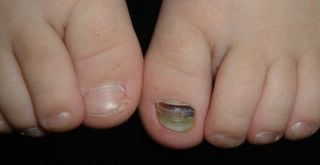
3 yo with nail dystrophy
This 3 yo boy has had a malformed left great toe nail since infancy. Recently, he traumatized it and may have had a subungual hematoma. His pediatrician tried to drain it with a hot paperclip, but was unsuccessful. His parents want to know what could be done to improve this nail.
Nail Dystrophy, Child
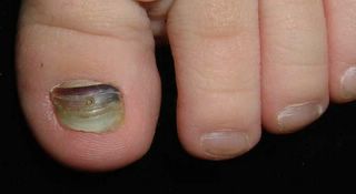
Nail Dyst - Closeup
Nail Dystrophy, Child
Nail Dyst - Closeup
Wednesday, March 16, 2005




Sunday, March 13, 2005
Atopic Infant
The patient is a one year old boy with an almost life-long history of atopic dermatitis. His father (age 30) has persistent facial eczema. His mother has significant food allergies (nuts and fruit cause angioedema and laryngeal edema). This child's facial eczema has proved difficult to control. Topical steroids have been of value (fluocinalone 0.25% ointment); but topical tacrolimus and pimecrolimus have not been effective. I suspect he may have food allergies. In addition, there is a cat at home. I think he needs to be tested for cat allergy. Food testing is controversial. Would serum IgE measurement be of value? Role of staph superinfection needs to be considered as staph, acting as a superantigen, may be driving this. I'd appreciate your thoughts. DJE
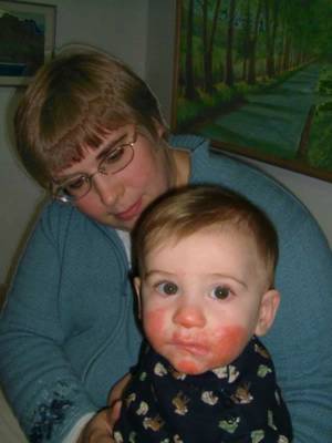
Child S.A.D.
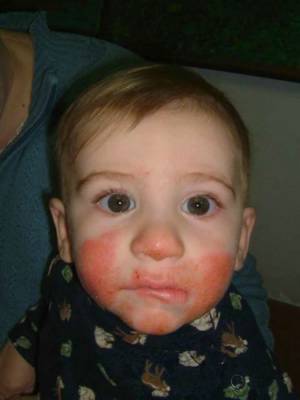
Child S.A.D.
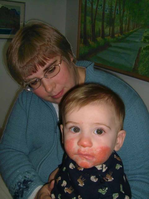
Child S.A.D.

Child S.A.D.
Saturday, March 12, 2005
Genital Papules in a 25 yo woman
Presented by:
Dr. Jayakar Thomas
KK CHILDS Trust Hospital, & Apollo Hospitals, Chennai, India
E mail: thomas_j@vsnl.net
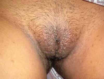
The patient is a 25 yo married woman from Chennai, India who presented with itchy papules over the vulval area for the last two years. There was no history suggestive of risk of acquiring any STD.
Physical Exam: Clinical examination revealed a healthy very good-looking young lady with multiple papules over her genital region.
Lab: Nil relevant
Histopathology : Not done
Diagnosis: Verruca plana
Comment: We see verruca vulgaris and condyloma usually in these site.
25 yo woman
Verruca plana is not common. This lady was in the habit of shaving her pubic hairs which has resulted in spread and persistence of lesions.
Topical 5% imiqumod should help if used with caution.
Question: What do the members feel would be the management strategy?
Dr. Jayakar Thomas
KK CHILDS Trust Hospital, & Apollo Hospitals, Chennai, India
E mail: thomas_j@vsnl.net
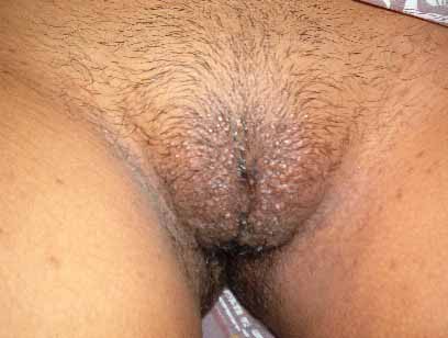
The patient is a 25 yo married woman from Chennai, India who presented with itchy papules over the vulval area for the last two years. There was no history suggestive of risk of acquiring any STD.
Physical Exam: Clinical examination revealed a healthy very good-looking young lady with multiple papules over her genital region.
Lab: Nil relevant
Histopathology : Not done
Diagnosis: Verruca plana
Comment: We see verruca vulgaris and condyloma usually in these site.
25 yo woman
Verruca plana is not common. This lady was in the habit of shaving her pubic hairs which has resulted in spread and persistence of lesions.
Topical 5% imiqumod should help if used with caution.
Question: What do the members feel would be the management strategy?
2o yo man with painful plaque
OFFICE VISIT
MARCH 07, 2005
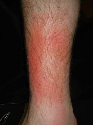
Sam D. was seen today on a same day basis. He has a few day's history of a tender, erythematous area on the right lower leg. This is at the site of a twine ankle bracelet that he had been wearing for two years. He feels a bit run down although he has not had a fever.
EXAMINATION: The examination shows an area of localized erythema and mild central scaling on the left lower leg just proximal to the ankle medially. It has a sharp margin. KOH prep was negative. There is definite heat at this site. The regional inguinal lymph nodes are enlarged.
IMPRESSION: Probable cellulitis. Doubt contact. Doubt tinea. Doubt phlebitis.
PLAN:
1. Warm compresses.
2. Dicloxacillin 500 mg q.i.d.
3. Return for follow-up in four days.
Follow-up visit March 11
S: Patient feels a bit better energy wise, but leg still red and tender.
He hasn't been able to rest - has classes, lots of responsibilities. Has not been able to do warm compresses more than twice in past 5 days.
No systemic symptoms.
O: No marked change in plaque
A: I still favor a diagnosis of cellulitis.
P: Post on ANAK VGRD for suggestions. How long should this take to resolve?
Clinically, this does not look like erythema nodosum. It began after prolonged microtrauma from a twine ankle bracelet.
20 yo man with painful plaque
MARCH 07, 2005
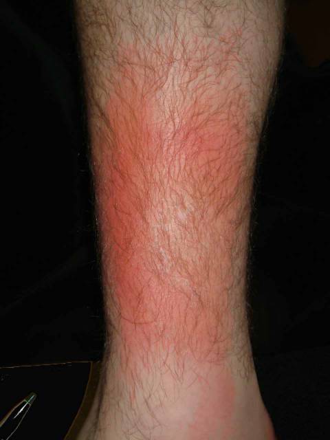
Sam D. was seen today on a same day basis. He has a few day's history of a tender, erythematous area on the right lower leg. This is at the site of a twine ankle bracelet that he had been wearing for two years. He feels a bit run down although he has not had a fever.
EXAMINATION: The examination shows an area of localized erythema and mild central scaling on the left lower leg just proximal to the ankle medially. It has a sharp margin. KOH prep was negative. There is definite heat at this site. The regional inguinal lymph nodes are enlarged.
IMPRESSION: Probable cellulitis. Doubt contact. Doubt tinea. Doubt phlebitis.
PLAN:
1. Warm compresses.
2. Dicloxacillin 500 mg q.i.d.
3. Return for follow-up in four days.
Follow-up visit March 11
S: Patient feels a bit better energy wise, but leg still red and tender.
He hasn't been able to rest - has classes, lots of responsibilities. Has not been able to do warm compresses more than twice in past 5 days.
No systemic symptoms.
O: No marked change in plaque
A: I still favor a diagnosis of cellulitis.
P: Post on ANAK VGRD for suggestions. How long should this take to resolve?
Clinically, this does not look like erythema nodosum. It began after prolonged microtrauma from a twine ankle bracelet.
20 yo man with painful plaque
Subscribe to:
Posts (Atom)