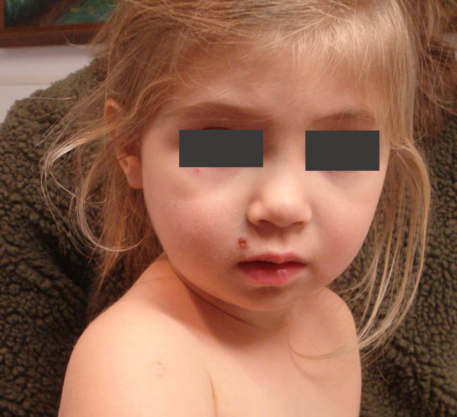
The patient is a 30 year-old woman in the 14th week of her first pregnancy. Her obstetrician asked her to see a dermatologist regarding a congenital nevus. She has noticed only slight change in the past decade.
O/E: There is a 3.0 cm bosselated nevus over the right costal margin. Some of the facets vary in color.
Clinical + Dermatoscopic Images:
Diagnosis: Congenital Nevus with a bosselated (pebbly, cobblestone) appearance. Does anyone have a comment? Presented for comments and to share the beautiful dermatoscopic appearance. Am I missing something?
Plan: I will reevaluate after she delivers.
Tuesday, June 05, 2018
Thursday, May 17, 2018
Painful Finger Tips Secondary to Opdivo
The patient is a 65-year-old woman with metastatic lung
cancer who presents for evaluation of painful fingertips. She has also had marked axillary
pruritus which she handles with Lotrisone spray.
Her medications include metformin, two types of insulin,
metoprolol, Lipitor, Lexapro, and irbesartan. Recently, she has been started on Opdivo (nivolumab) every two
weeks. Within a few months of
starting Opdivo, she developed intense axillary pruritus, and a month or so later, intensively painful fissuring of the fingertips. Each treatment of Opdivo re-exacerbates the fingertip
problem, which is unbearable. Her cancer has gone into remission and she feels well otherwise. She especially enjoys her grandkids.
Her oncologist put her on prednisone 20 mg q.i.d. for the
fingertip eczema. Because of the
patient's diabetes, her fasting blood sugars while on prednisone have gone up over 500
mg%.
EXAMINATION:
The examination shows erosions on a few of the fingertips without
erythema. Her feet and axillae are
normal.
Clinical Photos:
We did a literature search and there are a few early reports
of dermatitis with Opdivo but they do noot not affect the hands. It may be too early to say; but given the literature on painful hands and feet related to cancer therapies this is
probably a significant reaction.
PLAN: I would
try to keep this simple and avoid the prednisone. Start with wet soak for 20 minutes in warm water, followed
by clobetasol ointment, followed by gloves (a modified Soak and Smear
protocol). We will see how she
does with this. I have the
patient's permission to post her case on this site where we may
get ideas and/or be of help to others with a similar problem. There is a reference to
preventing Hand Foot reactions with chemotherapy. It seems that Celebrex (celecoxib) may be the best option. Note: Celecoxib has been reported to be associated with an increased incidence of heart attack and stroke. It's use should not be cavalier.
References:.
- Survey of cutaneous adverse reactions to targeted cancer therapies: value of dermatological advice. Damiani G OncoSkin working group. G Ital Dermatol Venereol. 2018 May 11. Abstract: From June to October 2012, 25 patients with cutaneous adverse reactions linked to targeted therapies were included in the study. The main prescribed drugs were cetuximab (52%) and erlotinib (20%) and the most common reactions were folliculitis/pustules (40%) and rash/erythema (40%). Hand-foot reaction syndrome was present in 8% of patients. A total of 30% of patients treated for a cutaneous reaction underwent a consultation by a dermatologist. In these patients the rate of oncologic therapy continuation without regimen modifications was higher (100%), while it was progressively lower in patients treated by oncologists (71%) or without any specific treatment (60%). Adverse reaction should be recognized by both dermatologists and oncologists and a multidisciplinary approach is mandatory.
2. Prevention strategies for chemotherapy-induced hand-foot
syndrome: a systematic review and meta-analysis of prospective randomised
trials.
Macedo LT, et.al. Support Care Cancer. 2014
Jun;22(6):1585-93.
Abstract
Amongst 295 studies identified, only ten met the inclusion
criteria. Celecoxib prevented both moderate to severe (odds ratio [OR] 0.39, 95
% confidence interval [CI] 0.20-0.73, P = 0.003) and all-grade HFS (OR 0.47, 95 % CI
0.29-0.78, P = 0.003), whereas pyridoxine and topical urea/lactic acid
formulations failed to prove efficacy. There were no proven benefits in mild
HFS. The use of topical antiperspirant has not been shown to improve results,
according to a single trial.
Friday, May 11, 2018
Generalized Pustular Eruption in a 27 yo woman
The patient is a 27 yo woman with a 2 week history of an
evolving, wide-spread eruption. The
initial lesions were on the popliteal fossae.
These were described as erythematous areas studded with pustules. Over a week or two these generalized. She has a history of mild psoriasis (scalp
and elbows) for over a decade. About
three months ago she was started on bupropion
75 mg a day for anxiety and restlessness. This was
increased to 150 mg per day ~ 2 week before the onset of the dermatitis. The patient has moderate cognitive impairment
and Type 2 diabetes. Here other
medications include thyroid supplementation, metformin and insulin. In the days since her initial office visit, the eruption has become more extensive and is taking on an erythrodermic appearance. She was admitted to hospital two days after her office appointment.
O/E: When seen on May 7, 2018, she had a
widespread eruption on arms, legs and torso,
The lesions were large arcuate patches with pustules at the periphery. She was experiencing considerable pain.
Clinical
Images:
Pathology: (courtesy of DR. Erin Tababa, Fellow in Dermatology, Boston University)
Lab:
The biopsy shows prominent, relatively large, subcorneal
pustules that are filled with a dense exudate of acute inflammatory. The
neutrophils extended into the underlying epidermis, which has evidence of
mild spongiosis. The papillary dermis is slightly oedematous, and there is a
moderately dense perivascular inflammatory infiltrate with a predominance of
neutrophils, although no eosinophils are noted.
Lab:
WBC:
29,000
Differential: Shift to left
Eosinophils:
normal
G6PD Normal
G6PD Normal
Chemistries
normal.
Wound
Culture: Pending
Dx: We are initially considering subcorneal pustulr dermatosis, but with more history, especially considering the recent prescription of bupropion a drug-induced annular
pustular psoriasis evolving into pustular and exanthematous psoriasis seemed more accurate. Histopathology supported that. Similar reactions have been reported to bupropion. Our patient had been on the bupropion for over a month when this began which is longer than the patients in the case report below. Reference 2 is a similar patient with a long latent period between initiation of drug and development of GPP.
Follow-up in hospital. The eruption continued to evolve. It became more exanthematous and desquamative. These pictures were sent us by her mother.
Plan:
Cyclosporin
3 – 4 mg per kg per day in divided doses
Wet
dressings followed by triamcinolone 0.1% ointment bid – tid
Adjunctive
secukinumab has been reported to be effective.
Follow-up:
Patient is doing very well. These photos were taken 5 days after starting cyclosporine 100 gm q.i.d. Her dose was dropped to 100 mg t.i.d. She also was treated with wet dressings and triamcinalone ointment 0.1% (although the hospital only gave her 15 mg tubes, so she could not cover most ot the lesions.
References:
1. Generalized pustular and erythrodermic psoriasis associated
with bupropion treatment. Cox NH, Gordon PM, Dodd H. Br J Dermatol. 2002
Jun;146(6):1061-3.
Abstract: Severe drug eruptions may cause diagnostic and
therapeutic difficulty when they mimic or provoke endogenous patterns of
dermatosis. We report three patients with known psoriasis in whom use of
bupropion (Zyban), prescribed to assist with cessation of smoking, led to
severe pustular or erythrodermic exacerbation of psoriasis within 3-5 weeks.
All patients were systemically unwell and required hospitalization to control
the disease flare.
.
2. A diagnostic challenge: acute generalized exanthematous
pustulosis or pustular psoriasis due to terbinafine
L. Duckworth et.al. Clin Exp Dermatol. 2012 Jan;37(1):24-7
Abstract: A
72-year-old man developed a generalized erythematous pustular eruption 11 weeks
after commencing terbinafine. Clinically and histologically, the appearance was
that of acute generalized exanthematous pustulosis (AGEP), and the disease was
managed with topical preparations. Initial improvement was marred by relapse of
acute pustulosis, now more in keeping with terbinafine-induced pustular
psoriasis (PP),which was successfully treated with acitretin. This case highlights
the difficulty of differentiating between AGEP and PP.
3. Acute generalized exanthematous pustulosis mimicking
toxic epidermal necrolysis in patients with psoriasis: a coincidence?
Worsnop F, et. a. Clin Exp Dermatol. 2015 Aug;40(6):688-9
Tuesday, May 08, 2018
An Infant Girl from Rwanda with a Congenital Dermatosis
presented by Caitlun Stiglmeier, M.D.
I was hoping to have some input on this case.The patient is an 11 month old female infant born via spontaneous vaginal delivery, to a G1P1 mom (young, but not sure of mothers age). No reported maternal history of perinatal infections. TORCH screening performed on the child during this admission is negative. The child has bilateral retinal detachment, but her hearing is retained. She has gross developmental delay, and the rash you see in the photos started after birth on the R forearm, and has migrated since to all areas of the body, including the chest, back, yo the upper and lower extremities, and scalp. Her mother states the rash begins as blisters (second photo), which then open (no drainage noted per mom) and leave areas of hypopigmentation (third photo) and finally hyper-pigmentation (first photo).
The child does not seem bothered by this rash, it is not pruritic, and she does not have any fevers. Despite negative TORCH screening, I'm still thinking along the lines of a congenital-type of disease. Your thoughts would be greatly appreciated!
Dr. Yoon Cohen wrote:
The clinical description and the images seems to go with incontinentia pigmenti in this her 11 month infant girl.
For management, the skin changes of IP usually do not requires any specific treatment other than wound care for blisters to prevent secondary skin infection. The baseline eye exam and close follow up by an ophthalmologist will be important, especially with her retinal detachment history. Neurological evaluation should be done for potential seizures, encephalopathy and ischemic stroke. Dental evaluation is recommended after teeth erupt for pegged or missing teeth. Other ectodermal abnormalities can be seen as alopecia and nail dystrophy. In countries with more resources, this infant would benefit from being seen at a center where the pediatric dermatologists have more experienc with genodermatoses.
References:
1. I.P. NORD site.
2. I.P. GARG (NIH Genetic and Rare Diseases information site)
3. I.P. World Community (some parts in French)
Friday, April 27, 2018
FADES
The patient is a 17 yo boy with a 10 year history of asymptomatic hyperkeratosis of both elbows. His knees are normal. He has no personal history of atopy and both he and his mother deny that he rubs or scratches the area. He's embarrassed by this and avoids sports as a result. He is moderately obese and does not exercise or do sports.
O/E: Symmetrical hyperkeratosis of both elbows. Knees perfectly normal as is the remainder of his cutaneous exam. He is moderately overweight.
Clinical Images:
Diagnosis: Asymptomatic Frictional Hyperkeratosis of the Elbows
Comment: I suppose we all see this entity on frictional sites, but rarely name it. The patient denies rubbing or scratching the area but was observed to lean on his elbows. A similar problem has been reported (see reference 1. below). I recommended starting with 5% Keralyt gel twice daily and may add tretinoic acid. I propose that this type of hyperkeratosis can follow pressure on bony prominences, such as is seen on the foreheads of devout Muslims who pray 5 times a day (2). Prayer marks in Muslims appear to be more common in diabetics (3). The patient described in this VGRD post could well have the metabolic syndrome. I wonder if this might be important. I'd appreciate your thoughts.
Reference:
O/E: Symmetrical hyperkeratosis of both elbows. Knees perfectly normal as is the remainder of his cutaneous exam. He is moderately overweight.
Clinical Images:
Comment: I suppose we all see this entity on frictional sites, but rarely name it. The patient denies rubbing or scratching the area but was observed to lean on his elbows. A similar problem has been reported (see reference 1. below). I recommended starting with 5% Keralyt gel twice daily and may add tretinoic acid. I propose that this type of hyperkeratosis can follow pressure on bony prominences, such as is seen on the foreheads of devout Muslims who pray 5 times a day (2). Prayer marks in Muslims appear to be more common in diabetics (3). The patient described in this VGRD post could well have the metabolic syndrome. I wonder if this might be important. I'd appreciate your thoughts.
Reference:
1. Frictional asymptomatic darkening of the extensor surfaces.
Krishnamurthy S, Sigdel S, Brodell RT. Cutis. 2005
Jun;75(6):349-55.
Abstract: Frictional asymptomatic darkening of the extensor
surfaces (FADES), also known as hyperkeratosis of the elbows and knees, is
commonly seen by dermatologists but has never been well characterized. Patients
present with uniform, asymptomatic, brown darkening over the extensor surfaces
of the elbows and knees with minimal scaling. Both frictional stress and family
history may play a role in the pathogenesis of this condition. The results of
cutaneous biopsy specimens typically reveal hyperkeratosis, acanthosis, and
mild papillomatosis with minimal inflammation. Keratolytic agents such as
lactic acid and urea cream along with avoiding frictional stress can be
effective in the management of this condition. We describe a series of cases of
FADES and its etiology and management options.
Comment in: Frictional asymptomatic darkening of the
extensor surfaces. [Cutis. 2007]
2. Prayer marks. Abanmi AA et. al. Int J Dermatol. 2002
Jul;41(7):411-4.
Prayer marks (PMs) are asymptomatic, chronic skin changes
that consist mainly of thickening, lichenification, and hyperpigmentation, and
develop over a long period of time as a consequence of repeated, extended
pressure on bony prominences during prayer. PubMed.
3. Prayer Marks in Immigrants from Bangladesh with Diabetes Who
Live in Greece. Papadakis G, et. al. J
Immigr Minor Health. 2016 Feb;18(1):274-6. PubMed.
Labels:
frictional dermatoses,
hyperkeratosis
Friday, April 20, 2018
Demodeciasis: One off
The patient is an 86 yo man was tarted on imbruvica for
a lymphoma in August of 2017. Around a
month later he developed a facial eruption that had the appearance of
rosacea. As it was mild, it was not
treated. The eruption has worsened over
the past few months.
Lab: A scraping from two papules revealed numerous (apparently happy) demodex mites.
O/E There are
erythematous papules and pustule on the right malar eminence and erythema and
mild swelling of the nose. The left
malar eminence and the remainder of the
head and neck are normal.
Clinical Photos:
Lab: A scraping from two papules revealed numerous (apparently happy) demodex mites.
Diagnosis:
Demodeciasis, most likely as a side-effect of imbruvica. This has not been reported in the literature
at present, but I suspect it will be soon.
Treatment was initiated with Sklice (topical
ivermectin). If this is not effective,
he will be offered oral ivermection. The
latter may have been a better strategy.
Follow-up 10 days after starting ivermectin Solution (Sklice); ~ 50% better.
Reference:
1. Parmar S, Patel K,
Pinilla-Ibarz J.
Ibrutinib
(imbruvica): a novel targeted therapy for chronic lymphocytic leukemia. P T.
2014 Jul;39(7):483-519. Free Full Text.
2. Patrizi A1,
Bianchi F, Neri I. Rosaceiform eruption
induced by erlotinib. Dermatol Ther. 2008 Oct. Suppl 2:S43-5.
Abstract:
Adverse events with
anti-epidermal growth factor receptor therapy mainly involve the skin. The most
common cutaneous adverse event is an acneiform eruption, which occurs in more
than 50% of cases. The aim of this paper is to report the case of rosaceiwform
eruption induced by erlotinib in an 81-year-old-man and to discuss the pathogeneic
role of Demodex folliculorum mites, found in the present patient, using skin
scraping.
Sunday, March 18, 2018
Granulomas in 62 year-old fisherman
Presented by Dr. Henry Foong
Ipoh, Malaysia
A 62 yr old man presented with 4 weeks history of pruritic papular lesions on the thighs bilaterally which then spread to the legs. The pruritus was intermittent and he felt feverish at times. No history of trauma. He is a retired fisherman and lives in Teluk Intan about 60 km south of Ipoh, Malaysia.
O/E: showed multiple indurated erythematous papules, non-tender distributed symmetrically over the inner thighs and legs. Superficial erosions and ulcerations were noted on the affected areas. There was no crepitus.
The referring physician suspected elephantiasis. The patient is on oral antibiotics ( metronidazole and unasyn) and albendazole. At one time he was on DEC (diethylcarbamazine) but it was withheld because of side effects.
Lab: Blood counts showed normal TWBC with 7% eosinophils. The physician did a colonoscopy last month and biopsy suggested underlying parasitic colon infection.
Culture of the skin lesions - Clostridium perfrigens.
A skin biopsy was performed on the skin papules on the thigh.
Pathology: Skin biopsy showed epidermis with marked spongiosis. the dermis show cluster and scattered granulomas with abundant surrounding neutrophils and plasma cells. There is a huge collection neutrophils with necrotic material. There are multinucleate gaints granulomas. The inflammatory cells involved the subcutaneous fat.
Clinical and Pathological Images:
Impression: Sporotrichosis?
Questions: What are your thoughts? What further studies would you consider.
Thursday, February 15, 2018
Dupilumab Conjunctivitis
The patient is a 55 year-old woman who has been on dupilumab (Dupixent) for two months. She had life-ling severe atopic dermatitis and states that her skin has never been this clear in her memory. She has, however, developed a conjunctivitis of her lid and bulbar conjunctivae. She is so happy that her skin is clear that she is not complaining, and her eyes are not uncomfortable.
There are no references to conjunctivitis on dupilumab, but the package insert indicates that 10% of patients have experienced this. If you have any recommendations, I would appreciate hearing them.
5/2/18. Two months after starting Protopic ointment 0.03%, there has been no improvement of her conjunctivitis. We have, therefore, prescribed fluoromethanlone 0.1% ophthalmic solution b.i.d. The patient says that having the cojunctivitis is acceptable as long as her skin is doing well for the first time in > 50 years. Photos taken before starting fluoromethalone ophthalmic.
Reference:
There are no references to conjunctivitis on dupilumab, but the package insert indicates that 10% of patients have experienced this. If you have any recommendations, I would appreciate hearing them.
![]() |
| Photos presented with patient permission |
5/2/18. Two months after starting Protopic ointment 0.03%, there has been no improvement of her conjunctivitis. We have, therefore, prescribed fluoromethanlone 0.1% ophthalmic solution b.i.d. The patient says that having the cojunctivitis is acceptable as long as her skin is doing well for the first time in > 50 years. Photos taken before starting fluoromethalone ophthalmic.
Reference:
Conjunctivitis occurring in atopic dermatitis patients
treated with dupilumab-clinical characteristics and treatment.
Wollenberg A J Allergy Clin Immunol Pract. 2018 Feb 9. pii:
S2213-2198(18)30089-8. Free
Full Text.
Here, we report our experience with this clinically relevant
complication of dupilumab treated AD seen in 25% and 50% of patients from our
two centers from April 2016 to February 2017, and give treatment
recommendations based on our personal experience with 13 moderate-to-severe
dupilumab-treated AD patients developing conjunctivitis as adverse event. This
conjunctivitis is reported in temporal association with dupilumab treatment,
but a causal relation is not established
.
However, in our limited experience, antihistamine eye drops
and artificial tears did not confer any alleviation in this type of
conjunctivitis. In 11 patients, dupilumab-related conjunctivitis was treated
with topical tacrolimus or steroids, leading to clinically significant
improvement or full recovery in all 11 patients treated.
Two treatment options were particularly effective.
In 5 patients conjunctivitis was treated with fluorometholone
0.1% eye drops, leading to significant improvement.
In four patients, conjunctivitis was
treated with tacrolimus 0.03% eye ointment. Signs and clinical symptoms improved
significantly in all patients, and in 2 patients, full recovery of
conjunctivitis was achieved.
Labels:
Atopic Dermatitis,
conjunctivitis,
dupilumab
Thursday, February 01, 2018
Mal Perforans Ulcer
The patient 74 year old divorcee who lives alone. He is an insulin-dependent
diabetic with peripheral neuropathy. He has had a plantar ulcer for > 6 months that began after a callosity was pared down by a podiatrist. He has been seen at a wound care clinic for six months where dressings are done. He is afraid he may lose his foot.
O/E: On the plantar aspect of the left foot, he has a clean. painless ulcer measuring about 1.4 cm in diameter. There was a thick callosity the ulcer's periphery. His pedal pulses are strong.
Clinical image:
Question: How would you approach this ulcer?
6/15/2018
The patient had an orthopedic procedure two months ago. This was intended to redistribute some of the pressure on the ulcerated area. It was quite successful. He has some mild foot edema presently, but the ulcer has healed completely. His A1C is normalizing, too.
Reference:
Lu SH, McLaren AM. Wound healing outcomes in a diabetic foot
ulcer outpatient clinic at an acute care hospital: a retrospective study. J
Wound Care. 2017 Oct 1;26(Sup10):S4-S11
Abstract
OBJECTIVE: Patients with diabetic foot ulcers (DFU) have an
increased risk of lower extremity amputation. A retrospective chart review of
patients with DFUs attending the Foot Treatment and Assessment chiropodist-led
outpatient clinic at an inner-city academic hospital was conducted to determine
wound healing outcomes and characteristics contributing to outcomes.
METHOD: We reviewed the complete clinical history of 279
patients with 332 DFUs spanning over a five-year period.
RESULTS: The mean age of patients was 61.5±12.5 years and
most patients (83.5%) had one DFU. The majority of wounds (82.5%) were in the
forefoot. Overall, 267/332 (80.5%) wounds healed. A greater proportion of
wounds healed in the forefoot (82.5%) and midfoot (87.1%) than hindfoot (51.9%;
p<0.001). Using a logistic regression model, palpable pedal pulse and use of
a total contact cast were associated with better wound healing.
CONCLUSION: Our findings are the first to demonstrate the
benefits of chiropodists leading an acute care outpatient clinic in the
management of DFUs in Canada and delivers wound healing outcomes equivalent to
or exceeding those previously published.
Labels:
diabetic foot ulcer,
mal perforans ulcer,
Ulcer
Thursday, January 25, 2018
Nail Dystrophy in 59 yo Woman
The patient is a healthy librarian who noticed a reddish area under the left thumb nail ~ 3 months ago. Shortly after that, the distal portion of the nail become yellowish. She has not history of trauma and the area is not painful
O/E: There is erythema noted medically in the nail bed and the nail plate is onycholytic. No abnormality of the nail plate is noted other than the yellowish area and possibly a Beau's line. The patient feels that the red area has migrated medially.
Clinical Image:
My thoughts: I am concerned that there may be a tumor under the nail plate. The fact that it is painless argues against glomus tumor. I welcome suggestions.
Thank you,
DJE
Saturday, January 13, 2018
Non-healing ulcer after surfing injury
Surfer's Sore
The patient is a 70 year-old surfer living in Hawaii. Two months ago, he sustained a cut over his shin bone on a lava rock/sandstone shelf of a reef on Kauai. It has not healed in spite of cleaning area daily with chlorhexidine scrub and applying Medihoney and triple anti bacteria ointment. He says, "What is weird and kinda creepy is it feels like something is crawling around in there from time to time, especially while sleeping?" The patient, a light-complected Caucasian, has a history of non-melanoma skin cancer. He is scrupulous about sun-protection, but has spent more than a half a century with significant sun-exposure.
This is the sandstone slab on which the injury took place:
What are your thoughts?
References
1.
Sea Ulcers Andrew
Nathanson, MD, Surfing Medicine (Journal
of Surfing Medical Association) Dec, 2014
Thursday, January 04, 2018
Two year-old with enanthem and exanthem for two weeks
Presented by Will Shepard, M.D.
Gillette, WY
The patient is a two year-old girl with a two week history of
oral and skin lesions. She has been well
and healthy otherwise and all of her milestones have been normal. The present
illness began with two ulcers on her tongue.
A few days later she started to develop skin lesions, first on the
arms. The new lesions start with
erythematous macules and became crusted after 12 – 18 hrs. She has continued to develop new lesions on
the torso, face and extremities. Throughout
this period she has been healthy, no fevers, appetite normal and in no discomfort.
O/E: The tongue
lesions have disappeared. The skin
lesions are few in number and measure 0.5 to 1 cm in diameter. They are scaly annular macules on an
erythematous base.
![]() |
| Rough area from Bandaid |
Impression: The onset of an acute problem with first oral and then skin lesions in an otherwise healthy toddler suggests a viral process.
References:
1
[Paraviral exanthems]. [Article in German]
Fölster-Holst R, Zawar V, Chuh A. Hautarzt. 2017
Mar;68(3):211-216.
Abstract: Paraviral exanthems are distinct skin diseases due
to infections with different viruses. Although no virus has been identified so
far in some exanthems, the main age of manifestation, the clinical course of
the exanthem, and the extracutaneous symptoms are suggestive for a viral
genesis. While many viral infections are a direct result of the infection,
paraviral exanthems reflect the response of the immune system to the infectious
pathogens. Viruses cannot be identified in the skin. Typical paraviral
exanthems include Gianotti-Crosti syndrome, pityriasis rosea, pityriasis
lichenoides, papular-purpuric gloves and sock syndrome, and asymmetrical
periflexural exanthema. Unilateral mediothoracic exanthem, eruptive
pseudoangiomatosis are rare and eruptive hypomelanosis has been described
recently.
Friday, November 03, 2017
Atypical Pigmented lesion in an 81 yo Man
The patient is a light-complected Caucasian with Type I skin and a personal history of non-melanoma skin cancer. His daughter has a history of melanoma.
He presented for a skin exam and was found to have an atypical pigmented lesion on his right shoulder. He remembers that this lesion had been biopsied years ago at another facility and he was told it was fine.
O/E: On the right shoulder there was a 1.2 mm in diameter irregularly pigmented macule with a play of color. The dermatoscopic picture was worrisome and he was scheduled for an excisional biopsy.
Pathology: The excisional biopsy showed a melanoma 0.3 mm thick, 0 mitoses per mm squared. Free margins, but narrow.
(The old biopsy report from 2007 was reviewed. This was a shave biopsy that showed a junctional nevus with mild to moderate atypia and margins were clear in the sections examined.)
Discussion: The patient has a thin melanoma arising in the site of a previous biopsy. This raises the question of whether shave biopsies of pigmented lesions are appropriate. At any rate, a thin melanoma, 0.3 mm thick just requires a wide-local excisison with one cm margins. Sentinel node biopsy is not indicated. The patient will have regular skin exams from this point on.
He presented for a skin exam and was found to have an atypical pigmented lesion on his right shoulder. He remembers that this lesion had been biopsied years ago at another facility and he was told it was fine.
O/E: On the right shoulder there was a 1.2 mm in diameter irregularly pigmented macule with a play of color. The dermatoscopic picture was worrisome and he was scheduled for an excisional biopsy.
![]() |
| Dermatoscopic image |
(The old biopsy report from 2007 was reviewed. This was a shave biopsy that showed a junctional nevus with mild to moderate atypia and margins were clear in the sections examined.)
Discussion: The patient has a thin melanoma arising in the site of a previous biopsy. This raises the question of whether shave biopsies of pigmented lesions are appropriate. At any rate, a thin melanoma, 0.3 mm thick just requires a wide-local excisison with one cm margins. Sentinel node biopsy is not indicated. The patient will have regular skin exams from this point on.
Subscribe to:
Posts (Atom)