Friday, November 03, 2017

Atypical Pigmented lesion in an 81 yo Man

The patient is a light-complected Caucasian with Type I skin and a personal history of non-melanoma skin cancer.  His daughter has a history of melanoma.

He presented for a skin exam and was found to have an atypical pigmented lesion on his right shoulder.  He remembers that this lesion had been biopsied years ago at another facility and he was told it was fine.

O/E:  On the right shoulder there was a 1.2 mm in diameter irregularly pigmented macule with a play of color.  The dermatoscopic picture was worrisome and he was scheduled for an excisional biopsy.
Dermatoscopic image
Pathology:  The excisional biopsy showed a melanoma 0.3 mm thick, 0 mitoses per mm squared.  Free margins, but narrow.

(The old biopsy report from 2007 was reviewed.  This was a shave biopsy that showed a junctional nevus with mild to moderate atypia and margins were clear in the sections examined.)

Discussion:  The patient has a thin melanoma arising in the site of a previous biopsy.  This raises the question of whether shave biopsies of pigmented lesions are appropriate.  At any rate, a thin melanoma, 0.3 mm thick just requires a wide-local excisison with one cm margins.  Sentinel node biopsy is not indicated.  The patient will have regular skin exams from this point on.


Monday, October 16, 2017

Linear Scleroderma in a 40 year-old Woman

Presented by Hamish McDougall
Cape Breton, Nova Scotia

The patient is a 37 year old woman with a four year history of a slowly progressively asymptomatic area of induration on the posterior aspect of the left thigh (photo).  A biopsy showed thickened collagen bundles in the reticular dermis and a sparse superficial and deep lymphoplasmacytic infiltrate consistent with morphea.

Strangely, her father-in-law is seriously ill with systemic sclerosis.  He lives far from the patient and her husband.  At this time, we have discovered no common exposures the patient and her father-in-law have.  The patient lives in an endemic area for Lyme Disease and serological testing will be offered.

Questions:
Is there any value in obtaining serologies, other than Lyme studies, on this woman?
What treatment might be of value? 
Do you have alternate diagnoses?


References:
1. Localized Scleroderma Review Article (Like a chapter in a text book)
2. Morphea Sculpted in Silica: A Case Report of Limited Cutaneous Systemic Sclerosis in a Woman with Long-Time Exposure to Silica Dust.

Pedro Gomes J, Shoenfeld Y.  Free Full Text.
3.  "Borrelia-associated early-onset morphea": a particular type of scleroderma in childhood and adolescence with high titer antinuclear antibodies? Results of a cohort analysis and presentation of three cases.

Prinz JC, et. al. J Am Acad Dermatol. 2009 Feb;60(2):248-55. CONCLUSION:B burgdorferi infection may be relevant for the induction of a distinct autoimmune type of scleroderma; it may be called "Borrelia-associated early onset morphea" and is characterized by the combination of disease onset at younger age, infection with B burgdorferi, and evident autoimmune phenomena as reflected by high-titer antinuclear antibodies. As exemplified by the case reports, it may take a particularly severe course and require treatment of both infection and skin inflammation.

Thursday, September 28, 2017

Metastatic melanoma in an elderly man

Presented by Henry Foong, M.D.
Ipoh, Malaysia

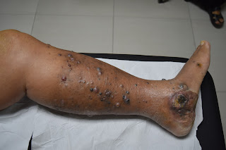
The patient is a 80-year-old man who presented with swelling of the left leg for 3 months. 

About 3 years ago he had a motor vehicle accident where he injured his left leg.  His attending doctor noticed a pigmented growth on the left foot associated with inguinal node swelling. A surgeon excised the pigmented growth on the left foot and removed some nodes from the left groin.  No histological reports were available at the moment.  The patient was well until recently when he noticed gradual swelling of the left leg with multiple pigmented nodules on the surface.  The leg was occasionally painful at night. He did not have any constitutional symptoms.  There was no family history of skin cancers. No significant other medical illness.

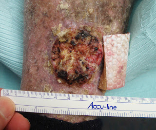
On examination his left leg was swollen and oedematous with many pigmented papules and nodules on the foot and lower 1/3 of the left leg.  A pigmented ulcerating tumour  5 x 5 cm was noted on the left foot which extended to the heel.  A firm matted lymph node swelling was noted on the left groin.  There was a surgical scar over the left groin.  No hepatosplenomegaly was present.

Biopsy of the pigmented papule on the foot was done and confirmed malignant melanoma.
Sheets and nests of malignant cells are seen invading the dermis. The
tumour cells show marked pleomorphism, have increased nucleo-cytoplasmic ratio, vesicular nuclei with prominent nucleoli and eosinophilic cytoplasm. Many of the cells contain melanin pigment. Numerous mitotic figures are seen. The tumour is seen at the margins. Masson Fontana stain is focally positive.
Skin biopsy Report: Features are consistent with malignant melanoma.

His work up included an oncology referral. CT scan of abdomen and pelvis which showed pelvic and para-aortic  lymph 
node metastasis.  CXR normal.  TWBC was 16,400. BRAF gene mutation studies pending 

He was being treated for concomitant cellulitis with IV antibiotics.

Questions:  What are the treatment options (targeted therapy) for his metastatic melanoma? Would oral vemurafenib and Anti PD-1 antibodies e.g.  pembrolizumab be useful?  They are very expensive though for most patients in Malaysia.
It's almost certain this patient will probably opt for palliative treatment. What local treatment of the in-trasit metastasis would be useful for him?

Thank you for your thoughts!


Wednesday, September 20, 2017

17 year-old girl with 8 year history of scalp dermatitis


The patient is a 17 year-old girl with an 9 year history of thick scales on her scalp.  She has used multiple medications without relief.  The patient has been bullied at school where she has been called “lice girl.”  Socially, this has been traumatic.

O/E:  She is a well-developed and well-nourished 17 yo with thick chestnut colored hair or normal intelligence.  There are no areas of alopecia. Thick, silvery adherent scales are present on the occipital, parietal and temporal scalp.  When these are removed, hair roots come out, too.  The remainder of the cutaneous examination is normal.  No nail dystrophy.
Clinical Images (July 2017)
Lab: Fungal culture negative.  Bacterial culture 3+ Staph aureus.

Failed Treatments (per mother):

"Every single otc dandruff shampoo
Every prescription medicated dandruff shampoo
Scalpicin
Prescribed scalp drops with and without coal tar
Every Tea Tree product you can find otc
Hot oil treatments
P & S Oil
Nutrogena T-gel and T-sal
Olive oil"
Terbinafine 250 mg p.o. x 1 month
Keflex 500 mg b.i.d. x 2 weeks

Scalp Biopsy read by Lynne Goldberg (Boston University Skin Path): was felt to be most compatible with psoriasis.  Seborrhea was in the differential diagnosis but less likely.

Diagnosis:  Working Dx:  Tinea amiantacea secondary to psoriasis.

Discussion: This 17 yo girl has suffered with what appears to be tinea aminatacea for almost a decade.  It appears unlikely that this is psoriasis. Tinea capitis has been ruled out by culture.  Her bacterial culture showed 3+ S. aureus but I suspect this is a secondary invader as she did not improve with cewplanexin.  Since the fungal  culture was negative and these approaches were not helpful, I may recommend isotretinoin.  The use of this has been reported for T. aminatacea only and in a Korean case report.

Dr. Goldberg's rotocol for Scalp Psoriasis, Tinea amiantacea and Related disorders:
1. Wet hair at night
2. Apply Dermasmoothe scalp oil liberally to scalp. Leave on overnight
3. Sleep with this overnight in a shower cap (to protect pillow)
4. Shampoo in the morning with T-Sal or other dandruff shampoo

Do this nightly at first if possible, but after a week or so she will be better and will not need to do it every night.


References:

1. Abdel-Hamid I et al. Pityriasis amiantacea: a clinical and etiopathologic study of 85 patients. Int J Dermatol. 2003 Apr;42(4):260-4.

2. Kwon JI.  Isotretinoin for Tinea amiantacea (A Case Report). Korean J Dermatol 2012;50(11):1002-1005 (In Korean)

3.  Mannino G, McCaughey C, Vanness E. A case of pityriasis amiantacea with rapid response to treatment WMJ. 2014 Jun;113(3):119-20.  Full Free Text.


4. Scalp psoriasis: European consensus on grading and treatment algorithm.  Ortonne J. J Eur Acad Dermatol Venereol. 2009 Dec;23(12):1435-44.

Monday, September 18, 2017

Cheilitis Query


September 2017
The patient is a 70 yo Caucasian who has lived on Moorea, French Polynesia, for the past 50 years.  She contacted us recently about her painful lips because there is no dermatologist available to her at present.  Here is her anamnesis:
In early July when I went to Montreal, my lips started to bother me. I thought maybe it was 18 hours on a plane, or even maybe it was a sunburn from being in the pool with my grandchildren in sunny Vancouver a couple weeks before. It didn't subside and I bought several lip therapies - cocoa butter, Vaseline, Aquaphor. When I came home I used a mild steroid ointment for a couple weeks, but to no effect. I now carry Aquaphor with me all the time and apply it constantly. Chapstick with SPF (from a friend in the States) stings my lips, as does toothpaste. My lips are not chapped, as in flaky or peeling, but they feel and look burnt, even blistery sometimes, and they can feel severely tight, dry and very sore. Actually, my upper lip is not as involved as my lower lip, and the corners are not affected. 

Photo sent by patient to VGRD

Diagnosis: This appears to be actinic cheilitis or possibly allergic/irritant cheilitis.  Strangely,  the patient got more sun in Vancouver than she does in French Polynesia!

What are your thoughts?

Update (April 2018):
(from the patient) I want to share with you the results of a recent experiment I conducted unwittingly. Our daughter was visiting Tahiti for the last week, and we went to the beach several days in a row. Although I wore a hat and Vanicream lip sunscreen, I got too much sun and my lower lip has been on fire. I think actinic cheilitis was an early guess last year, and I have NO DOUBT that it was correct. I'm using Vaseline, of course, and the betamethasone dipropionate ointment after nothing else worked. I'm happy not to be puzzled, confused and freaked out this time around.  Note:  I think we may be dealing with a case of actinic prurigo of the lips (see references 3 and 4 below)


References:

1. Actinic cheilitis: a treatment review.
Shah AY, Doherty SD, Rosen T.
Abstract:  All other factors being equal, the presence of actinic cheilitis, a pre-invasive malignant lesion of the lips, doubles the risk of squamous cell carcinoma developing in this anatomic area. Various forms of local ablation, immunomodulation and surgical extirpation have been proposed as therapeutic interventions. This paper critically evaluates the available medical literature to highlight the evidence-based strength of each recommended therapy for actinic cheilitis. Vermilionectomy remains the gold standard for efficacy; trichloroacetic acid application is easy and convenient, but the least efficacious overall.

2. Contact allergy in cheilitis.
O'Gorman SM, Torgerson RR. Int J Dermatol. 2016 Jul;55(7):e386-91.
BACKGROUND: Recalcitrant non-actinic cheilitis may indicate contact allergy.
CONCLUSIONS: Contact allergy is an important consideration in recalcitrant cheilitis. Fragrances, antioxidants, and preservatives dominated the list of relevant allergens in our patients. Nickel and gold were among the top 10 allergens. Almost half (45%) of these patients had a final diagnosis of ACC. Patch testing beyond the oral complete series should be undertaken in any investigation of non-actinic cheilitis.

3. Actinic Prurigo Cheilitis: A Clinicopathologic Review of 75 Cases.
Plaza JA, et al. Am J Dermatopathol. 2016 Jun;38(6):418-22.

4. Actinic prurigo of the lip: Two case reports.
Miranda AM. World J Clin Cases. 2014 Aug 16;2(8):385-90. Free Full Text.

Tuesday, September 05, 2017

Man from India with Wide-Spread Vesicular Eruption


Presented by Dr. Bassem Ghali
Jagadguru Sri Shivarathreeswara University
Mysuru, India


The patient is a 60 yo man with a  pmhx of COPD who noted a recent eruption of vesicles on trunk, as well as his forehead and scalp. No fever. No other pertinent history. No itch. No pain. No new meds.
It started as blisters on the trunk, slightly itchy but no other symptoms, and not painful. The lesions opened up with clear fluid being expressed and leaving shallow ulcers. They have started become generalized with new lesions on the scalp and genitalia.

O/E:  Lesions appear like small bullae/vesicles on chest and abdomen, with clear fluid. In the center is what appears to be a black point, probably the hair follicle. There is no erythema on or around these lesions. 

Clinical Photo:

 Diagnosis:  What are your thoughts?

Friday, September 01, 2017

Hailey-Hailey Disease

The patient is a 38 year-old cook with a 14 year history of a pruritic, occasionally painful rash on his torso.  He used triamcinalone cream in the past without relief.  No family history aof a similar process.

O/E.:  There are plaques in axillary folds, on the chest, back, and popliteal fossae.Some advancing borders appear vesicular on close observation.  There is mild post-inflammatory hyperpigmentation in healed sites.

Clinical Photos from L. Axilla:
Laboratory:  An incisional biopsy was taken from the crusted/vesicular edge.

Histopathology:  Courtesy Dr. Lynne Goldberg. Boston University Skip Path


Tentative Diagnosis:  Benign Familial Pemphigus (Hailye-sHaily)

Postscript:  After one month of doxycycline 100 mg b.i.d. and betamethasone 0.1% cream the patient was > 90% resolved.  He is pain and itch free.  He was asked to cut back on the cream to prn and will continue the doxycycline.  It will be tapered in a month.


Thursday, August 03, 2017

Labial Melanocytic Macule


The patient is a 62 yo woman who presents with an 18 month history of a pigmented macule on the lower lip.  She has noted no change over time.  No history of skin cancer.

O/E:  There is a 6 mm in diameter dark tan macule on the lower lip.  Dermatocopic exam shows pigmented dots.

Photos:
Diagnosis:  This appears to be a Labial Melanocytic Macule.

Discussion:  There are only very few references that describe the dermatoscopic appearance of these lesions.  Those I found were solitary case reports.  The brown dots may indicate the lesion is in the active growth phase.  I offered to biopsy the lesion, but  told the patient it was likely a LMM and is shepreferred, we could follow her up in 3 months.

Thursday, July 20, 2017

Tinea amiantacea


Tinea amiantacea

Abstract:  17 yo girl with 10 year history of thick adherent scales over scalp

HPI:  The patient is a 17 yo girl who has suffered with wide-spread thick adherent scaly concretions over the scalp.  She has been bullied and teased at school for many years, often being called “lice girl” and similar epithets. She has tried many tar shampoos, ketoconazole shampoo, olive oil, and P&S liquid; all without effect.  There is no personal or family history of psoriasis or atopy.

O/E:  She is a well-developed and well-nourished 17 yo with thick chestnut colored hair or normal intelligence.  There are no areas of alopecia. Thick, silvery adherent scales are present on the occipital, parietal and temporal scalp.  When these are removed, hair roots come out, too.  The remainder of the cutaneous examination is normal.  No nail dystrophy.
Photo:
Laboratory: 
CBC, Chemistries normal.
Fungal Culture:Negative at 1 month
Bacterial Culture: 3+ Coag positive Staph aureus (sensitivities pending)

Scalp Biopsy read by Lynne Goldberg (Boston University Skin Path): was felt to be most compatible with psoriasis.  Seborrhea was in the differential diagnosis but less likely.

Diagnosis: Tinea amiantacea, aka Pityriasis amiantace.  In this case, the cause of T. aminatacea was most likley psoriasis.

Discussion:  This 17 yo girl has suffered with what appears to be tinea aminatacea for a decade.  It appears unlikely that this is psoriasis. Tinea capitis has not been ruled out.  I have found KOH preps from the scalp difficult, so did a fungal culture.  Her bacterial culture showed 3+ S. aureus but I suspect this is a secondary invader.  My plan at this time is to treat with two weeks of an antibiotic based on sensitivities, and start on terbinafine pending fungal culture.  If culture negative and if these approaches are not helpful, I may recommend isotretinoin.  The use of this has not been reported for T. aminatacea; but it makes some sense.  The other question I have is whether a scalp biopsy may be helpful.


Dr. Goldberg's rotocol for Scalp Psoriasis, Tinea amiantacea and Related disorders:
1. Wet hair at night
2. Apply Dermasmoothe scalp oil liberally to scalp. Leave on overnight
3. Sleep with this overnight in a shower cap (to protect pillow)
4. Shampoo in the morning with T-Sal or other dandruff shampoo

Do this nightly at first if possible, but after a week or so she will be better and will not need to do it every night.

References:
1. Pityriasis amiantacea: a clinical and etiopathologic study of 85 patients.
Abdel-Hamid IA. Int J Dermatol. 2003 Apr;42(4):260-4.
Abstract
RESULTS: A total of 85 PA patients were collected and studied. Pathological diagnosis of scalp psoriasis was confirmed in 35.3% of cases. Eczematous features suggesting a diagnosis of seborrheic and atopic dermatitis were detected in 34.2%. Diagnosis of tinea capitis, diagnosed by potassium hydroxide preparation, fungal culture, and periodic-acid Schiff staining, was detected in 12.9% of the PA patients. Staphylococcus isolates were detected in 96.5% of the PA patients compared with 15% in healthy persons as the control (P > 0.00001).
CONCLUSIONS: Pityriasis amiantacea represents a particular reaction pattern of the scalp to various inflammatory scalp diseases. The most frequent skin diseases associated with PA are psoriasis and seborrheic dermatitis. It is important to keep the diagnosis of tinea capitis in mind when evaluating PA patients. Staphylococci on the scalp could participate in the pathogenesis of PA.

2. Tinea capitis favosa misdiagnosed as tinea amiantacea.
Anane S, Chtourou O. Med Mycol Case Rep. 2012 Dec 28;2:29-3

Friday, July 07, 2017

10 cm annular plaque in a 62 yo globe trotter

The patient is an otherwise healthy 62 yo man with an eight month history of an anympromatic plaque on his right arm.  His work takes him to places like the Atacama desert of Chile, Baghdad and Mosul, Japan, and Indonesia where he repairs specialized equipment in the field.  He was treated in a number of clinics with prednisone, various topical azoles and 40 days of grizeofulvin to no effect.

O/E:  The is a solitary 10 cm ring-shaped plaque on the right arm.  Sensaton to pinprick is normal.

Clinical Image:

Pathology:  A 4 mm punch biopsy was taken from the border of the ring. 
The specimen exhibits a superficial and deep interstitial proliferation of variably plump to spindle-shaped cells with mildly increased dermal mucin and a superficial and deep perivascular lymphocytic infiltrate with occasional plasma cells. The findings support the diagnosis of Interstitial Granulomatous Dermnatitis associated with a systemic disease.

Diagnosis:  Interstitial Granulomatous Dermnatitis (IGH) vs. Granuloma annulare.

The patient lives in an endemic region for Lyme disease.  His work takes him to Asia, South America, the Mid-East, and all parts of North America. Presently, he is in Iraq and will have a Lyme serology when he returns.  Certainly, the clinical picture could suggest erythema migrans, but an eight month history would be unusual.  There are a host of case reports of IGH in association with arthritis, collagen vascular disease, inflammatory bowel disease and even Lyme disease; but they are hard to make sense of.  (Guilt by association?) Although we see erythema chronicum migrans with regularity in New England, this lesion did not jump out as typical to me; but in retrospect, it could be the interface between IGH, GA and ECM. An old ECM?   Food for thought.  How long will the primary lesion of Lyme disease remain without treatment.  Eight months seems a long time.

References:
1. The expanding spectrum of cutaneous borreliosis.
Eisendle K1, Zelger B. G Ital Dermatol Venereol. 2009 Apr;144(2):157-71.
Abstract:  The known spectrum of skin manifestations in cutaneous Lyme disease is continuously expanding and can not be regarded as completed. Besides the classical manifestations of cutaneous borreliosis like erythema (chronicum) migrans, borrelial lymphocytoma and acrodermatitis chronica atrophicans evidence is growing that at least in part also other skin manifestations, especially morphea, lichen sclerosus and cases of cutaneous B-cell lymphoma are causally related to infections with Borrelia. Also granuloma annulare and interstitial granulomatous dermatitis might be partly caused by Borrelia burgdorferi or similar strains. There are also single reports of other skin manifestations to be associated with borrelial infections like cutaneous sarcoidosis, necrobiosis lipoidica and necrobiotic xanthogranuloma. In addition, as the modern chameleon of dermatology, cutaneous borreliosis, especially borrelial lymphocytoma, mimics other skin conditions, as has been shown for erythema annulare centrifugum or lymphocytic infiltration (Jessner Kanof) of the skin.

2. Interstitial granulomatous dermatitis: a distinct entity with characteristic histological and clinical pattern.  Peroni A et. al.  Br J Dermatol. 2012 Apr;166(4):775-83.
Abstract
BACKGROUND:
Interstitial granulomatous dermatitis (IGD) is a rare disease for which a clinical-pathological correlation is essential to establish diagnosis.
RESULTS:
All cases showed a predominant CD68-positive macrophage infiltrate distributed between collagen bundles of the mid- and deep dermis. Macrophages were also surrounding degenerated collagen fibres. A few neutrophils and/or eosinophils were also present. No vasculitis or significant mucin deposition was observed. Of the 62 cases of IGD reported since 1993, 53 fulfilled stringent diagnostic criteria. Erythematous papules and plaques on the trunk and proximal limbs were the dominant manifestation. Approximately 10% of patients had cord-like lesions. More than 50% of patients with IGD had arthralgia or arthritis, and less commonly other rheumatic disorders. Disease duration is months to years, but long-term prognosis seems favourable.
CONCLUSIONS:
IGD is a distinct entity with a typical histological and clinical pattern. The importance and the nature of the association with extracutaneous diseases remains to be clarified. Patients should be screened for rheumatic and autoimmune diseases.
 
3. Erythema migrans: a spectrum of histopathologic changes.
Wilson TC et. al. Am J Dermatopathol. 2012 Dec;34(8):834-7.
Abstract: Early cutaneous Lyme disease, erythema migrans, manifests as a gyrate erythema at the site of a tick bite. The standard histopathologic description is that of a superficial and deep perivascular lymphocytic infiltrate in which plasma cells are identified at the periphery of the lesion and eosinophils in the center. Deviation from these commonly accepted histopathologic findings may lead to an erroneous diagnosis. Herein, we describe 4 cases of erythema migrans, all biopsied at the periphery of the lesion and confirmed by serologic studies, demonstrating a variety of unconventional histopathologic patterns. These findings include eosinophils and neutrophils at the periphery of the expanding annular plaque of erythema migrans, focal interface change, spongiosis, involvement of the superficial vascular plexus alone, and an absence of plasma cells in all cases. These cases highlight the varied and nonspecific histopathologic changes that can be seen in erythema migrans, including the absence of plasma cells and the presence of focal interface change. Based on these findings, the dermatopathologist should always consider erythema migrans as a diagnostic possibility in a biopsy specimen from an expanding gyrate or annular erythema despite the presence of unusual features. In atypical clinical cases, serologic confirmation may be required for diagnosis in the presence of histopathologic findings considered unconventional for erythema migrans.


Saturday, July 01, 2017

Single Digit Leuconycia

The patient is a 64 yo school teacher who presents for a white nail.
Anamnesis: "The nail has been white for at least 6 months.  No other fingernails or toenails are white.  I had a ring on that finger that got super tight. I have finally removed the ring.  Sometimes the finger hurts, even now, like it may be arthritic."
The patient's questions are:
Is there something I can do to get it back to being a normal color?
Will it ever go back on its own to being normal - no longer white?

O/E:  The nail of the 4th finger of the left hand is uniformly white.  There is no flaking or friability.  Other than the color, the nail plate looks normal. All other finger- and toenails are normal

Photo:

Diagnosis:  Ring finger Leuconycia totalis.  This does not look like superficial white onycholysis.  At the time I saw the patient, I did not think to do a KOK prep since the nail plate looked so normal.

Thoughts: Single-digit leuconychia totalis has not been reported in the medical literature.  It is likely that prolonged ischemia may have played a role.
Dr. Eckart Haneke's comments were helpful: "Single digit leukonychia is just a description of the condition, not a diagnosis. I think this is due to the long-term poor blood supply of the finger. There is no similarity to superficial white onychomycosis. I cannot predict the future development. It may get slowly better, but this will take some more time. If you want to know something more about the situation you need a comparative demonstration of the blood circulation, e.g. by thermography or another similar method, of this and the contralateral finger. Diffuse leukonychia is sometimes seen in Raynaud's and other disturbances of blood supply."

Reference:
Leuconychia in reflex sympathetic dystrophy:
a chance association? Vanhooteghem O, et.al.
Br J Dermatol. 1998 Aug;139(2):355-6.
PDF of reference.


Leprosy, New York Times, August 31, 2108

We have come a long way since 1908; yet we still have a long ways to go.

Wednesday, June 28, 2017

Primary Lyme Disease

The patient is a 67 yo woman who presents for evaluation of a 14 cm diameter annular patch on her right lower abdomen,  No history of a tick bite, but there is an erythematous papule eccentrically placed in the patch.  She is a gardener who lives in an endemic area (Northeast U.S.A.)

This appears to be a good example of early localized Lyme disease


She was started on doxycycline, and if she tolerates it, will take it for two to three weeks.

Labs were ordered, mostly because she has a history of Lyme a few years ago, and I wanted to make sure that there was no likely co-infection.  

From Dermnet: Erythema migrans, a red expanding patch of skin, is the most typical sign of Lyme disease and is present in 70–80% of cases. It usually appears 7–14 days (range 3–33 days) after the infected tick bite. It starts at the site of the tick bite as a red papule or macule that gradually expands. The size of the rash can reach several dozens of centimetres in diameter. A central spot surrounded by clear skin that is in turn ringed by an expanding red rash (like a bull's-eye) is the most typical appearance. Erythema migrans may also present as a uniform erythematous patch or red patch with central hardening and blistering. The redness can vary from pink to very intensive purple.

Sunday, June 25, 2017

Black Dermographism

The patient, a 75 year-old man noted a blackish under his right sideburn for a couple of weeks.  He googled black marks on the skin and became anxious regarding the possibly of skin cancer, in particular melanoma.

O/E:  Dark greenish-black area with ill-defined borders.
When asked to see his eyeglasses:
an alcohol prep pad was rubbed over the area:
and the pigment disappeared:
The patient was embarrassed and left the office smiling and profoundly relieved.

Diagnosis:  Thisis an example of "black dermographism."
Cyberchondria exacerbated the patient's anxiety prompting the office visit.  Cyberchondria is a 21st Century illness syndrome that we see daily in our practices..


Comment:  From Visual Diagnosis on Black Dermographism
The term "black dermographism" designates the fact that under certain conditions a well defined black line appears where the skin is stroked with certain metals.This phenomenon was first reported by the Russians Emdin and Kusmenko1 in 1925. They contrasted the well known red and white dermographisms with black dermographism and stated that only the latter was to be regarded as a true one, i. e. "actual writing on the skin," since both the white and red dermographisms are due to stimulation of the nerve and muscle fibers of the blood vessels of the skin.According to these authors, the black "writing" which appears on the skin following application of certain blunt metallic implements with moderate pressure represents not a chemical but a physical process due to the particles of the metal rubbed off by friction and remaining on the skin. While they originally assumed that a hysterical condition.

References:
1. Black Dermographism
Erin Lowe, DO; Scott Lim, DO
JAMA Dermatol. 2017;153(3):352-353

Friday, June 16, 2017

The Fung Shui Nevus

Presented by: Micah Ashkenazi
Root Town, Ohio

The patient is a 19 year-old American-born, Chinese college student living in Ohio.  Her mother brings her in for an office visit regarding a nevus..  The 2 mm in diameter lesion has been present near the bulb of the nose for years with no worrisome changes.

Diagnosis:  Acquired melanocytic nevus.  Possibly a blue nevus.

Discussion:  I reassured the patient's mother that this is a benign lesion and can be safely observed.  The mother only speaks Cantonese.  She and her daughter have a longish conversation in Chinese.  The daughter tells me that her mother believes that moles on this part of the nose have bad fung shui.  In that case, I acquiesced and agreed to remove the mole which can be done with a 3 mm punch.  A shave may leave some residual pigment and that would be unacceptable to them.

The patient and her mother take fung shui seriously and want the lesion removed.  This will be scheduled at a propitious time as decided by their astrologer in Chinatown.

Clearly, this is a cultural issue, not a medical one.  Failure to consider that might have led to a disconnect between the patient, her family and the physician. How many other similar scenarios have I missed over the years?

Post 3 mm punch bx


Follow-up at Six Months

Reference:
PubMed is strangely silent on Feng Shui; however there are many references on Google.

Face Reading Feng Shui in Chinese Five Arts.  It says, " If there is a mole on the nose, it will be bad luck between 40 to 50 years old."


Wednesday, June 07, 2017

Between a Rock and a Hard Place

This is the saga of a man whose care poses therapeutic dilemmas.

HPI:  The patient is a 69 yo man with a greater than 40 year history of severe psoriasis.  He was a research subject at a prestigious university hospital for many years.  Over that time, he has received PUVA, methotrexate and an investigational drug, Rapamycin.  He has been treated with NB UVB and acitretin with some improvement.  As a result of his therapies, he has developed numerous squamous cell carcinomas.  These pose therapeutic challenges They are painful, foul smelling and difficult for the patient to care for.  An additional problem is occasionally disabling trigeminal neuralgia.  He has had many well-differentiated SCCs excised over the past decade.  He has atrial fibrillation, Factor 5 deficiency and is on warfarin and a host of cardiac medication.

O/E:  Wide-spread erythema and scale covering entire integument except head and neck.  There is a 3 cm exophytic tumor of the left pre-tibial area and smaller similar tumors on right leg, back and chest.

Clinical Photos (6.6.17)
L. Pretibial
Diagnosis:  Generalized psoriasis, Squamous cell carcinomas

Questions:  Should these lesions be excised? Should we consider  one of the new targeted therapies?  Do any of our members have expertise with them?  This man has a number of co-morbidities which may make therapy more difficult.

References:

Oral therapy for nonmelanoma skin cancer in patients with advanced disease and large tumor burden: a review of the literature with focus on a new generation of targeted therapies.
Rudnick EW, Thareja S, Cherpelis B. Int J Dermatol. 2016 Mar;55(3):249-58
Author information
Abstract: This review of the literature aims to describe previous and current treatment options for oral therapy in locally advanced and metastatic NMSC otherwise unamenable to standard treatment. Oral Smoothened (Smo) inhibitors Vismodegib, Sonidegib, and Taladegib have shown to be effective in several trials. Oral tyrosine kinase inhibitors Erlotinib and Gefitinib, which target epidermal growth factor receptor (EGFR), have early supporting data and are currently undergoing large multicenter trials. Oral therapy in NMSC is useful in high risk patients with recurrent and aggressive disease who may not tolerate other systemic therapies.