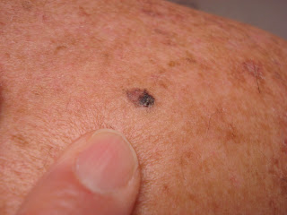
This 64 yo light-complected carpenter noticed a few rough spots on his chest. Since he vacations in Mexico two to three times a years, he is worried re: skin cancer.
A skin exam showed a 4 mm nodular BCC on his chest and a few hypertrophic AKs on the chest. An incidental finding (that he was unaware of) was a 6 mm in diameter irregularly pigmented papule on the right upper back. The dermatoscopic image is ugly and worrisome.
He is scheduled for an excisional biopsy.
This case is very similar to that presented on VGRD last week. Therefore, this patient probably has a SSM that is < 1 mm thick. We will see.
Thursday, December 29, 2016
Tuesday, December 20, 2016
New Pigmented Lesion
Thiss 56 year-old man was brought into the office by his wife who has noticed a new pigmentedlesion on his mid-back for around a year. He did not see the point in coming in, but she made the appointment and came in with him.
The exam showed a 1.1 cm pigmented nodule with a play of color and irregular borders. The dermatoscopic exam shows whitish areas, black areas, and pigment dots (among other things).
Clinically, I thought this was a superficially spreading melanoma. I could not be 100% sure it was not a seborrheic keratosis.
An excisional biopsy was done. Pathology will be added in a few days.
Pathology: Superficial spreading melanoma, at greatest, 0.68 mm thick.
I will recommend a WLE. Not further studies.
Note: Often, a wife will make the appointment for her husband who is reluctant to see the physician. This patient insisted on going to work as a pipe-fitter after the excision. When a wife finds a lesion on her skin and asks her husband to take a look; he often says to her, "If your worried, see a doctor."
Thursday, December 08, 2016
A Case for Diagnosis
History: The patient is an otherwise healthy 67 year-old writer with a three month history of an intensely pruritic papular
and pustular dermatitis in an otherwise. He’s been on Welbutrin, HCTZ and Lipitor for
years. Previously treatments
with triamcinolone 0.1% ointment and prednisone for two weeks were not helpful.
O/E: There are
hundreds of 2 – 3 mm erythematous papules on arms, legs, torso, scalp. Face spared.
No lesions on hands, feet or genitalia.
Clinical Images:
Pathology:
Lab: CBC, Chemistries normal. Wound culure grew 2+ SAUR sensitive to everything.
The patient was treated with Keflex 250 mg qid for a week and Prednisone starting at 40 mg a day. He cleared quickly, but when he stopped the prednisone after ~ 3 weeks, the eruption and pruritus recurred. The new lesions are distinct erythematous papules mostly on the torso. Background looks normal.
Thoughts: Could this be "subacute prurigo" othewise known as Itchy Red Bump Disease? I will have slides reviewed and offer another biopsy to the patient.The patient was treated with Keflex 250 mg qid for a week and Prednisone starting at 40 mg a day. He cleared quickly, but when he stopped the prednisone after ~ 3 weeks, the eruption and pruritus recurred. The new lesions are distinct erythematous papules mostly on the torso. Background looks normal.
Subscribe to:
Posts (Atom)


