This 30 yo woman has noticed an enlarging pigmented lesion in her right axilla for almost a year. Her mother, a registered nurse, asked her to see a dermatologist. Her husband did not notice it.
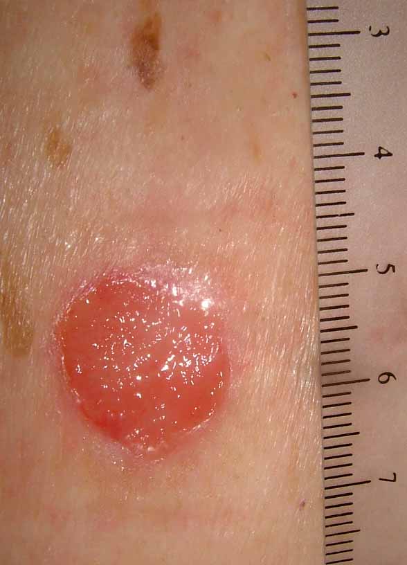
The patient has Type II skin. The general cutaneous exam was unremarkable save for a 1.2 cm in diameter barely elevated plaque. There is a play of pigment and outline is irregular.
An excisional biopsy was performed.
Pathology: Malignant melanoma in situ , superficial spreading type. (Read by H. Byers of BU Skin Path who took photomicrographs) "The specimen exhibits a marked nested and lentiginous melanocytic proliferation with large severely atypical epithelioid cells. There is irregular nesting, focal confluent lentiginous melanocytic proliferation and cellular dyshesion."


She is scheduled for a wider excision, 0.5 cm on either side. No further work-up other than regular follow-up visits.
Your thoughts are appreciated.