Friday, July 29, 2005

84 yo man with Pemphigus


This 84 yo man was seen around weeks ago with an erosive bullous process on torso, head and neck of 4 months duration. He was on no meds by mouth and in good general health.
My initial impression was pemphigus vulgaris vs. impetigo. The skin culture showed many coag + staph, and the bx showed an acantholytic bulla. The DIF was positive for intracellular IgG.
He was treated with dicloxacillin 250 mg qid and prednisone 20 mg tid.
He cleared quickly. At present he is on 30 mg per day of prednisone and tapering by 5 mg every two weeks. He's had no new lesions since therapy was initiated and his itch has disappeared.
Questions:
1) Value of adjuvent therapy? I am thinking of starting in a benign manner with minocycline/
2) Should I try to get on alt. day steroid first?
3) Any suggestions?
Follow-up (August 6, 2005) The patient continues to do well. He is now on Prednisone 20 mg per day. No symptoms and no new lesions. His prednisoe will be dropped by 5 mq every 10 days until 10 mg per day - then we will convert to alt day therapy. I may add minocycline 100 mg bid.

40 yo woman with scarring alopecia


The patient is a 40 yo woman with an eight year history of scarring alopecia.
Her ANA is + at 1:160 - all other labs are normal.
No other cutaneous findings or systemic disease other than "fibromyualgia."
She has been treated with 200 mg of plaquenil b.i.d. for a number of years. This has not been of much help.
The patient is quite concerned about her scalp. She's going through a divorce and worries about her appearance.
Questions?
1) Any value to a biopsy?
2) Would scalp reduction be an option?
3) Role for hari transplantation? Should a biopsy be done first

Tuesday, July 12, 2005

Melanonychia in a Six Year-Old Child

Presented by
Dr. Chee Meng Loh
RMG
Singapore
E mail: bliss88@singnet.com.sg

The patient is a six year old Chinese girl who developed a dark pigmented longitudinal band on the nail of her right index finger about 1 year ago. It started as a narrow, light brown band and grew in darkness (black in centre of band, and lighter shade of brown at edge of band) and width. The band is now about 3 mm in width. It has not changed in the past 3 months.

There is no history of trauma to the right index finger. There is also no complaint of pain in that finger.

The girl suffers from eczema and is on steriod medication (hydrocortisone 1% cream). She is otherwise healthy. There is no history of melanoma in the girl, her parents or grand parents. Neither her parents have pigmented bands on their nails.

Physical Exam: Dark longitudinal pigmented band on right index finger.
Colour ranges from black to brown.
Width is about 3 mm.
(Pictures attached)

Biopsy not performed.

Diagnosis: Melanonychia of right index finger.

Questions:
What are the likely causes of melanonychia in this child?
What is the likelihood that the cause is melanoma?
Should biopsy be performed to rule out melanoma?
If yes, what biopsy procedure is appropriate and what is the risk of permanent damage to the nail?
If a wait and see approach is appropriate, what other signs should the parents watch out for and within what time frame?


Melanonychia
Originally uploaded by David Elpern.


Melanonychia
Originally uploaded by David Elpern.

Friday, July 01, 2005

16 yo girl with papulonecrotic lesions

The patient is a 16 yo girl with a 2 year history of papulonodular lesions on the extremities and face. There is no evidence that these are excoriations. Biopsy 9 months ago from the arm was read as prurigo nodularis, but lesions look atypical and have now started to appear on the face. She is well otherwise.


A repeat biopsy on June 27. 2005 showed:
DIAGNOSIS: Skin - Left Temple:

Epidermal necrosis with s cale crust containing neutrophils , sub-epidermal abundant neutrophils and fibrin deposition, superficial and deep perivascular lymphohistiocytic infiltrate with focal neutrophil ic microabscesses, septal and lobular panniculitis with mixed inflammatory cell infiltrate of abundant neutrophils , lymphocytes , histiocytes, and eosinophils and numerous activated endothelial cells, surrounding a medium-sized vessel with marked mixed inflammatory cell infiltrate of neutrophils , histiocytes and occasional eosinophils .

NOTE : These changes are suggestive of a medium-sized vasculitis with overlying necrosis. Elastic tissue stain (EVG) does not reveal the vessel in the deeper sections, therefore, arterial or venular distinction cannot be made. The differential diagnosis includes a large vessel vasculitis such as periarteritis nodosa or early Wegener's granulomatosis. P.A.S. stain is negative for fungal organisms. Fite stain is negative for mycobacteria . However, an infectious vasculitis cannot be entirely excluded . If the clinical suspicion persists, culture studies may be of help . The differential diagnosis also includes , in the appropriate clinical setting , factitial panniculitis with secondary vascular involvement. These are not the changes of lupus erythematosus , pityriasis lichenoides et varioliformis acuta or prurigo nodularis . Serologic studies may be helpful. Clinico-pathologic correlation is suggested.

Wednesday, June 22, 2005

Bullous Eruption after Surgery

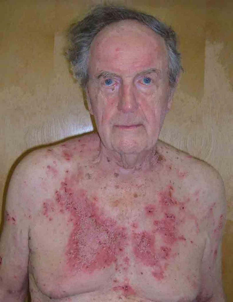
This 72 yo man had a laparoscopic cholecystectomy four months ago in Florida. Within a few weeks, he developed a bullous eruption around the area of surgery. His surgeon and primary care physician have treated with cephalosporins, topical mupirocin and triamcinalone cream. All without avail.
A culture obtained at my office showed: coagulase negative staph resistant to everything except tetracyclines and rifampin. This is probably not significant.
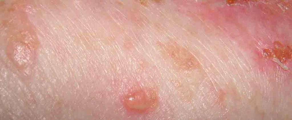
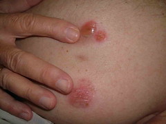
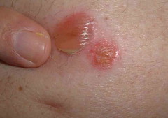
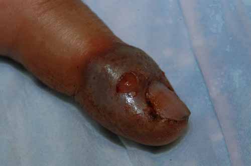
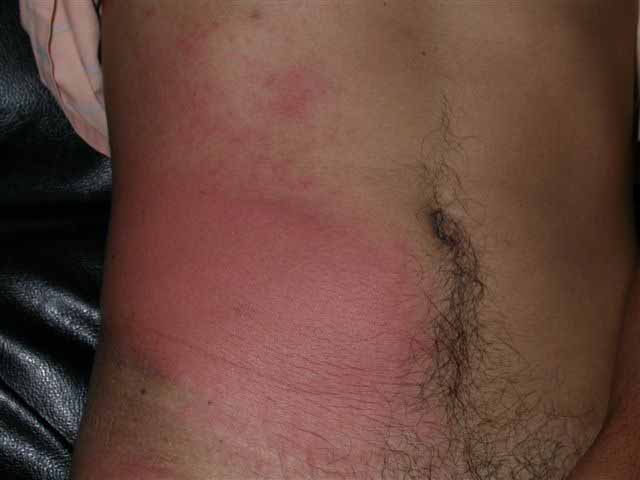
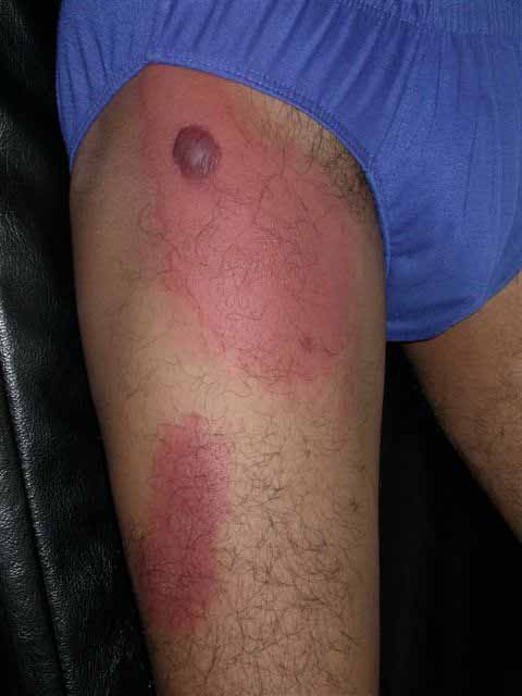
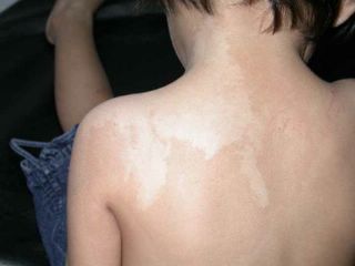
The exam shows flaccid bullae, some with fluid levels of pus. See photos of right subcostal area.
I suspect this is a localized variant of pemphigus that followed surgical trauma.
A biopsy for DIF is planned.
Is there any value iin IIF?
I started him on minocycline for the staph on the odd chance that this is an unusual pathogenic coag negative staph; but the more I think about this, themore convinced I am that this is a benign variant of P.V.
I may start superpotent topical corticosteroids since the disease manisfestation is so localized.
Not sure if localized PV has been described after laparascopic surgery.


Bullous process
Originally uploaded by David Elpern.
Note laparoscopy scar between two bullae


Close-up
Originally uploaded by David Elpern.
Note fluid level of wbcs


Pathology
DIAGNOSIS: Skin - (A) Abdomen:
Intra-and sub-epidermal blister with numerous eosinophils , focal re-epithelialization, eosinophil ic spongiosis and mild s uperficial perivascular lymphocytic infiltrate with numerous eosinophils and papillary dermal fibrosis .
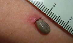
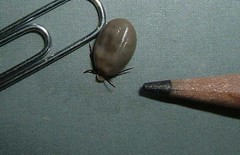
NOTE : These findings are suggestive of re-epithelialized bullous pemphigoid . The differential diagnosis includes a bullous arthropod bite reaction or bullous drug eruption . These are not the changes of pemphigus vulgaris.

DIRECT IMMUNOFLUORESCENCE RESULTS : Perilesional skin sections were incubated with 1:10-1:20 dilutions of antisera specific for IgG, IgM, IgA and C3. "Immunostaining" was not observed.

NOTE : These findings do not support the diagnosis of bullous pemphigoid ; however, they do not exclude it as some rare cases may be negative for these immunoreactants. I f the clinical suspicion persists, an additional biopsy may be of help . Clinico-pathologic correlation is suggested.

Reference:
J Am Acad Dermatol. 1989 Mar;20(3):437-40
Direct immunofluorescence in bullous pemphigoid: effects of extent and location of lesions.
Weigand DA, Clements MK.

Dermatology Service, Veterans Administration Medical Center, Oklahoma City, OK.

We have reevaluated the previously reported conclusion that direct immunofluorescence in bullous pemphigoid is often negative in biopsy specimens from the legs. Duplicate tests from the trunk and legs were generally of equal intensity in a prospectively evaluated series of eight patients with generalized bullous pemphigoid. Also, in 36 patients evaluated retrospectively, the intensity of the direct immunofluorescence reaction correlated roughly with extent of disease, rather than with specific anatomic region. Localized disease predictably required less vigorous treatment to achieve control, but the intensity of the immunofluorescence reaction was not similarly predictive. Direct immunofluorescence is a less useful diagnostic test in localized bullous pemphigoid than in generalized bullous pemphigoid.

Rapidly Growing Lesion in a 57 yo woman

The patient is a 57 yo woman with a 6 month history of a papule on the right arm.
The lesion measures 6 mm in diameter and had not diagnostic features.
An excisional biopsy done since this was a rapidly growing lesion which was not clearly benign.
Pathology shows a malignant melanoma, 2.3 mm thick, Level IV.
Signed out as superficial spreading, but clinically looks more like a nodular melanoma.
She has been referred to a dermatologic oncologist for wide local excision, SLN and other studies.


Bozek2
Originally uploaded by David Elpern.

Sunday, June 12, 2005

78 year old woman with unusual tumor

This 78 yo woman presented with a two week history of a dark growth on the posterior aspect of her shoulder. She had myalgias and felt unusually tired. The exam showed a tick engorged with blood. I could not tell if this was the deer tick that may carry Lyme Disease. Lyme antibidoes were ordered and she was started on amoxicillin 500 mg. tid for three weeks. She's a light complected Caucasian and doxycycline may cause photosensitivity in this season.
The tick was removed by gently grasping it with a forceps and rotating it. It appeared intact and was sent to the lab for identification.


Wynant 004
Originally uploaded by David Elpern.


Wynant 003
Originally uploaded by David Elpern.

Wednesday, June 01, 2005

Cold Urticaria


1 month history
Originally uploaded by David Elpern.
MAY 31, 2005

This 17-year-old high school student presented for evaluation of hives after exposure to cold air or products. This may have followed an upper respiratory infection. It has been present for only about a month.

EXAMINATION: Dermographia is negative. After applying a cold soda can to her arm for two minutes, she developed an urticarial area exactly in the shape of the can five minutes after the can was removed.

IMPRESSION: This is probably the common form of cold urticaria. Some of these cases follow upper respiratory infection. This is idiopathic.

PLAN: She was warned against swimming in very cold water and she was given hydroxyzine to take half an hour before exposure if she knows that she is going to be in a cold environment or be exposed to cold products. I will do a literature search to see what I can come up with for her.

Thoughts??

55 yo woman with new facial lesion

This 55 yo woman has has a one week history of a painful necrotic lesion on her left temple. She suffers from chronic pain and depression but is on no new meds. Her health is otherwise good. Her hemogram is unremarkable. Two days ago, she developed a similar lesion on her left shoulder. At present, it is just an erythematous papule with a surface erosion. He internist started her on cephalexin 500 mg bid before I saw her.
Both lesions, by history, began with a vesicle superimposed on an erythematous papule. She feels well otherwise and has had no fever.


1 week history
Originally uploaded by David Elpern.


In the differential disgnosis I am considering:
Ecthyma
Ecthyma gangrenosum (normal hemogram is against this)
Brown Recluse Spider bite - but these are usually not so symmetrical

A bacterial culture was done but may not be helpful since she was on cephalexin.
I doubt biopsy will help; but will be done if she continues to develop new lesions.

Tuesday, May 24, 2005

23 yo woman with facial erythema

This 23 yo woman has had facial erythema and acuminate micropapular lesions since childhood. She has extensive keratosis pilaris of her arms and thighs in addition. She has not gone out in public without thick make-up for 10 years. Her fiance has never seen her without make-up. She is desperate to haved this treated. I suspect this is a variant of KP rubra facei, possibly with ulerythema oopryogenes


KPRF 005
Originally uploaded by David Elpern.


KPRF 006
Originally uploaded by David Elpern.

Saturday, May 07, 2005

Seven Year Old Boy with Chronic Dermatitis

A.D. is a 7 year-old boy who was adopted from Siberia by a family in western Massachusetts at 21 months of age. Since adoption he has had recurrent dermatitis on torso and extremities. The lesions are mostly nummular by history. They have responded to systemic antibiotics on occasion. He was seen here yesterday for the first time for a second opinion.

The child appears normal otherwise. He has no evidence of atopy. This is the largest lesion. All are plaques, all covered with some crust, intensely pruritic. I applied some pressure to the large plaque with a cotton tipped applicator and a small amount of creamy pus was extruded.

My working diagnosis is nummular eczema driven by hypersensitivity to staph. A culture was taken and I'll wait for results before treating. This has been going on for 5 years. I may do a biopsy, but am not sure it will be helpful.

Any thoughts would be appreciated. The parents are at their wits end. He has also been treated with mupirocin cream in past with some success. Tacrolimus was not helpful.

The culture grew out coagulase positive staph sensitive to everything; even Penicillin G. This is unusual in the U.S. where most Saph is resistant to penicillin. One wonders if this is a strain he brought over from Russia when he was adopted. I started him on Pen VK 250 mg qid. I will add a topical corticosteroid and mupirocin - the latter for nares and crural folds. Will give follow-up after a couple of weeks. If he continues to have staph infections like this, I will look into his Ig status.


Right leg

Wednesday, April 27, 2005

A Change of Pace

The patient accompanied her "mother" to the office. She is a 12 yo chihuaha with autoimmune thyroid disease and extensive alopecia areata. I was aksed what could be done and injected large areas of her skin with intralesional triamcinalone. Unfortunately, there is no billing code for this - the dog lacked health insurance.
(Actually, I deferred treatment until I hear from my august colleagues)

Note tail alopecia in Fig. 3


Fig 1


Fig 2


Fig. 3

Ref:
A natural canine homologue of alopecia areata in humans.
Tobin DJ, Gardner SH, Luther PB, Dunston SM, Lindsey NJ, Olivry T.
Department of Biomedical Sciences, University of Bradford, Bradford, UK.
Br J Dermatol. 2003 Nov;149(5):938-50.

BACKGROUND: Alopecia areata (AA) is suspected to be an autoimmune disease directed preferentially against hair follicles (HF) affecting both humans and various mammalian species. Recently, two rodent models of AA were described, namely the ageing C3H/HeJ mouse and the DEBR rat. Despite several case reports of canine AA in the literature, there has been no systematic assessment of the disease in these companion animals, and it is also not known whether dogs with AA could be useful as an outbred homologue of this disease in humans. OBJECTIVES: To evaluate the clinical, histopathological and immunopathological features of 25 dogs with AA and compare these data with those found in the human disease. PATIENTS/METHODS: Twenty-five client-owned dogs exhibiting macroscopic alopecia with peri- or intrabulbar lymphocytic infiltrates were selected for study. Biopsies and sera were obtained and assessed by histopathology, direct immunofluorescence of immunoreactant deposition, immunohistochemistry for lymphocyte markers, indirect immunofluorescence and immunoblotting analysis of circulating serum IgG, selective immunoprecipitation of HF proteins by serum IgG, and passive transfer of purified canine IgG into naive C57BL/10 mice. RESULTS: Clinical signs including alopecia, skin hyperpigmentation and leucotrichia usually developed during adulthood and were first seen on the face, followed by the forehead, ears and legs. Spontaneous remission of alopecia occurred in 60% of dogs and regrowing hair shafts were often non-pigmented. Histological examination of skin biopsy specimens revealed peri- and intrabulbar mononuclear cell infiltrates affecting almost exclusively anagen HF. Direct immunofluorescence analysis detected HF-specific IgG in 73% of dogs, while indirect immunofluorescence revealed circulating IgG autoantibodies to the HF inner and outer root sheaths, matrix and precortex. Immunoblotting analysis revealed IgG reactivity to proteins in the 45-60 kDa molecular weight range and with a 200-220 kDa doublet. The latter was identified as trichohyalin by selective immunoprecipitation. Purified HF-reactive IgG, pooled from AA-affected dogs, was injected intradermally to the anagen skin of naive mice where it was associated with the local retention of HFs in an extended telogen phase in AA-treated skin compared with that seen in controls. CONCLUSIONS: These findings are very similar to those reported for human AA patients; therefore, they support the consideration of dogs with AA as a useful homologue for the study of the pathogenesis of this common autoimmune disease of humans.

Sunday, April 24, 2005

Asymptomatic Blanching Palmar Eruption


Asymptomatic non blanching eruption Posted by Hello

A 14-year-old, otherwise healthy boy presented with an asymptomatic red blanching eruption on the palms that developed about one week earlier. Soles were not involved. No inciting factor including drugs or illness could be recognized. Rest of the physical examination and routine blood and urine tests were unremarkable. What conditions can give rise to this eruption and how should we work it up ?
Shahbaz A Janjua MD

Case for diagnosis (Alberta, Canada)

Day #1 this guy helped deliver a breech calf by c-section which was so traumatic that the calf was still-born and then the following day he helped butcher a cow. On Day #3 he noted swelling of his left index finger and by
Day #4 it blew up to look like these pictures. The more I read about orf the more I feel that this is likely the case. The risk factors are there and the time of year and setting is classic. Anyway, what do the viewers think?

(The break in the skin/expelled fluid in the pictures is my doing. There
were no vesicles or absesses and the tissue was tough and non-fluctuent.)

Duncan


Sunday, March 27, 2005

20 yo man with cutaneous larva migran


The patient is a 20-year-old man who noticed multiple intensely pruritic creeping lesions on his abdominal wall for more than a month. He is a body building enthusiast.

Examination showed multiple 2-3mm wide serpiginous raised erythematous tunnels on the left periumbilical and right upper quadrant of the abdominal wall.
Treatment was with freezing 1cm distal to the end of the trail with liquid nitrogen and adding oral mebendazole (zentel) 200mg bd for 3 days. It was successfully eradicated within a week of treatment.

Henry

 Posted by Hello

Wednesday, March 23, 2005

10 year old girl with endogenous cheilitis


This patient presented with recurrent dry and fissured lips for more than 2 years. She has a strong family history of atopy. Her brothers and sisters have asthma and allergic rhinitis. Fortunately she didn't use any lipstick.

Examination showed fissures and dryness on both the upper and lower lips. There were crusts formations on the lips

Clinically she has endogenous (atopic) cheilitis

There are few factors that need to be considered here. is there a contact allergen element here? Lipstick would be the biggest culprit here. Is there a need to do a patch test?

Irritants can be an aggravating factor too. Lip smacking and hot spicy food can aggravate the eczema. Even toothpaste with strong mint flavour can be a factor too. I have seen several patients whose cheilitis was aggravated by our local "Darlie" toothpaste.

Moisturisers eg vaselin would be very helpful and they can be applied 4-5 times daily. Mild topical corticosteroids eg 1% hydrocortisone ointment or cutivate ointment helps. I prefer ointment to cream base in this situation.

Henry
 Posted by Hello

34 year old man with multiple erythematous plaques

Recently I saw this interesting patient: a 34 yr old factory worker presented with redness over the thigh and lower abdomen for a week. Started as "burning discomfort" over the left knee and then spread to the right knee, thigh and lower abdomen. He has no fever. No history of diabetes. He denied any insect bite. There was no preceding drug history.
Examination showed diffuse and glistening redness on the thighs and lower abdomen. On the right thigh, there was a single non hemorrhagic blister. Regional nodes were not enlarged. The lesion was not particularly tender. He was afebrile.


Impression: cellulitis - right thigh. Unusual site for cellulitis though. what is your take on this patient?

Henry

Tuesday, March 22, 2005

18 yo girl just returned from Costa Rica

This 18 year old girl just returned from a school trip to Costa Rica. Her group explored streams and swamps. She developed asymptomatic papules and pustules on the dorsae of her feet after being in stagnant muddy water and a running stream. I am wonderind if this is a gram negative organism or a parasite. Any thoughts?

36 yo man with lesion on back

This 36 yo man presented with a 1.6 cm in diameter pigmented plaque on his back for approximately 2 years. Barely elevated. Has enlarged over past few months. Dx: Melanoma vs. Seborrheic keratosis. An excisional bx was performed today.


Path Report
DIAGNOSIS: Skin - Right Mid Back:

Malignant melanoma.

Type: Superficial spreading
Greatest thickness: 0.90 mm.
Anatomic level: II
Margins: Complete excised
Radial growth phase: Present
Vertical growth phase: Absent
Mitoses: None
Tumor infiltrating lymphocytes: Present, non-brisk
Ulceration: Absent
Regression: Present
Microsatellites: Absent
Vascular invasion: Absent
Precursor lesion: Not identified

NOTE: The lesion represents a severely atypical compound melanocytic neoplasm characterized by a predominantly intra-epidermal component with marked confluent lentiginous and nested melanocytic hyperplasia , pagetoid spread, extension into adnexae, and by a severely atypical dermal component with papillary dermal regression.

Sunday, March 20, 2005

2 yo girl with segmental hypopigmentation

This is a 2 year old girl who has a patch of hypopigmentation on her left shoulder since birth. It extends from back of neck to the left shoulder. It was asymptomatic. Examination showed a hypopigmented macule 5cm by 10cm on the back of left shoulder. It is segmental in distribution and has an irregular border. There was no central hypoaesthesia. She has no ophthalmic or CNS defects.


Clinically she has nevus depigmentosus or some form of pigmentary mosaicism. Biopsy of the lesion was not done.

This is usually a benign skin disorder and is caused by the functional defects of melanocytes and the morphologic abnormalities of melanosomes.