Thursday, September 28, 2017

Metastatic melanoma in an elderly man

Presented by Henry Foong, M.D.
Ipoh, Malaysia

The patient is a 80-year-old man who presented with swelling of the left leg for 3 months. 

About 3 years ago he had a motor vehicle accident where he injured his left leg.  His attending doctor noticed a pigmented growth on the left foot associated with inguinal node swelling. A surgeon excised the pigmented growth on the left foot and removed some nodes from the left groin.  No histological reports were available at the moment.  The patient was well until recently when he noticed gradual swelling of the left leg with multiple pigmented nodules on the surface.  The leg was occasionally painful at night. He did not have any constitutional symptoms.  There was no family history of skin cancers. No significant other medical illness.

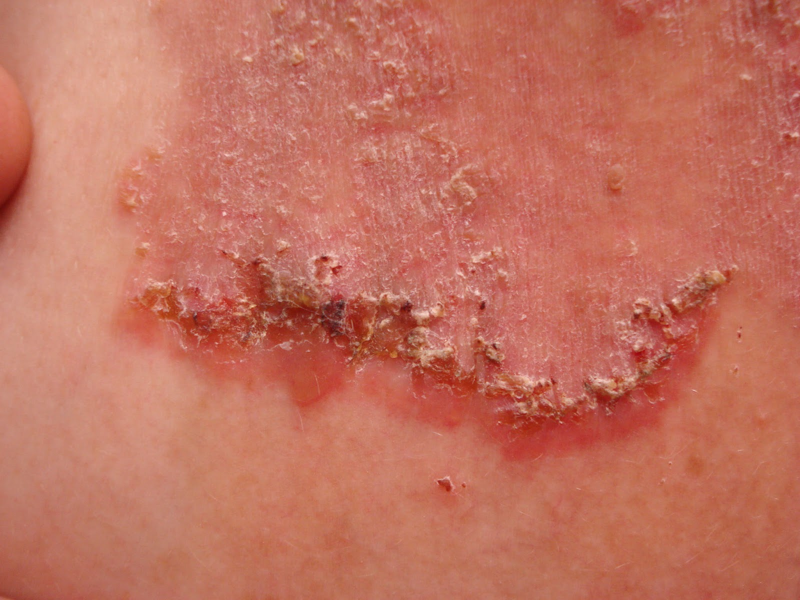
On examination his left leg was swollen and oedematous with many pigmented papules and nodules on the foot and lower 1/3 of the left leg.  A pigmented ulcerating tumour  5 x 5 cm was noted on the left foot which extended to the heel.  A firm matted lymph node swelling was noted on the left groin.  There was a surgical scar over the left groin.  No hepatosplenomegaly was present.

Biopsy of the pigmented papule on the foot was done and confirmed malignant melanoma.
Sheets and nests of malignant cells are seen invading the dermis. The
tumour cells show marked pleomorphism, have increased nucleo-cytoplasmic ratio, vesicular nuclei with prominent nucleoli and eosinophilic cytoplasm. Many of the cells contain melanin pigment. Numerous mitotic figures are seen. The tumour is seen at the margins. Masson Fontana stain is focally positive.
Skin biopsy Report: Features are consistent with malignant melanoma.

His work up included an oncology referral. CT scan of abdomen and pelvis which showed pelvic and para-aortic  lymph 
node metastasis.  CXR normal.  TWBC was 16,400. BRAF gene mutation studies pending 

He was being treated for concomitant cellulitis with IV antibiotics.

Questions:  What are the treatment options (targeted therapy) for his metastatic melanoma? Would oral vemurafenib and Anti PD-1 antibodies e.g.  pembrolizumab be useful?  They are very expensive though for most patients in Malaysia.
It's almost certain this patient will probably opt for palliative treatment. What local treatment of the in-trasit metastasis would be useful for him?

Thank you for your thoughts!


Wednesday, September 20, 2017

17 year-old girl with 8 year history of scalp dermatitis


The patient is a 17 year-old girl with an 9 year history of thick scales on her scalp.  She has used multiple medications without relief.  The patient has been bullied at school where she has been called “lice girl.”  Socially, this has been traumatic.

O/E:  She is a well-developed and well-nourished 17 yo with thick chestnut colored hair or normal intelligence.  There are no areas of alopecia. Thick, silvery adherent scales are present on the occipital, parietal and temporal scalp.  When these are removed, hair roots come out, too.  The remainder of the cutaneous examination is normal.  No nail dystrophy.
Clinical Images (July 2017)
Lab: Fungal culture negative.  Bacterial culture 3+ Staph aureus.

Failed Treatments (per mother):

"Every single otc dandruff shampoo
Every prescription medicated dandruff shampoo
Scalpicin
Prescribed scalp drops with and without coal tar
Every Tea Tree product you can find otc
Hot oil treatments
P & S Oil
Nutrogena T-gel and T-sal
Olive oil"
Terbinafine 250 mg p.o. x 1 month
Keflex 500 mg b.i.d. x 2 weeks

Scalp Biopsy read by Lynne Goldberg (Boston University Skin Path): was felt to be most compatible with psoriasis.  Seborrhea was in the differential diagnosis but less likely.

Diagnosis:  Working Dx:  Tinea amiantacea secondary to psoriasis.

Discussion: This 17 yo girl has suffered with what appears to be tinea aminatacea for almost a decade.  It appears unlikely that this is psoriasis. Tinea capitis has been ruled out by culture.  Her bacterial culture showed 3+ S. aureus but I suspect this is a secondary invader as she did not improve with cewplanexin.  Since the fungal  culture was negative and these approaches were not helpful, I may recommend isotretinoin.  The use of this has been reported for T. aminatacea only and in a Korean case report.

Dr. Goldberg's rotocol for Scalp Psoriasis, Tinea amiantacea and Related disorders:
1. Wet hair at night
2. Apply Dermasmoothe scalp oil liberally to scalp. Leave on overnight
3. Sleep with this overnight in a shower cap (to protect pillow)
4. Shampoo in the morning with T-Sal or other dandruff shampoo

Do this nightly at first if possible, but after a week or so she will be better and will not need to do it every night.


References:

1. Abdel-Hamid I et al. Pityriasis amiantacea: a clinical and etiopathologic study of 85 patients. Int J Dermatol. 2003 Apr;42(4):260-4.

2. Kwon JI.  Isotretinoin for Tinea amiantacea (A Case Report). Korean J Dermatol 2012;50(11):1002-1005 (In Korean)

3.  Mannino G, McCaughey C, Vanness E. A case of pityriasis amiantacea with rapid response to treatment WMJ. 2014 Jun;113(3):119-20.  Full Free Text.


4. Scalp psoriasis: European consensus on grading and treatment algorithm.  Ortonne J. J Eur Acad Dermatol Venereol. 2009 Dec;23(12):1435-44.

Monday, September 18, 2017

Cheilitis Query


September 2017
The patient is a 70 yo Caucasian who has lived on Moorea, French Polynesia, for the past 50 years.  She contacted us recently about her painful lips because there is no dermatologist available to her at present.  Here is her anamnesis:
In early July when I went to Montreal, my lips started to bother me. I thought maybe it was 18 hours on a plane, or even maybe it was a sunburn from being in the pool with my grandchildren in sunny Vancouver a couple weeks before. It didn't subside and I bought several lip therapies - cocoa butter, Vaseline, Aquaphor. When I came home I used a mild steroid ointment for a couple weeks, but to no effect. I now carry Aquaphor with me all the time and apply it constantly. Chapstick with SPF (from a friend in the States) stings my lips, as does toothpaste. My lips are not chapped, as in flaky or peeling, but they feel and look burnt, even blistery sometimes, and they can feel severely tight, dry and very sore. Actually, my upper lip is not as involved as my lower lip, and the corners are not affected. 

Photo sent by patient to VGRD

Diagnosis: This appears to be actinic cheilitis or possibly allergic/irritant cheilitis.  Strangely,  the patient got more sun in Vancouver than she does in French Polynesia!

What are your thoughts?

Update (April 2018):
(from the patient) I want to share with you the results of a recent experiment I conducted unwittingly. Our daughter was visiting Tahiti for the last week, and we went to the beach several days in a row. Although I wore a hat and Vanicream lip sunscreen, I got too much sun and my lower lip has been on fire. I think actinic cheilitis was an early guess last year, and I have NO DOUBT that it was correct. I'm using Vaseline, of course, and the betamethasone dipropionate ointment after nothing else worked. I'm happy not to be puzzled, confused and freaked out this time around.  Note:  I think we may be dealing with a case of actinic prurigo of the lips (see references 3 and 4 below)


References:

1. Actinic cheilitis: a treatment review.
Shah AY, Doherty SD, Rosen T.
Abstract:  All other factors being equal, the presence of actinic cheilitis, a pre-invasive malignant lesion of the lips, doubles the risk of squamous cell carcinoma developing in this anatomic area. Various forms of local ablation, immunomodulation and surgical extirpation have been proposed as therapeutic interventions. This paper critically evaluates the available medical literature to highlight the evidence-based strength of each recommended therapy for actinic cheilitis. Vermilionectomy remains the gold standard for efficacy; trichloroacetic acid application is easy and convenient, but the least efficacious overall.

2. Contact allergy in cheilitis.
O'Gorman SM, Torgerson RR. Int J Dermatol. 2016 Jul;55(7):e386-91.
BACKGROUND: Recalcitrant non-actinic cheilitis may indicate contact allergy.
CONCLUSIONS: Contact allergy is an important consideration in recalcitrant cheilitis. Fragrances, antioxidants, and preservatives dominated the list of relevant allergens in our patients. Nickel and gold were among the top 10 allergens. Almost half (45%) of these patients had a final diagnosis of ACC. Patch testing beyond the oral complete series should be undertaken in any investigation of non-actinic cheilitis.

3. Actinic Prurigo Cheilitis: A Clinicopathologic Review of 75 Cases.
Plaza JA, et al. Am J Dermatopathol. 2016 Jun;38(6):418-22.

4. Actinic prurigo of the lip: Two case reports.
Miranda AM. World J Clin Cases. 2014 Aug 16;2(8):385-90. Free Full Text.

Tuesday, September 05, 2017

Man from India with Wide-Spread Vesicular Eruption


Presented by Dr. Bassem Ghali
Jagadguru Sri Shivarathreeswara University
Mysuru, India


The patient is a 60 yo man with a  pmhx of COPD who noted a recent eruption of vesicles on trunk, as well as his forehead and scalp. No fever. No other pertinent history. No itch. No pain. No new meds.
It started as blisters on the trunk, slightly itchy but no other symptoms, and not painful. The lesions opened up with clear fluid being expressed and leaving shallow ulcers. They have started become generalized with new lesions on the scalp and genitalia.

O/E:  Lesions appear like small bullae/vesicles on chest and abdomen, with clear fluid. In the center is what appears to be a black point, probably the hair follicle. There is no erythema on or around these lesions. 

Clinical Photo:

 Diagnosis:  What are your thoughts?

Friday, September 01, 2017

Hailey-Hailey Disease

The patient is a 38 year-old cook with a 14 year history of a pruritic, occasionally painful rash on his torso.  He used triamcinalone cream in the past without relief.  No family history aof a similar process.

O/E.:  There are plaques in axillary folds, on the chest, back, and popliteal fossae.Some advancing borders appear vesicular on close observation.  There is mild post-inflammatory hyperpigmentation in healed sites.

Clinical Photos from L. Axilla:
Laboratory:  An incisional biopsy was taken from the crusted/vesicular edge.

Histopathology:  Courtesy Dr. Lynne Goldberg. Boston University Skip Path


Tentative Diagnosis:  Benign Familial Pemphigus (Hailye-sHaily)

Postscript:  After one month of doxycycline 100 mg b.i.d. and betamethasone 0.1% cream the patient was > 90% resolved.  He is pain and itch free.  He was asked to cut back on the cream to prn and will continue the doxycycline.  It will be tapered in a month.