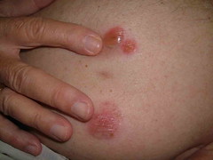
This 72 yo man had a laparoscopic cholecystectomy four months ago in Florida. Within a few weeks, he developed a bullous eruption around the area of surgery. His surgeon and primary care physician have treated with cephalosporins, topical mupirocin and triamcinalone cream. All without avail.
A culture obtained at my office showed: coagulase negative staph resistant to everything except tetracyclines and rifampin. This is probably not significant.
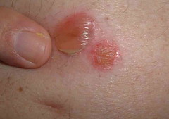
The exam shows flaccid bullae, some with fluid levels of pus. See photos of right subcostal area.
I suspect this is a localized variant of pemphigus that followed surgical trauma.
A biopsy for DIF is planned.
Is there any value iin IIF?
I started him on minocycline for the staph on the odd chance that this is an unusual pathogenic coag negative staph; but the more I think about this, themore convinced I am that this is a benign variant of P.V.
I may start superpotent topical corticosteroids since the disease manisfestation is so localized.
Not sure if localized PV has been described after laparascopic surgery.
Note laparoscopy scar between two bullae
Note fluid level of wbcs
Pathology
DIAGNOSIS: Skin - (A) Abdomen:
Intra-and sub-epidermal blister with numerous eosinophils , focal re-epithelialization, eosinophil ic spongiosis and mild s uperficial perivascular lymphocytic infiltrate with numerous eosinophils and papillary dermal fibrosis .
NOTE : These findings are suggestive of re-epithelialized bullous pemphigoid . The differential diagnosis includes a bullous arthropod bite reaction or bullous drug eruption . These are not the changes of pemphigus vulgaris.
DIRECT IMMUNOFLUORESCENCE RESULTS : Perilesional skin sections were incubated with 1:10-1:20 dilutions of antisera specific for IgG, IgM, IgA and C3. "Immunostaining" was not observed.
NOTE : These findings do not support the diagnosis of bullous pemphigoid ; however, they do not exclude it as some rare cases may be negative for these immunoreactants. I f the clinical suspicion persists, an additional biopsy may be of help . Clinico-pathologic correlation is suggested.
Reference:
J Am Acad Dermatol. 1989 Mar;20(3):437-40
Direct immunofluorescence in bullous pemphigoid: effects of extent and location of lesions.
Weigand DA, Clements MK.
Dermatology Service, Veterans Administration Medical Center, Oklahoma City, OK.
We have reevaluated the previously reported conclusion that direct immunofluorescence in bullous pemphigoid is often negative in biopsy specimens from the legs. Duplicate tests from the trunk and legs were generally of equal intensity in a prospectively evaluated series of eight patients with generalized bullous pemphigoid. Also, in 36 patients evaluated retrospectively, the intensity of the direct immunofluorescence reaction correlated roughly with extent of disease, rather than with specific anatomic region. Localized disease predictably required less vigorous treatment to achieve control, but the intensity of the immunofluorescence reaction was not similarly predictive. Direct immunofluorescence is a less useful diagnostic test in localized bullous pemphigoid than in generalized bullous pemphigoid.
Wednesday, June 22, 2005
Rapidly Growing Lesion in a 57 yo woman
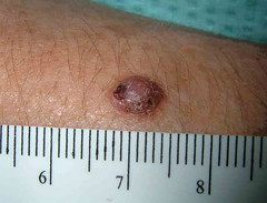
The patient is a 57 yo woman with a 6 month history of a papule on the right arm.
The lesion measures 6 mm in diameter and had not diagnostic features.
An excisional biopsy done since this was a rapidly growing lesion which was not clearly benign.
Pathology shows a malignant melanoma, 2.3 mm thick, Level IV.
Signed out as superficial spreading, but clinically looks more like a nodular melanoma.
She has been referred to a dermatologic oncologist for wide local excision, SLN and other studies.
Sunday, June 12, 2005
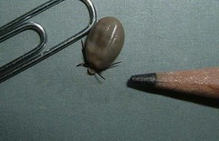
78 year old woman with unusual tumor
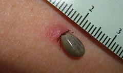
This 78 yo woman presented with a two week history of a dark growth on the posterior aspect of her shoulder. She had myalgias and felt unusually tired. The exam showed a tick engorged with blood. I could not tell if this was the deer tick that may carry Lyme Disease. Lyme antibidoes were ordered and she was started on amoxicillin 500 mg. tid for three weeks. She's a light complected Caucasian and doxycycline may cause photosensitivity in this season.
The tick was removed by gently grasping it with a forceps and rotating it. It appeared intact and was sent to the lab for identification.
The tick was removed by gently grasping it with a forceps and rotating it. It appeared intact and was sent to the lab for identification.
Wednesday, June 01, 2005
Cold Urticaria
MAY 31, 2005
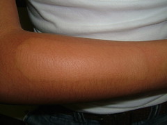
This 17-year-old high school student presented for evaluation of hives after exposure to cold air or products. This may have followed an upper respiratory infection. It has been present for only about a month.
EXAMINATION: Dermographia is negative. After applying a cold soda can to her arm for two minutes, she developed an urticarial area exactly in the shape of the can five minutes after the can was removed.
IMPRESSION: This is probably the common form of cold urticaria. Some of these cases follow upper respiratory infection. This is idiopathic.
PLAN: She was warned against swimming in very cold water and she was given hydroxyzine to take half an hour before exposure if she knows that she is going to be in a cold environment or be exposed to cold products. I will do a literature search to see what I can come up with for her.
Thoughts??
55 yo woman with new facial lesion
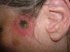
This 55 yo woman has has a one week history of a painful necrotic lesion on her left temple. She suffers from chronic pain and depression but is on no new meds. Her health is otherwise good. Her hemogram is unremarkable. Two days ago, she developed a similar lesion on her left shoulder. At present, it is just an erythematous papule with a surface erosion. He internist started her on cephalexin 500 mg bid before I saw her.
Both lesions, by history, began with a vesicle superimposed on an erythematous papule. She feels well otherwise and has had no fever.
In the differential disgnosis I am considering:
Ecthyma
Ecthyma gangrenosum (normal hemogram is against this)
Brown Recluse Spider bite - but these are usually not so symmetrical
A bacterial culture was done but may not be helpful since she was on cephalexin.
I doubt biopsy will help; but will be done if she continues to develop new lesions.
Subscribe to:
Posts (Atom)