Dear Colleagues,
Please help me with a 4 year-old lad who was referred today by his GP for genital warts. A court-appointed foster parent brought him in for evaluation. His 21 year-old mother left him recently with relatives. She is an inattentive parent with an alcohol and possibly a drug problem. The child was in pre-school but was suspended a few days ago because he was using sexually explicit language, being physically abusive to his peers, discussing French kissing and saying he was going to shoot himself.
The exam showed a quiet, calm and health 4 year-old. The only findings were lobulated and verrucous flesh-coloured papules grouped on the shaft of the penis. There were no bruises or evidence of trauma.
Diagnosis: Condyloma acuminata.
Photos were removed at advice of a pediatrician. Apparently, FBI may moniter some sites. Gets my Canadian soul anxious!
Discussion:
Although genital warts in children can occur in the absence of child sexual abuse, this case is suspicious. The child's broken home, the fact that he has been left with numerous teenage girls as baby sitters, his sexually explicit language, his antisocial behavior and language all are red flags for a child at risk.
Here, in Nova Scotia, we are mandated to report such children to the authorities. I would like to ask wo questions:
1) Your opinion as to whether reporting this child is appropriate.
2) How should he be treated?
a) electrosurgery under general anesthesia
b) cryo therapy
c) imiquimod or podophyllin.
Your thoughts and comments are most welcome.
Hamish Dunwoodie, M.D. FRCPC - Paediatrics
Moncton, NS, Canada
Saturday, December 30, 2006
Thursday, December 28, 2006
Unusual Case
This 53 yo man presented with a three week history of a pustular eruption on the left small finger. It began as a papule and slowly enlarged.
History reveals that he has a farm with 500 sheep. He has also been exposed to cats and dressed a deer around a month ago.
Diagnostic tests were done.


Follow-up: The KOH prep was positive. The fungal culture is positive at ten days. Thus, this is inflammatory tinea manus. Await fungal culture for typing of dermatophyte. Resolved with dilute Clorox soaks and 2% ketoconazole cream in one week.
12 days after initial visit.

Saturday, December 16, 2006
Unilateral Bullae
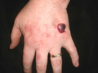
History
The patient is a 28 yo man referred on a walk-in basis with an eight month history of a bullous eruption on the dorsum of his left hand. He gets one or two painless lesions a month. No history was sent over with the patient and he is a poor historian. His medications include Welbutrin, Clozeril and Soma (carisprodol/aspirin)
Exam:
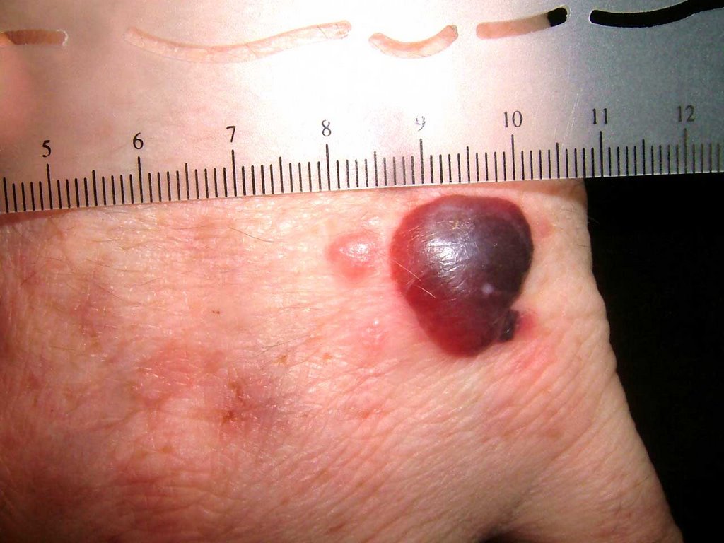
The only findings are on the dorsum of the left hand. Here, there is a hemorrhagic bulla, a vesicle and areas of mild erythema at sites of previous lesions.

Biopsy:
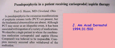
I suspect fixed drug or pseudoporphyria cutanea tarda. I biopsied the small papule and the edge of the bullae and made a follow-up appointment.
Path Report:
A subepidermal separation, individually necrotic keratinocytes , and a sparse superficial perivascular lymphocytic infiltrate with occasional neutrophils .
NOTE : (A and B). Amphophilic globular material is seen deposited in the dermis and around blood vessels in both specimens. This material is P.A.S. stain positive, and stains negative for amyloid and elastic tissue. The differential diagnosis could include porphyria cutanea tarda , although this is usually less inflammatory. A subepidermal autoimmune bullous disorder could also be considered. An additional biopsy for direct immunofluorescence may be of help.
Laboratory Studiess:
CBC normal, LFTs normal, ferritin normal. Hep B and C negative, All urinary porphyrins well within normal levels. Uroporphyrin: 9.6 ug (nl < 30.0) Coproporphyrin 40 ug (normal < 65/24 hr)
Discussion: This is likely pseudoporphyria cutanea tarda. A similar case has been reported. Your comments are welcomed.
The patient is a 28 yo man referred on a walk-in basis with an eight month history of a bullous eruption on the dorsum of his left hand. He gets one or two painless lesions a month. No history was sent over with the patient and he is a poor historian. His medications include Welbutrin, Clozeril and Soma (carisprodol/aspirin)
Exam:
The only findings are on the dorsum of the left hand. Here, there is a hemorrhagic bulla, a vesicle and areas of mild erythema at sites of previous lesions.

Biopsy:
I suspect fixed drug or pseudoporphyria cutanea tarda. I biopsied the small papule and the edge of the bullae and made a follow-up appointment.
Path Report:
A subepidermal separation, individually necrotic keratinocytes , and a sparse superficial perivascular lymphocytic infiltrate with occasional neutrophils .
NOTE : (A and B). Amphophilic globular material is seen deposited in the dermis and around blood vessels in both specimens. This material is P.A.S. stain positive, and stains negative for amyloid and elastic tissue. The differential diagnosis could include porphyria cutanea tarda , although this is usually less inflammatory. A subepidermal autoimmune bullous disorder could also be considered. An additional biopsy for direct immunofluorescence may be of help.
Laboratory Studiess:
CBC normal, LFTs normal, ferritin normal. Hep B and C negative, All urinary porphyrins well within normal levels. Uroporphyrin: 9.6 ug (nl < 30.0) Coproporphyrin 40 ug (normal < 65/24 hr)
Discussion: This is likely pseudoporphyria cutanea tarda. A similar case has been reported. Your comments are welcomed.

Subscribe to:
Posts (Atom)