Saturday, August 13, 2005

Hemangioma of Auricle


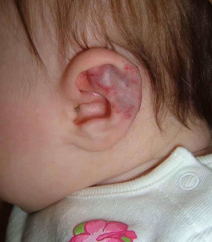
This 4 month infant girl was born with hemangiomas of the chin and left ear.
The lesion on the chin (not pictured) measures 1 cm in diameter and is typical of a congenital hemangioma.

The abnormality of the left ear involves the triangular fossa and the helix. I am concerned that as this involutes it could cause disfigurement. At the same time, I wonder if anyone has experience handling similar lesions in this site. A PubMed site found only a paucity of pertinent references.

Ref:
Cavernous hemangioma of the external ear canal.
Reeck JB, Yen TL, Szmit A, Cheung SW.
Laryngoscope. 2002 Oct;112(10):1750-2. Related Articles, Links
Division of Otology, Neurotology and Skull Base Surgery, Department of Otolaryngology-Head and Neck Surgery, Veterans Administration Medical Center, San Francisco, California, USA.

OBJECTIVE: To document the occurrence of a cavernous hemangioma of the external ear canal and to review the relevant literature.STUDY DESIGN Case report and literature review. METHODS: Review of a patient chart, imaging studies, operative report, and histologic findings. RESULTS: A cavernous hemangioma of the external ear canal not involving the tympanic membrane was surgically excised without complication. This is the third documented cavernous hemangioma of the external ear canal without tympanic membrane involvement in the English literature. Computed tomography scan is invaluable to narrow the differential diagnosis. Complete removal is curative. CONCLUSIONS: Cavernous hemangioma of the external ear canal with or without tympanic membrane involvement is a rare otologic entity amenable to surgical treatment. Temporal bone computed tomography scan imaging is an important preoperative diagnostic tool.

Wednesday, August 10, 2005

Nevus, Congenital


This 20 yo college student presented in consultation for removal of a congenital nevus.
The lesion is located just distal to her left knee. She's been embarrassed about this since childhood and for the past six or seven years won't let anyone see it. Indeed she covers it at all times with bandaids.
The lesion measures 5.2 cm in diameter.
I have referred her to a surgeon for removal since I think that's better than psychotherapy. To minimize the scar it may need staged excision.
The young woman appears normal in all other respects - she is very upset about the lesion and the questions she gets about it.
I wonder if this has been reported. I would hardly call this Body Dysmprphic Syndrome.
Your thoughts are appreciatged.

Not much written about this area -- here's a ref that may touch on it:


Congenital melanocytic nevi. Evaluation and management.
Marghoob AA., Dermatol Clin. 2002 Oct;20(4):607-16, viii.
Department of Medicine, Dermatology Division, Memorial Sloan-Kettering Cancer Center, 1275 York Avenue, New York, NY 10021, USA. marghooa@mskcc.org

This article discusses the care of patients with CMN, who often require
a multidisciplinary approach involving pediatricians, family physicians,
internists, dermatologists, psychologists, plastic surgeons,
neurologists, and radiologists. The cosmetic and psychosocial issues,
combined with the knowledge of the increased risk of developing
melanoma or NCM, is a huge burden that many of these patients
and their families have to carry. This article describes the importance
for physicians to help these patients and families come to terms
with these issues, as well as remind their patients and their family
members that although melanoma, NCM, or other complications can
develop, most affected individuals do not develop any complications.
The article mentions that there are many healthy, happy, functional
adults with large, small, and multiple CMN alive today.

Monday, August 08, 2005

Imiquimod Side-Effect?


This 66 yo woman began topical imiquimod on July 27, 2005 for a biopsy proven superficial BCC of the right upper lip. One week later, she developed aphthous ulcers on the mucous membrane surface of the area just below the site treated. A literature search retrieved a reference to his occurence. This is another unexpected adverse effect. It is unclear how common this is.

Aphthous ulcers associated with imiquimod and the treatment of
actinic cheilitis.
Chakrabarty AK, Mraz S, Geisse JK, Anderson NJ.
J Am Acad Dermatol. 2005 Feb;52(2 Suppl 1):35-7.
Solano Clinical Research, Davis, California, USA. chakak3@yahoo.com

Our case series report is the first documented depiction of the
appearance of aphthous ulcers secondary to imiquimod
application. This case series presentation discusses the
underlying pathophysiology of aphthous ulcer development and
imiquimod therapy in terms of the stimulation of pro-
inflammatory cytokines, such as tumor necrosis factor alpha
(TNF-alpha). The literature review suggests more than just a
mere coincidence for the development of aphthous ulcers
subsequent to the treatment of actinic cheilitis with imiquimod
application.

Sunday, August 07, 2005

46 year old woman with indurated depressed lesion


A 46-year-old woman presented with 7 years history of depressed lesion just above the left side of mouth. It was asymptomatic. She has seen few dermatologists and a plastic surgeon but none really helped her lesion. She did not have polyarthalgia or other constitutional symptoms. Drug history was nil significance.
Examination showed 2 areas of depression, almost tethered to the underlying dermis just above the left side of the mouth and another a bit above the angle of mouth. There is no induration on deep palpation. The inner buccal mucosa appeared normal.
Serology for lupus including ANA, anti Ro, Anti La, and other extractable nuclear antigens were negative. Possible diagnoses would include lupus profundus and morphoea. Biopsy with immunofluorescence studies should help. Anyone has any therapeutic pearls for this lady??

Basal Carcinoma - Deconstructed

August 9, 2006.

LATE BREAKING!! BIOPSY HERE SHOWED THE LESION TO BE AN INTRADERMAL NEVUS.
I should have paid more attention to the history.

MORE... The patient underwent an excision - and the final report was a Basal Cell + an intradermal nevus. Quite unusual. The initial clinical impression was more accurate that the incisional biopsy. This is sobering.


This 57 yo man presented with an 8 mm in diameter papule that has bled since he started to wear glasses a year or so ago. He has been aware of a slowly growing lesion in this area since age 19 (38 years ago).
The lesion has been biopsied and I await the results. It appears to be a BCC. The long history underscores the benign behavior of many of these lesions. While we have all seen case reports of aggressive BCCs that have caused loss of eyes, ears, nose - these are likely in the very small minority. It is the growth characteristics and behavior of these lesions which is likely key, not their appearance micorscopically. Our therapy for these indolent tumors may be too aggressive based on the slow growth and lack of metastatic potential.