Friday, December 28, 2012

A Challenging Case of Cystic Acne

Abstract:  28 yo woman with 3.5 month history of cystic acne

HPI:  The patient's mother contacted us regarding her daughter, a 28 yo woman with severe cystic acne for over three months.  She has had acne in the past, but has also enjoyed long acne free periods as well. No recent changes in medications. The patient has a complicated medical and psychiatric history.  She has had a diagnosis of bipolar illness disease (BPD) for greater than ten years.  She has two children but her pregnancies were possibly complicated with hypercoagulation secondary to Protein S (history vague).  She has been hospitalized for psychiatric disease.  Presently, shes care for her children and is in school hoping to get a degree in social work.  She is on a host of medications which include lithium, a mini OCP (progesterone only because estrogen is contraindicated due to Protein S).  She is understandably depressed as a result of her acne.

O/E: The patient is a sad looking woman who sat in the waiting room with a baseball cap low on forehead and head bowed. She has cores of small to moderate cysts covering face.  Back and chest clear.  She is aesthenic.

Phtotos:


Labs:  None yet

Questions:
It seems that periodically something triggers her acne.  What are your thoughts?
Workup:  Which serum androgens should be ordered?  I am considering Total and Free Testosterone and DHEA-S. 
Management:  Since estrogenic OCPs are contraindicated and tetracyclines have interactions with lithium, what is best approach?
Lithium and progesterone both can cause acne flares.  Do they act synergistically?
I started her on amoxicillin 500 mg tid until I get some better ideas.
There is a worrisome article on the use of isotretinoin in patients with BPD. (Ref 1)  Do you believe this?

References:
1. Psychiatric reactions to isotretinoin in patients with bipolar disorder.
Schaffer LC, Schaffer CB, Hunter S, Miller A.
J Affect Disord. 2010 May;122(3):306-8. doi: 10.1016/j.jad.2009.09.005.
Sutter Community Hospitals, United States. schafferpsych@sbcglobal.net
Conclusions: These results indicate that BD patients treated with isotretinoin for acne are at risk for clinically significant exacerbation of mood symptoms, including suicidal ideation, even with concurrent use of psychiatric medicines for BD. The clinical implications of this study are especially relevant to the treatment of patients with BD because acne usually occurs during adolescence, which is often the age of onset of BD and because a common side effect of lithium (a standard treatment for BD) is acne.  URL

2. [Retinoids: drug interactions]. [Article in French]
Berbis P.  Ann Dermatol Venereol. 1991;118(4):271-2.
Abstract There is little available literature on possible drug interactions involving retinoids despite their widespread use. Unlike some other molecules, the retinoids regardless of their generation do not entail a high risk of interference with other medications. A current study found that concomitant administration of etretinate did not significantly modify the timing or value of the peak serum level of 8 methoxy sporalene. Isotretinoin seems to have an inhibiting effect on certain microsomal hepatic and cutaneous oxydases. An isolated observation has been reported of reduced serum concentration of the antiepileptic Carbamazepine in a patient treated with isotretinoin for severe acne. The report, through unconfirmed, should prompt intensified monitoring of patients receiving antiepileptics and retinoids. Among potential pharmacodynamic interactions, studies with the most evident practical importance have assessed possible interference of orally administered retinoids with the efficacy of oral contraceptives (OCs). 1 study of isotretinoin and OCs concluded on the basis of serum levels of progesterone on the 21st or 22nd cycle day that there was no interference. Another study using the same evaluation criteria concluded that there is no interaction between the aromatic retinoids etretinate or acitretin and OCs. The use of low-dose progestins is however not recommended. A recent study on healthy volunteers demonstrated the absence of influence of acitretin on the efficacy of the antivitamin K agent phenprocoumon. The combination of cyclines with isotretinoin can cause intracranial hypertension and is formally contraindicated. Intracranial hypertension has also been reported with aromatic retinoids, which are not recommended. The combination of lithium and retinoids should also be avoided. Because of the additive effect of undesirable side effects, the combination of retinoids and potentially hepatotoxic molecules especially methotrexate and of isotretinoin and potentially photosensitizing molecules should be avoided..  URL.

3. Is thrombophilia testing useful?
Middeldorp S.
Hematology Am Soc Hematol Educ Program. 2011;2011:150-5. doi: 10.1182/asheducation-2011.1.150\
Department of Vascular Medicine, Academic Medical Center, Amsterdam, The Netherlands.
Abstract: Thrombophilia is found in many patients presenting with venous thromboembolism (VTE). However, whether the results of such tests help in the clinical management of such patients has not been determined. Thrombophilia testing in asymptomatic relatives may be useful in families with antithrombin, protein C, or protein S deficiency or homozygosity for factor V Leiden, but is limited to women who intend to become pregnant or who would like to use oral contraceptives. Careful counseling with knowledge of absolute risks helps patients in making an informed decision in which their own preferences can be taken into account. Observational studies show that patients who have had VTE and have thrombophilia are at most at a slightly increased risk for recurrence. In an observational study, the risk of recurrent VTE in patients who had been tested for inherited thrombophilia was not lower than in patients who had not been tested. In the absence of trials comparing routine and prolonged anticoagulant treatment in patients testing positive for thrombophilia, testing for such defects to prolong anticoagulant therapy cannot be justified. Diagnosing antiphospholipid syndrome (APS) in women with recurrent miscarriage usually leads to treatment with aspirin and low-molecular-weight heparin (LMWH), although the evidence to support this treatment is limited. Because testing for thrombophilia serves a limited purpose, this test should not be performed on a routine basis.  Free Full Text.

Tuesday, December 25, 2012

Psoriasis vs. PRP: Your Thoughts

Presented by Dr. Henry Foong, Ipoh, Malaysia
Abstract: A 65 year old man presented with a history of erythroderma for 3 weeks.

History: A 65 year old man presented with pruritic scaly erythematous patches on the chest about 3 weeks ago.  Then he noticed the rashes spreading to the abdomen and back.  Now it has spread to the face and both upper and lower extremities. He had no fever or other constituional symptoms.  He denied taking any previous medication or health supplements.There was no past history of psoriasis or eczema.

Examination showed extensive multiple scaly erythematous patches on the abdominal wall, entire back and thighs.  His scalp was scaly too.  Over the lower back and abdominal wall, the erythema was confluent and generalised with multiple erythematous patches with islands of white in between. The lesions has an irregular margin. There is no follicular hyperkeratosis.  The nails were normal. Both palms and soles appeared normal and not hyperkeratotic.
Photos:


Pathology: 

The epithelium shows parakeratosis. The squamous epithelium does not show spongiosis or basal layer degeneration. The upper dermis shows mild perivascular and interstitial infiltrates of lymphocytes and eosinophils. The deep dermis is normal. Features do not suggest psoriasis. 

INTERPRETATION

Features are compatible with pityriasis rubra pilaris without follicular involvement.


Diagnosis: Erythroderma - Extensive psoriasis vs pityriasis rubra pilaris
Reason for posting: This is an interesting diagnostic problem as whether erythroderma is due to PRP or psoriasis.  Though there are features of psoriasiform changes over the extremioties and lower back, the presence of "islands of white" within patches of erythema is suggestive of PRP. As our experience with generalised PRP is limited, it is difficult to make a definitve diagnosis.  A biopsy was done to help to make a more definitive diagnosis.  In terms of treatment, however, oral retinoids such as acitretin would be useful in both conditions.

Wednesday, December 19, 2012

Editorial Board


Yoon Cohen, D.O.
Dermatology Resident
Mesa, Arizona
( yoon@yooncohen.com )

David Elpern M.D.
The Skin Clinic
Williamstown, MA, USA
( kauai@bcn.net )

Henry Foong FRCP (Edinburgh)
Foong Skin Specialist Clinic
Ipoh, Malaysia
( bbfoong@gmail.com )

Stephanie Hu, M.D.
Department of Dermatology
New York University
New York, New York
( stephwhu@gmail.com )

Barry Ladizinski, M.D.
School of Public Health
Johns Hopkins University
Baltimore, Maryland
( barryladizinski@gmail.com )

Sunday, December 16, 2012

Lichen Nitidus

Presented by Yoon Cohen, D.O.

Abstract: 13 year-old boy with a few years history of lichen nitidus

HPI:  This is a 13 year-old Hispanic boy who presented for treatment of warts. During the exam, multiple small white shiny dots were noticed. The patient has been aware of these asymptomatic lesions for the past few years.  He had been given triamcinolone 0.1% ointment bid, but forgot to apply it because it did not bother him. 

O/E:  There  are multiple pinhead-sized 1 mm white shiny flat-topped papules in an oval shape on the right knee.  

Dermatoscopic Photo:


Multiple small white circles
The dermoscopic image taken with the Canfield Dermatoscope with iPhone attachment

Diagnosis:

Lichen nitidus

Discussion:
While there are many dermoscopic studies on the skin cancers, there is a growing interest of dermoscopic features on other skin diseases such as inflammatory diseases, connective tissue disorders, or even psychogenic related skin diseases. Lichen nitidus is easily recognized clinically with experienced eyes. With my own interest in dermoscopy, I report an interesting dermoscopic finding of lichen nitidus. There are 1-2 mm multiple small white circles which represent subepidermal infiltrate of lymphocytes, histiocytes, and multinucleated giant cells in the dermal papillae. 

Lichen nitidus was first described by Pinkus in 1907. Lichen nitidus, which means shiny papules, is a relatively uncommon, asymptomatic, chronic eruption, consisting of minute sharply demarcated skin-colored papules. It has a predilection for males (4:1) and the mean age of onset was 7 yrs for males and 13 yrs for females. Typically, patients present for assessment with an asymptomatic or mildly pruritic eruption. The eruption may be localized to one or more areas or generalized in distribution. The pathogenesis of lichen nitidus is unknown. It was originally believed to be a form of tuberculid and more recently considered to be a variant of lichen planus. Histologially, lichen nitidus is characterized by a circumscribed collection of inflammatory cells in the papillary dermis that abuts the overlying epidermis. The inflammatory infiltrate consists of lymphocytes, histiocytes, and multinucleated giant cells. DIF negative.  Most cases require no treatment due to the asymptomatic nature of the eruption and tendency for spontaneous resolution. For symptomatic cases, moderately potent or potent topical steroid therapy may help. There are a number of anecdotal reports of improvement or clearance with narrow band UVB phototherapy, extensive sunlight exposure, oral astemizole, oral cetirizine-levamisol combination, topical dinitrochlorobenzene, itraconazole and oral cyclosporin. Oral retinoids have been used successfully in the treatment of palmoplantar lichen nitidus.

Reference:
Schachner LA, Hansen RC. Pediatric Dermatology. 4th edition. 2011
L. Nitidus eMedicine (free open access)

Comment: By DJ Elpern.  This is the first report of the dermatoscopic appearance of L. nitidus.  Yoon Cohen deserves credit for this observation.


Sunday, December 09, 2012

Mycosis Fungoides

Abstract: 65 year-old man with 14 year history of mycosis fungoides (MF).

HPI:  This 65 yo man has had a dermatitis around his hips since childhood.  In 1999, a biopsy was done that was reported as "very suggestive of MF."  He was treated with PUVA in 1999  and since then with topical corticosteroids (initially clobetasol and more recently fluocinonide). His disease is either quiescent or has not progressed since diagnosis.

O/E:  There are patches and plaques on the left and to a lesser extent right hips.  Some atrophy and "cigarette paper changes" are present on the left hip.  There is no to minimal involvement elsewhere.  He has no lymphadenopathy.

Clinical Photos:


Pathology:  Photomicrographs courtesy of Deon Wolpowitz, M.D., Boston University SkinPath
Focal parakeratosis, flattened epidermis, lymphocytic exocytosis and "haloed lymphocytes" lining up along the  dermal epidermal junction and forming small intraepidermal aggregates, and intermittent moderate band-like lymphohistiocytic infiltrate.  These changes are suggestive of CTCL, MF type.


Diagnosis
:  CTCL, MF type. Patient has had a rash in the area since childhood.  Since the pathological diagnosis was first made in 1999 his disease has not progressed much and is still probably Stage IA.

Questions:  Do you think this is early M.F.?  Could the poikilodermatous change be due to proponged use of topical corticosteroids? Will treatment make a difference?  He has read about new treatments.  We discussed imiquimod. Does anyone have experience with that?  How would you approach this patient?

References:
1.  [This valuable reference was kindly suggested by Barry Ladizinski, M,D.]  Defining early mycosis fungoides.
Pimpinelli N, Wood GS; et. al. International Society for Cutaneous Lymphoma.
J Am Acad Dermatol. 2005 Dec;53(6):1053-63. Review.
Abstract: This editorial review summarizes the results of 5 meetings sponsored by the International Society for Cutaneous Lymphoma at which the clinicopathologic and ancillary features of early mycosis fungoides were critically examined. Based on this analysis, an algorithm was developed for the diagnosis of early mycosis fungoides involving a holistic integration of clinical, histopathologic, immunopathologic, and molecular biological characteristics. A novel aspect of this algorithm is that it relies on multiple types of criteria rather than just one, for example, histopathology. Before its finalization, the proposed diagnostic algorithm will require validation and possibly further refinement at multiple centers during the next several years. It is anticipated that a more standardized approach to the diagnosis of early mycosis fungoides will have a beneficial impact on the epidemiology, prognostication, treatment, and analysis of clinical trials pertaining to this most common type of cutaneous lymphoma. 
2.  Treatment of cutaneous T cell lymphoma: current status and future directions.  Apisarnthanarax N, Talpur R, Duvic M.  Am J Clin Dermatol. 2002;3(3):193-215. Abstract: The treatment of cutaneous T cell lymphoma (CTCL), which includes mycosis fungoides and Sezary syndrome, has been in a state of continual change over recent decades, as new therapies are constantly emerging in the search for more effective treatments for the disease...  Read full abstract to give an idea of how complicated this is.

3. Imiquimod in mycosis fungoides. Free Open Access
Martínez-González MC, et. al.  Eur J Dermatol. 2008;18(2):148-52.
Abstract:  Imiquimod is a topically active imidazoquinoline immunomodulator agent. It works as an indirect antiviral and antitumoral and stimulates the production of INF-alpha and various other cytokines. We assayed topical imiquimod in treating early stages of mycosis fungoides. We applied imiquimod 5% cream in four patients with multi-treatment resistant plaques of MF (stages IA and IIB). We applied it on one patient in association with systemic INFalpha-2a. We observed a complete clinical clearance of the lesions in all four patients. In three cases we achieved a complete histopathological clearance and in one case a partial histopathological clearance. The patient treated with imiquimod and systemic INFalpha-2a showed the most spectacular improvement with a rapid total response. We ascribe this improvement to a synergic effect of imiquimod and systemic INFalpha-2a treatment. Before the introduction of imiquimod, this patient had been treated for 2 years with systemic INFalpha-2a alone, without any evidence of clinical response. Imiquimod could be an effective therapy for early-stage disease of CTCL, used alone or in combination with systemic immunomodulatory therapy.

Saturday, December 08, 2012

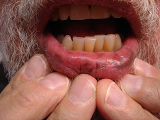
Pigmented Macules of the Lips

The patient is a 70 year-old architect who presented for another problem.  Incidental hyperpigmented macules were noted on the lower lip.  No family members are similarly affected nor does he have a family hisrory of colon cancer.  He has had unremarkable colonoscopies.  The pigmentation began ~ 20 years ago.

O/E:  Hyperpigmented macules of lower lip.

Clinical Photo:

Diagnosis: Probable Laugier Hunziker syndrome is favored.

References:
1. Laugier Hunziker syndrome. Jabbari A, et. al. Dermatol Online J. 2010;16(11):23.
Abstract: Laugier Hunziker syndrome is a rare disorder that is characterized by adult-onset hyperpigmented macules of the lips, oral cavity, and fingertips. Longitudinal melanonychia is present in the majority of cases. We present a 45-year-old woman with adult-onset hyperpigmented macules of the oral cavity as well as linear melanonychia that involved multiple fingernails. The history, clinical examination, and paucity of laboratory abnormalities or systemic findings support a diagnosis of Laugier Hunziker syndrome.  Free Full Text.


2. Peutz-Jager Syndrome eMedicine.
Peutz-Jeghers syndrome (PJS) is an autosomal dominant inherited disorder characterized by intestinal hamartomatous polyps in association with a distinct pattern of skin and mucosal macular melanin deposition. Patients with PJS have a 15-fold increased risk of developing intestinal cancer compared with that of the general population.

Monday, December 03, 2012

Acanthosis Nigricans in an 8 year old girl

Abstract:  Eight year-old girl with acanthosis nigricans

Presented by: 
Dr. Soheila Sotoudeh
Children’s Medical Center
Tehran, Iran

History:  This 8- year-old girl, born of nonrelative parents, presented
with a one year history of darkening and thickening of body folds especially neck and axilla.  It began with pruritic, hyperpigmented and corrugated
plaques on her neck, axilla, groin and perioral and periumbilical area.
Family history is negative for any skin disease. Drug history: levothyroxine
Her disease is gradually progressive.

O/E:  She is otherwise healthy. Her weight and height is in 50 percentile. There are hyperpigmented and corrugated plaques on her neck, axilla, groin and perioral and periumbilical area.

Photos:


Lab:  Routine hematologic and biochemical parameters (including
blood glucose and insulin level) were normal. Chest x-ray and abdominopelvic sonography were normal. Clinical screening for evidence of internal malignancy was negative.

Histopathology:
Skin biopsy showed hyperkeratosis, papillomatosis and
acanthosis, mild pigment incontinence and a sparse perivascular
inflammatory dermal infiltrate.

Diagnosis:
Acanthosis nigricans

Comments and Questions: My diagnosis was Acanthosis Nigricans. I have prescribed Acitretin 0.5 mg/kg for more than 3 months but without remarkable response.

1-Do you agree with the diagnosis of acanthosis nigricans?
2-What type of acanthosis nigricans does she have?
3-Do you recommend genetic testing?
4-What other treatment do you recommend?

References:
1. Remission of acanthosis nigricans, hypertrichosis, and Hashimoto's thyroiditis with thyroxine replacement.
Dix JH, Levy WJ, Fuenning C.  Pediatr Dermatol. 1986 Sep;3(4):323-6.
Abstract: Hypothyroidism is not commonly associated with acanthosis nigricans (AN). We examined a 13-year-old girl with AN, hypertrichosis, and Hashimoto's thyroiditis. Overt biochemical hypothyroidism, thyroid enlargement, and positive titers of antimicrosomal and antithyroglobulin antibodies confirmed Hashimoto's thyroiditis. Both AN and hypertrichosis resolved with thyroid hormone replacement. There was no evidence of insulin resistance, polycystic ovarian disease, lipoatrophy, or other endocrine dysfunction, or of malignancy. In two patients from the literature with AN and hypothyroidism, AN was attributed to associated thyroid carcinoma or insulin resistance, rather than coexisting hypothyroidism. Since the skin lesions improved with thyroid hormone therapy in those two patients and in ours, hypothyroidism appears to be directly involved in the pathogenesis of AN.

2. A Case of Generalized Acanthosis Nigricans with Positive Lupus Erythematosus-Related Autoantibodies and Antimicrosomal Antibody: Autoimmune Acanthosis Nigricans?  Y. Kondo,et. al. Case Rep Dermatol. 2012 Jan-Apr; 4(1): 85–91.  Free Full Text Open Access.

3. Acanthosis nigricans: A practical approach to evaluation and management
Steven P Higgins MD1, Michael Freemark MD2, Neil S Prose MD3
Dermatology Online Journal Volume 14  September 2008.  Free Open Access,  This is a good overview.

Saturday, November 24, 2012

Facial Lesion in a Child

Case presented by Dr Munqithe M Jabir, Addiwaniya, Iraq who writes:
"Can I please have your opinion about this problematic case.
The patient is a female child with a swelling on her left cheek for the past three months.  It discharges pus through many openings (carbuncle-like!!).  There has been no change although many courses of different antibiotics, but, recently it has responded slowly to Rifampicin 150mg twice daily and Clarithromycin 250 mg twice daily.  Complete blood picture and ESR are normal."


Here is a Video. 
References:
1. Embedded toothbrush foreign body in cheek - report of an unusual case.
Sathish R, Suhas S, Gayathri G, Ravikumar G, Chandrashekar L, Omprakash TL.  Eur Arch Paediatr Dent. 2011 Oct;12(5):272-4.
Source: Oral and Maxillofacial Surgery, Sri Siddhartha Dental College, India. drsathish75@gmail.com [This was suggested by Brian Maurer's comment]

2. Pediatr Dermatol. 2010 Jul-Aug;27(4):410-1.
Cutaneous facial sinus tract of dental origin.Mardones F, Oroz J, Muñoz C, Alfaro C, Soto R.
Dermatology Department, Hospital Clínico, Universidad de Chile, Santiago, Chile. fmardonesv@yahoo.com
Abstract: Cutaneous sinus tract on the head and neck area in a child may originate from dental disease. A high degree of clinical suspicion and complementary tests are often needed, as the diagnosis is usually not straight forward. Anatomical correlation is also useful in tracing the affected tooth or teeth. We present the case of a boy with a facial sinus tract that originated from periapical abscesses of maxillary molars.


Saturday, November 17, 2012

The Rudolph Sign

Abstract: 81 year old man with new onset of a red nose

HPI :  This 81 yo man presented with a one day history of a painful inflammatory process of his nose.  He had been in hospital recently and a routine throat culture grew MRSA but since he was asymptomatic, it was not treated. He has atrial fibrillation and meds include warfarin.  Here is the history in his own words.


O/E:  The examination shows an erythematous, slightly indurated area around the bulb of the nose.  

IMPRESSION:  With a history of MRSA and the clinical appearance this looks like nasal vestibular furunculosis as described recently in the dermatologic literature by Dahle andSontheimer.  

Course: He was treated with mupirocin ointment applied intranasally, but after three days there was no change and the process was somewhat worse.  Initially he graded the pain in the nose as a "7" and after three days as a "9" on the Pain Scale of 0 - 10. A culture was taken from the nares and he was placed on minocycline as Bactrim is contraindicated with warfarin.  He was admitted to hospital later that day for uncontrolled atrial fibrillation and  treated with i.v. vancomycin for two days until the preadmission culture came back negative.  Discharged home after two days a papule appeared on the bulb of the nose which drained serosanguinous material and the process started to resolve.  Repeat culture was taken (no pathogens).  When seen at Day 14, he showed marked improvement and he rated his pain as a "O."

Comments:  Nasal vestibular furunculosis (NVF) was described by Dahle and Sontheimer.  They recommended intranasal application of mupirocin with Q-tip applicators.  Our patient did not respond to that which suggests that NVF may need more aggressive therapy in some cases.  We did not perform an initial culture since he'd had one before, but in retrospect we should have done that.  For the clinician, one needs to consider the rare occurrence of cavernous sinus thrombosis with infections of the central face.  The literature on NVF is sparse and most articles lack abstracts.  This area needs more attention as NVF may not be as uncommon as the literature suggests.

Clinical Photos:  
Day i
Day 4

Day 7
Day 14
Day 21

Reference:
1. Dahle KW, Sontheimer RD. The Rudolph sign of nasal vestibular furunculosis: questions raised by this common but under-recognized nasal mucocutaneous disorder.  Dermatol Online J. 2012 Mar 15;18(3):6.  Free Open Access


2. Laupland KB, Conly JM. Treatment of Staphylococcus aureus colonization and prophylaxis for infection with topical intranasal mupirocin: an evidence-based review.
Clin Infect Dis. 2003 Oct 1;37(7):933-8. Epub 2003 Sep 8.  Email: laupland@calgaryhealthregion.ca
Abstract: Most Staphylococcus aureus infections are endogenously acquired, and treatment of nasal carriage is one potential strategy for prevention. We critically appraised the published evidence regarding the efficacy of intranasal mupirocin for eradication of S. aureus nasal carriage and for prophylaxis of infection. Sixteen randomized, controlled trials were appraised; 9 trials assessed eradication of colonization as a primary outcome measure, and 7 assessed the reduction in the rate of infection. Mupirocin was generally highly effective for eradication of nasal carriage in the short term. Prophylactic treatment of patients with intranasal mupirocin in large trials did not lead to a significant reduction in the overall rate of infections. However, subgroup analyses and several small studies revealed lower rates of S. aureus infection among selected populations of patients with nasal carriage treated with mupirocin. Although mupirocin is effective at reducing nasal carriage, routine use of topical intranasal mupirocin for infection prophylaxis is not supported by the currently available evidence. Free Open Access.

3. Dr. Richard Sontheimer sent us this article which may explain why our patient did not respond to mupirocin.  This is a sobering article -- one wonders if resistance patterns elsewhere are as high or whether this was uniqueto the burn center in Tehran.
Burns, 2012 vol. 38(3) pp. 378-82
A high prevalence of mupirocin and macrolide resistance determinant among Staphylococcus aureus strains isolated from burnt patients.
Shahsavan, et. al.  (Tehran University of Medical Sciences)
Abstract: Infections due to Staphylococcus aureus have become increasingly common among burn patients. The antibiotic resistance profile of S. aureus isolates and inducible resistance against clindamycin were investigated in this study. The presence of mecA gene, mupA gene and macrolide resistance genes were detected using PCR and multiplex-PCR. The resistance rate to methicillin, erythromycin and mupirocin were 58.5%, 58% and 40%, respectively. The prevalence of constitutive and inducible resistance among macrolide resistant isolates was 75% and 25%, respectively. Ninety five percent of the isolates were positive for one or more erm genes. The most common genes were ermA (75%), ermC (72%) and ermB (69%), respectively. The ermA gene predominated in the strains with the inducible phenotype, while ermC was more common in the isolates with the constitutive phenotype. The msrA gene was only found in one MRSA isolate with the constitutive phenotype. A total of 27 isolates (25%) carried the mupA gene. All the mupirocin resistant isolates and almost all the erythromycin resistant isolates were also resistant against methicillin which may indicate an outbreak of MRSA isolates with high-level mupirocin and erythromycin resistance in the burn unit assessed.

Thursday, November 08, 2012

Hypopigmented Rings

18 year old college coed with two month history of two incompletely hypopigmented rings on abdomen.  No contacts to area recalled.  On no meds.

O/E:  On right and left abdomen, there are two relatively subtle 5 cm in diameter rings.  No scale.  Possibly a few small incompletely hypopigmented macules on abdomen as well.

Clinical Photo:
Labs:  N/A

Path: Will offer to biopsy

Diagnosis:  Hypopigmented Contact Dermatitis?

Question:  Has anyone seen anything like this?

Reference:
Chemical leukoderma
Kathryn E O’Reilly MD PhD, Utpal Patel MD PhD, Julie Chu MD, Rishi Patel MD, Brian C Machler MD
Dermatology Online Journal 17 (10): 29  OA Full Text
Department of Dermatology, New York University, New York, New York
Abstract: Chemical leukoderma is defined as an acquired, hypopigmented dermatosis that results from repeated cutaneous application of an agent that destroys epidermal melanocytes in genetically susceptible patients. Chemical leukoderma may develop both at the site of contact with the chemical as well as remotely from the exposure. Avoidance of the causative agent may lead to spontaneous repigmentation, but treatments commonly used in vitiligo, such as narrow-band ultraviolet B phototherapy, PUVA photchemotherapy, or topical immunosuppressants, often are necessary. We present a case of chemical leukoderma secondary to pyrethroid insecticides that has progressed despite avoidance of the agent for over ten years.


Friday, November 02, 2012

Atypical Acneiforn Eruption

Abstract: 18 yo man with two month history of papules and pustules on chin

HPI:  The patient describes mild acne before leaving for college in late August 2012.  He did being a new electric razor with him.  Shortly after arriving at school, he developed an inflammatory process on his chin.  He was treated with Keflex for > one month without relief.

O/E:  Papules + pustules on the chin.  Rest of exam unremarkable.

Photos:  11/2/12


Lab:  Culture grew "Few Serratia marsencens"  plus Staph epidermitis and alpha hemolytic strep

Diagnosis: Gram Negative Acne is favored over Pyoderma faciale

Discussion:  This young man had acne which had been treated with cephalexin for one to two months.  It has not improved and the presentation with pustules suggested gram negative acne.  His college health center's provider wisely performed a bacterial culture which grew Serratia.  Initially, I was thinking of prescribing ciprofloxicillin, but the literature suggests that isotretinoin may be the treatment of choice.

Followup after five months isotretinoin:


References:
1. James WD, Leyden JJ. Treatment of gram-negative folliculitis with isotretinoin: positive clinical and microbiologic response. J Am Acad Dermatol. 1985 Feb;12(2 Pt 1):319-24
Abstract: Thirty-two patients with gram-negative folliculitis were treated with 0.47 to 1.0 mg/kg/day of isotretinoin. Serial microbiologic evaluations demonstrated rapid clearing of the face and nasal mucosa of gram-negative rods. The clinical response was rapid, complete, and induced prolonged remissions. Twenty-six of thirty-two patients developed Staphylococcus aureus nasal carriage by the end of the 20-week treatment course. Isotretinoin has decided advantages over previously reported therapies for gram-negative folliculitis.
Photo from James and Leyden's article, above
2.  Böni R, Nehrhoff B.  Treatment of gram-negative folliculitis in patients with acne.  Am J Clin Dermatol. 2003;4(4):273-6.
Department of Dermatology, University Hospital, Zürich, Switzerland. rboeni@derm.unizh.ch
Abstract:  Gram-negative folliculitis may be the result of long-term antibacterial treatment in acne patients. It is caused by bacterial interference and replacement of the Gram-positive flora of the facial skin and the mucous membranes of the nose and infestation with Gram-negative bacteria. These Gram-negative bacteria include Escherischia coli, Pseudomonas aeruginosa, Serratia marescens, Klebsiella and Proteus mirabilis. The occurrence of Gram-negative folliculitis should be considered in acne patients in whom oral treatment with tetracyclines has not resulted in a significant improvement of acne lesions after 3-6 months' treatment. The occurrence of Gram-negative folliculitis in acne patients is believed to be generally underestimated, since correct sampling and bacteriology is rarely performed by clinicians. Gram-negative folliculitis in acne and rosacea patients is best treated with isotretinoin (0.5-1 mg/kg daily for 4-5 months).

3.  Massa MC, Su WP.  Pyoderma faciale: a clinical study of twenty-nine patients.  J Am Acad Dermatol. 1982 Jan;6(1):84-91.
Abstract:  Pyoderma faciale is a distinctive entity. Twenty-nine patients with this process were seen in the Mayo Clinic from 1969 to 1980. Twenty-seven patients had follow-up that ranged from 1 month to 11 years, and twenty-two had follow-up of 3 years or more. Clinical features that characterize the patients were (1) female predominance, (2) onset later than teenage acne vulgaris, generally at 19 to 40 years of age, (3) rapid onset and progression, (4) facial involvement with sparing of the back and chest, (5) cysts, swelling, and purulent drainage with a lack of comedones, and (6) paucity of systemic complaints. Patients were treated with multiple forms of therapy simultaneously, often including Vleminckx packs, oral antibiotics, incision and drainage, ultraviolet B, and intralesional steroids. Of twenty-five patients available for follow-up at 1 year, twenty-three had achieved remission, though fifteen patients required ongoing treatment to maintain optimal control. Twenty-three patients had scarring as a sequela. Patients with pyoderma faciale represent a subset of patients with acne in whom the outlook is favorable with appropriate therapy.

Wednesday, October 31, 2012

Nevus Depigmentosis

The patient is an 8 yo girl with a congenital lesion on the left arm.  She was seen for another reason.  There are no other significant skin lesions and the child is normal in all respects.

O/E:  3.5 cm diameter hypopigmented macule with irregular border,


Diagnosis: This is probably a nevus depigmentosis.  Nevus anemicus is less likely as is ash leaf macule of tuberous sclerosis.  (I did not do dermoscopy of this lesion, but that would have been a good idea.

Reference:
1. Oiso N, Kawada A. The diagnostic usefulness of dermoscopy for nevus depigmentosus. Eur J Dermatol. 2011 Jul-Aug;21(4):639-40.  Free full text

2. Abdullah L, Abbas O.  Dermacase. Can you identify this condition? Nevus depigmentosus. Can Fam Physician. 2011 Jun;57(6):682, 684.
Department of Nursing, American University of Beirut, Lebanon. Free Full Text


Saturday, October 20, 2012

Toxic Erythema


Presented by Dr. Henry Foong,
Ipoh, Malaysia

Abstract:  41 yo man with short history of toxic erythema
HPI: The patient is a 41 yr old restaurant worker who presented with 5 day history of bilateral and symmetrical erythema over the loins, groins and knees.
About a week ago, he had a furunculosis on upper back and was prescribed Augmentin. Two days later he developed high fever and was treated with ciprofloxacin by a physician. A day later he noticed itchy rashes on both thighs and since then, the macular erythema spread symmetrically to the loins, upper shoulders and knees.  He has no fever.
His past medical history was non-contributory.
Examination showed bilateral and symmetrical diffuse erythema on the thighs, knees, loins and upper shoulders. Over the loins, there was symmetrical and bilateral edematous areas  with vesicles over a background of erythema.  His scalp, oral cavity and genitalia were clear.
Clinical Photos:


Lab: Culture and a skin biopsy has been done. Culture negative.  Biopsy pending.
 TWBC 3200 (N 85.1%  L 10.3%  E0 .5%  M3.4%  B 0.8%)  ESR 36

Diagnosis: Toxic erythema
Differentials:  Streptococcal/Staph cellulitis 

Questions and Comments:  The presence of blisters /edema on the loin is worrying.  In the meantime what would you recommend for this patient? Would you use corticosteroids in this patient? I have stopped both augmentin and ciprofloxacin. In terms of dressing, I used wet compress with dil. KMNO4.Have I missed anything?

References:
1.  Miyahara A, et. al.  A new proposal for a clinical-oriented subclassification of baboon syndrome and a review of baboon syndrome. Asian Pac J Allergy Immunol. 2011 Jun;29(2):150-60
Source: Department of Pediatrics, Tokyo Medical University. miyahara.pediatrics@gmail.com
Abstract
OBJECTIVE: To review baboon syndrome (BS). Data Sources: Date sources were obtained from PubMed and Google Scholar: Photographs of baboon syndrome were obtained from our patient.
STUDY SELECTIONS: PubMed and Google Scholar were searched up to June 30, 2010. The search terms were "baboon syndrome", "SDRIFE" and "thimerosal allergy". Reverse references from relevant articles and Google Scholar were also used. As BS is a classical disease and cases of offending agents were relatively old, some references were more than five years old. In order to gather as many cases of offending agents as possible, more than 50 references were collected.
RESULTS AND CONCLUSION: We divided BS into as 4 groups; classical baboon syndrome, topical drug-induced baboon syndrome, systemic drug-induced baboon syndrome and symmetrical drug-related intertriginous and flexural exanthema (SDRIFE). The pathomechanism of BS is still unknown. A delayed type of hypersensitivity reaction, a recall phenomenon, pharmacologic interaction with immune-receptors and anatomical factors may be involved in the causation of BS.  This valuable article is available in Free full text

2. Tan SC, Tan JW.  Symmetrical drug-related intertriginous and flexural exanthema. Curr Opin Allergy Clin Immunol. 2011 Aug;11(4):313-8.
Department of Rheumatology, Allergy and Immunology, Tan Tock Seng Hospital, Singapore, Singapore. Sze_Chin_Tan@ttsh.com.sg
Abstract
PURPOSE OF REVIEW: Symmetrical drug-related intertriginous and flexural exanthema (SDRIFE), previously termed drug-related baboon syndrome, is a benign and self-limiting type IV hypersensitivity reaction characterized by symmetrical erythema involving the gluteal and intertriginous areas in the absence of systemic involvement. It may also occur in the absence of previous drug exposure.
RECENT FINDINGS: Antibiotics, in particular beta-lactams, comprise the majority of causes of SDRIFE. Other drugs which have been implicated include antihypertensives, radiocontrast media, chemotherapeutic agents, and biologics. Histology of lesional skin is variable with predominance of superficial perivascular inflammatory cell infiltrates. Outcomes of allergy tests are variable with positive delayed intradermal tests reported for penicillin V, allopurinol; positive patch tests for erythromycin, mitomycin, nystatin, pseudoephdrine; positive lymphocyte transformation tests for erythromycin; and positive drug provocation tests for clindamycin, cimetidine, corticosteroids, terbinafine, and valacyclovir.
SUMMARY: Diagnosis of SDRIFE is dependent upon recognition of the clinical morphology and distribution of the rash, and its temporal relationship to the use of the suspected drug. Outcomes of in-vivo and in-vitro tests have been inconsistent, and thus may not be useful in the identification of the putative drug.