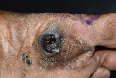
HPI: An 72-year-old Indian housewife present with a pigmented growth on the right sole for a year. Started as a small growth and gradually increased in size. She saw a GP earlier and was advised to remove it. However she was not keen then. Recently she felt pain when she walked and this prompted her to seek medical attention again.
O/E: An ulcerated pigmented warty growth 2 x 3 cm on the sole of the right foot with surrounding pigmented satellite lesions. Her regional nodes
(popliteal and inguinal) were not enlarged.
Clinical Image:
Pathology:
Nests of atypical cells are seen in the epidermis and dermis. Most of the cells contain melanin pigment. They show pleomorphism, have vesicular nuclei and eosinophilic cytoplasm. These features are suggestive of malignant melanoma. Suggest wide excision for definite diagnosis.
Diagnosis: Malignant melanoma, acral lentiginous type with nodular component.
Questions:
How would you approach this patient?
Do you think Sentinel Lymph Node Biopsy is important in her case?
What would give her the best quality of life?
After surgery, is there a role for topical imiquimod?
Reference:
1) Kanzler MH. Sentinel node biopsy and standard of care for melanoma: a re-evaluation of the evidence. J Am Acad Dermatol. 2010 May;62(5):880-4.
2) No survival benefit for patients with melanoma undergoing sentinel lymph node biopsy: critical appraisal of the Multicenter Selective Lymphadenectomy Trial-I final report. Sladden M1, Zagarella S, Popescu C, Bigby M. Br J Dermatol. 2015 Mar;172(3):566-71. PubMed.
3) Aral lentiginous melanoma treated with topical imiquimod cream: possible cooperation between drug and tumour cells.
Clin Exp Dermatol. 2015 Jan;40(1):27-30.
Savarese I, et. al.
Abstract: An 85-year-old woman presented with a lesion on
the sole of her right foot, which was histologically confirmed as acral
lentiginous melanoma. Because of the large field involved and because the
patient refused any invasive or painful treatment, topical treatment with
imiquimod was commenced. At the 20-month follow-up, the patient was still
continuing treatment with topical imiquimod, and no metastases to the lymph
nodes or viscera were found, either clinically or in imaging studies. We
believe that the success of the treatment cannot be explained only by the
stimulation of the immune system induced by imiquimod. A possible explanation
might be 'tumour dormancy', where a tumour grows very slowly because of a
balance between the neoplasia and the immune (and nonimmune) mechanisms of
tumour control. The use of imiquimod has so far allowed our patient to avoid
surgery, and perturbation of the mechanisms of tumour regulation, such as local
immunity and angiogenesis, has not taken place.


