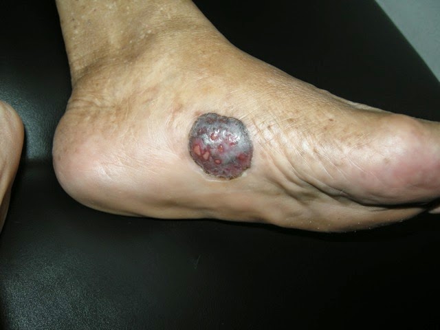
Abstract: 70 yo woman with acral melanoma
Presented by Dr. Henry Foong, Ipoh Malaysia
HPI: The patient is a
70-year-old Malay woman who presented with a one-year history of a pigmented
lesion on the left foot. She has seen at
least 4 doctors and I am sure all have advised her to have a biopsy done. It
was occasionally painful but otherwise asymptomatic. The lesion had been gradually increasing in
size.
Her medical history
includes diabetes, hypertension and hypercholesterolemia. She is on glibenclamide, metformin,
perindopril, aspirin, hydrochlorothiazide and lovastatin.
She lives in a
rural area south of Ipoh, Malaysia. She has 10 children. There was no family history of skin cancer.
O/E: shows a localised pigmented tumor 3 x 3 cm
with superficial ulcerations on the medial aspect of the sole of left
foot. It has an irregular margin but was
well circumscribed. The nodule is firm
on deep palpation.
Clinical Images:
Skin Biopsy
Nests of
melanoma cells are seen invading the dermis. The tumour cells are pleomorphic, have
vesicular nuclei and eosinophilic cytoplasm. There is increased mitotic
activity. Many of the cells contain melanin pigment.
Diagnosis: Left foot biopsy
Malignant melanoma, acrolentiginous type, nodular
Discussion and
Questions: We rarely see melanoma here in Malaysia. The prevalence rate is reported to be about
0.4 per 100 000 population. I have not
had a single case of melanoma the whole of last 2 years. This patient waited for a year before a
diagnosis was made. What has gone
wrong?
The histopath
report unfortunately did not indicate the thickness of the tumor neither is
there any mitotic rate or Clark’s level of invasion. In a study in Malaysia most of the cases are
located on the sole of the foot as in this patient. (12/24 cases) Histologically majority are of
the nodular type. I think based on the
report, our patient has a nodular type of melanoma.
Plan:
Dermatologists
in Malaysia don't manage malignant melanoma.
Instead, they are referred to surgeons for excision. Sentinal node biopsy and CT scan abdomen and
chest would be useful for staging of the tumor.
Would PET scan give more useful information for this patient? Immunotherapy and BRAF inhibitors are
probably too expensive for her.
References:
1. Malaysian J
Pathol 2012; 34(2) : 97 – 101
Cutaneous malignant melanoma: clinical
and histopathological
review of cases in a Malaysian tertiary
referral centre
Jayalakshmi
PAILOOR, Kein-Seong MUN and Margaret LEOW*
Departments
of Pathology and *Surgery, Faculty of Medicine, University of Malaya
Abstract
Melanoma is a
lethal skin cancer that occurs predominantly among Caucasians. In Malaysia, the
incidence of melanoma is low. This is a retrospective study of clinical and
histopathological features of patients with cutaneous melanoma who were seen at
the University Malaya Medical Centre from 1998 to 2008. Thirty-two patients
with cutaneous melanoma were recorded during that period. Of these, 24 had
sought treatment at the onset of disease at our centre. Chinese patients
constituted the largest group (19 cases). The median age of these 24 patients
at the time of presentation was 62 years. 16 patients had melanoma involving
the lower limb with 12 affecting the sole of the foot. None had melanoma
arising from the face. Histopathology showed nodular melanoma in 22 cases
(91.6%), with superficial spreading and acral lentiginous melanoma diagnosed in
1 case each. The majority of patients (62.5%) were found to be in Stage III of
the disease at the time of diagnosis.
2.
Whiteman DC1, Pavan WJ,
Bastian
BC. The melanomas: a synthesis of epidemiological, clinical,
histopathological, genetic, and biological aspects, supporting distinct
subtypes, causal pathways, and cells of origin. Pigment Cell Melanoma Res. 2011
Oct;24(5):879-97.
Free Full Text
Abstract: Converging
lines of evidence from varied scientific disciplines suggest that cutaneous
melanomas comprise biologically distinct subtypes that arise through multiple
causal pathways. Understanding the respective relationships of each subtype
with etiologic factors such as UV radiation and constitutional factors is the
first necessary step toward developing refined prevention strategies for the
specific forms of melanoma. Furthermore, classifying this disease precisely
into biologically distinct subtypes is the key to developing mechanism-based
treatments, as highlighted by recent discoveries. In this review, we outline
the historical developments that underpin our understanding of melanoma
heterogeneity, and we do this from the perspectives of clinical presentation,
histopathology, epidemiology, molecular genetics, and developmental biology. We
integrate the evidence from these separate trajectories to catalog the emerging
major categories of melanomas and conclude with important unanswered questions
relating to the development of melanoma and its cells of origin.

