Tuesday, July 31, 2007

Buttock Necrosis

The patient is a 50 yo nurse who presented to my office on July 31, 2007. Here is the history in her words:

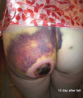
"On June 28th I fell off of my horse. Large dark hematomas covered left buttocks cheek, vagina, and coccyx. Per doctors orders warm, moist heat applied. Thw first picture, taken by my daughter ten days after the fall, shows where the circular area of skin fell off as a result of the compress. I then applied collagenase santyl cream for two weeks but was still unable to debride. One month later, in addition to area on left cheek, new symptoms have arrived which include headaches, 10 - 15 pound weight loss due to diarrhea, burning sensation in face and ears, chest pain, pain between shoulder blades and in neck. There are currently blood tests being done to confirm the existence of what is believed to be a blood disorder. [CBC and Comprehensive chem profile normal except for a slightly elevated BUN of 24 mg%]

O/E: I know this patient well as a care giver and was surprised to see how thin and pale she looked. There is a 9 cm escar on the left buttock. The tissue is necrotic and can not be debrided at this time.

Lab: A culture of the exudate under the eschar grew out many Pseudomonas aeroginosa -- sensitive to Cipro and levofloxacillin.


Discussion:
The trauma seems to have caused skin necrosis. Pseudomonas may be related as well since this looks like echthyma gangrenosum, but the patient has a presumably normal immune system and feels well otherwise. It's unclear if there was fat or muscle necrosis in addition or whether there was a compartment syndrome. The patient is now going to a wound clinic where she can get this are properly debrided.
I have not seen necrosis like this from trauma before. Similar (but more irregular necrosis) can follow brown recluse spider bite. Here, I suspect trauma was the cause; however at one month out the patient has systemic symptoms. One wonders if a CT of the buttock might be of any value.

One week later:
Wound started to drain and was explored at wound clinic. A large cavity was found under the gluteus maximus muscle (around seven cm in diameter). it was irrigated and packed. I am not sure if the cavity contained blood or pus. Today, it was clean with no drainage. The area will likely need surgical intervention as it will take months to heal by the appearance. We will seek surgical opinions.

Thursday, July 26, 2007

A Textbook Case

A 76 year old woman presented to the dermatologist with a four month history of darkening of the skin of her neck, axillae, inframammary areas and groin. She had vague G.I. symptoms and had seen her primary care doctor around four times with these complaints. Although her abdomen was "bloated" she'd lost 5 - 10 pounds over the past month. Her sister had died of pancreatic cancer. (Her daughter who works at a neurosurgery office had tried to get her seen by a dermatologist but none had time. It took two months to get her seen at my office which I see as a personal failure)

The patient is a pleasant outgoing woman who appears about her stated age. She has velvety hyperpigmentation of her skin folds.


In addition, she has developed around 10 verrucous tumors measuring 8 - 10 mm in diameter on arms and legs. One of these was biopsied.

A C.T. scan performed on the day of the dermatology visit revealed what appeared to be metastatic tumors in the peritoneum. The workup is in progress.

Most cases of acanthosis nigricans (AN) are benign and related to ethnicity, obesity or endocrinopathy. AN developing in an elderly individual should be a red light to pursue a work-up for malignancy. AN is an easy diagnosis to make and a four month delay in diagnosis is unfortunate. In all likelihood, this tumor had metastasized before the AN became manifest, but it seems unfortunate that the delay in diagnosis occurred.

A good review of AN can be found on eMedicine.

Saturday, July 14, 2007

Fingertip Eczema

The patient is a 52 yo warehouseman with a two year history of a painful fingertip eczema of the first three fingers of his right hand and first four fingers of his left (dominant) hand. He handles cardboard boxes and drives a fork lift. He is also an avid golfer. The dermatitis is not seasonal.


He has been only minimally helped by super-potent topical corticosteroids.

Patch testing is scheduled but all we have available here is T.R.U.E. Test. If not helpful, he will be sent to an occupational dermatology department for further testing.

I assume this is an occupational contact dermatitis and have recommended that he use cotton gloves at work if possible.

Your ideas as to etiology and treatment pending definitive patch testing will be appreciated.

Tuesday, July 10, 2007

Turtles, Birds and Bears

Dermatologists in the U.S. spend a fair bit of time screening patients for skin cancers. In spite of this, there is little evidence that this screening decreases the mortality from skin cancer (mainly melanoma).

"Cancers follow three basic patterns: turtles, birds and bears.
1. Turtles move so slowly that you can still capture them while they’re moving slowly along;
2. The birds fly away so quickly that you can’t catch them in time;
3. The bears can escape if you ignore them, but if you catch them in time, you can capture them."

This simple but brilliant formula comes from a superb article by Christie Aschwanden on sunscreens and skin cancer that appeared in the July 10, NY Times.

Doctors Balk at Cancer Ad, Citing Lack of Evidence See Permalink.

This is a reasoned piece which quotes the best evidence-based information we have. Please read the article and draw your own conclusions.

Friday, July 06, 2007

Atypical Cheilitis

The patient is a 58 yo gynecologist who has had a scaly lower lip for over seven months. When it began, she was wearing orthodontic braces. These were removed around four months ago, but the scaling on the lower lip has persisted. She is a non-smoker. She has treated patients with genital warts with a laser for years and had questions about virus in the plume.

O/E: Hyperkeratosis of lateral portions of the lower lip. This is not the picture of leukoplakia usually seen with lichen planus or actinic cheilitis.


She was treated with hydrocortisone valerate 0.2% cream which was marginally helpful and then clobetasol ointment which was not effective.

Please tell me your thoughts. I am planning a biopsy. Most likely a 3 mm punch -- would you punch or shave?

Thank you.