Friday, July 29, 2005

84 yo man with Pemphigus


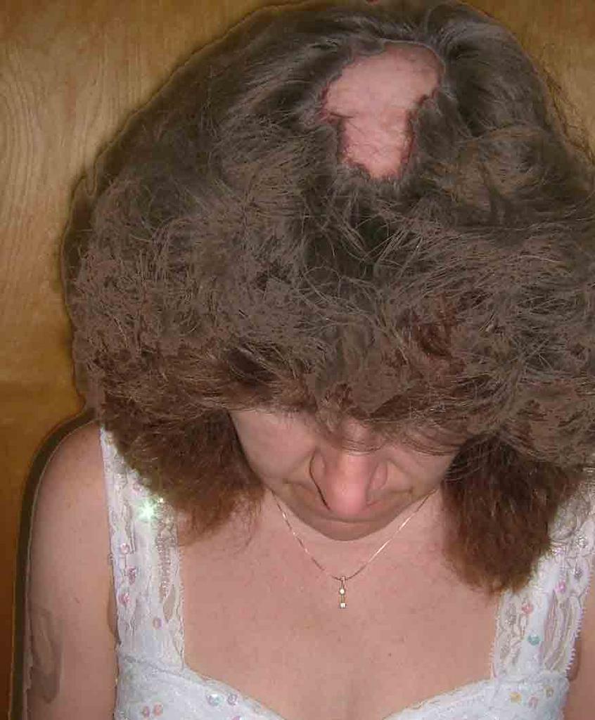
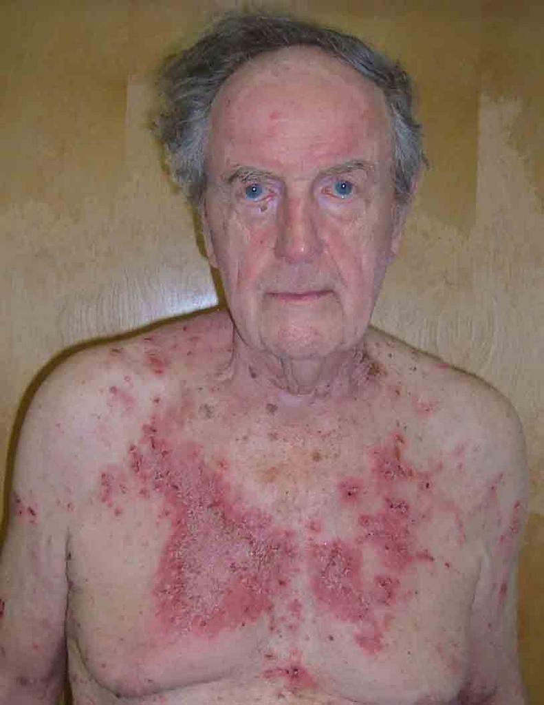
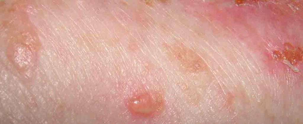
This 84 yo man was seen around weeks ago with an erosive bullous process on torso, head and neck of 4 months duration. He was on no meds by mouth and in good general health.
My initial impression was pemphigus vulgaris vs. impetigo. The skin culture showed many coag + staph, and the bx showed an acantholytic bulla. The DIF was positive for intracellular IgG.
He was treated with dicloxacillin 250 mg qid and prednisone 20 mg tid.
He cleared quickly. At present he is on 30 mg per day of prednisone and tapering by 5 mg every two weeks. He's had no new lesions since therapy was initiated and his itch has disappeared.
Questions:
1) Value of adjuvent therapy? I am thinking of starting in a benign manner with minocycline/
2) Should I try to get on alt. day steroid first?
3) Any suggestions?
Follow-up (August 6, 2005) The patient continues to do well. He is now on Prednisone 20 mg per day. No symptoms and no new lesions. His prednisoe will be dropped by 5 mq every 10 days until 10 mg per day - then we will convert to alt day therapy. I may add minocycline 100 mg bid.

40 yo woman with scarring alopecia


The patient is a 40 yo woman with an eight year history of scarring alopecia.
Her ANA is + at 1:160 - all other labs are normal.
No other cutaneous findings or systemic disease other than "fibromyualgia."
She has been treated with 200 mg of plaquenil b.i.d. for a number of years. This has not been of much help.
The patient is quite concerned about her scalp. She's going through a divorce and worries about her appearance.
Questions?
1) Any value to a biopsy?
2) Would scalp reduction be an option?
3) Role for hari transplantation? Should a biopsy be done first

Tuesday, July 12, 2005

Melanonychia in a Six Year-Old Child

Presented by
Dr. Chee Meng Loh
RMG
Singapore
E mail: bliss88@singnet.com.sg

The patient is a six year old Chinese girl who developed a dark pigmented longitudinal band on the nail of her right index finger about 1 year ago. It started as a narrow, light brown band and grew in darkness (black in centre of band, and lighter shade of brown at edge of band) and width. The band is now about 3 mm in width. It has not changed in the past 3 months.

There is no history of trauma to the right index finger. There is also no complaint of pain in that finger.

The girl suffers from eczema and is on steriod medication (hydrocortisone 1% cream). She is otherwise healthy. There is no history of melanoma in the girl, her parents or grand parents. Neither her parents have pigmented bands on their nails.

Physical Exam: Dark longitudinal pigmented band on right index finger.
Colour ranges from black to brown.
Width is about 3 mm.
(Pictures attached)

Biopsy not performed.

Diagnosis: Melanonychia of right index finger.

Questions:
What are the likely causes of melanonychia in this child?
What is the likelihood that the cause is melanoma?
Should biopsy be performed to rule out melanoma?
If yes, what biopsy procedure is appropriate and what is the risk of permanent damage to the nail?
If a wait and see approach is appropriate, what other signs should the parents watch out for and within what time frame?


Melanonychia
Originally uploaded by David Elpern.


Melanonychia
Originally uploaded by David Elpern.

Friday, July 01, 2005

16 yo girl with papulonecrotic lesions

The patient is a 16 yo girl with a 2 year history of papulonodular lesions on the extremities and face. There is no evidence that these are excoriations. Biopsy 9 months ago from the arm was read as prurigo nodularis, but lesions look atypical and have now started to appear on the face. She is well otherwise.


A repeat biopsy on June 27. 2005 showed:
DIAGNOSIS: Skin - Left Temple:

Epidermal necrosis with s cale crust containing neutrophils , sub-epidermal abundant neutrophils and fibrin deposition, superficial and deep perivascular lymphohistiocytic infiltrate with focal neutrophil ic microabscesses, septal and lobular panniculitis with mixed inflammatory cell infiltrate of abundant neutrophils , lymphocytes , histiocytes, and eosinophils and numerous activated endothelial cells, surrounding a medium-sized vessel with marked mixed inflammatory cell infiltrate of neutrophils , histiocytes and occasional eosinophils .

NOTE : These changes are suggestive of a medium-sized vasculitis with overlying necrosis. Elastic tissue stain (EVG) does not reveal the vessel in the deeper sections, therefore, arterial or venular distinction cannot be made. The differential diagnosis includes a large vessel vasculitis such as periarteritis nodosa or early Wegener's granulomatosis. P.A.S. stain is negative for fungal organisms. Fite stain is negative for mycobacteria . However, an infectious vasculitis cannot be entirely excluded . If the clinical suspicion persists, culture studies may be of help . The differential diagnosis also includes , in the appropriate clinical setting , factitial panniculitis with secondary vascular involvement. These are not the changes of lupus erythematosus , pityriasis lichenoides et varioliformis acuta or prurigo nodularis . Serologic studies may be helpful. Clinico-pathologic correlation is suggested.