Wednesday, December 24, 2014

Congenital Gluteal Lesion


The patient is an 18 year-old boy with a congenital lesion in the gluteal cleft.
His mother says it has been slowly enlargely.  He is in excellent health and the lesion is asymptomatic.  The patient is an athlete, plays basketball.

O/E:  There is a 1 cm in diameter flesh-colored tumor just in the gluteal cleft.  No other cutaneous findings.

Clinical Photo:


Investigations:  None at this time

Diagnosis:  Soft tissue tumor of gluteal cleft.  Fibroma vs. lipoma

Question:  Midline lesions of the lubbosacral area may be a sign of spinal dysraphism.  Usually, the findings are more impressive than this.  It is unclear if a banal lesion such as this requires a work-up.  What are your thoughts.  If the reference below is to be believed, this patient's risk of OSD is low and workup may be of questionable value.

Reference:
Skin markers of occult spinal dysraphism in children: a review of 54 cases.
Guggisberg D et. al.  Arch Dermatol. 2004 Sep;140(9):1109-15.  Free Full Text.
Abstract
OBJECTIVES: To verify the diagnostic value of lumbosacral midline cutaneous lesions in asymptomatic children to detect occult spinal dysraphism (OSD) and to propose a practical approach for clinical investigations with respect to the type of cutaneous lesions observed.
DESIGN:Retrospective study of 54 children referred to the Department of Pediatric Dermatology between 1990 and 1999 for congenital midline lumbosacral cutaneous lesions.
SETTING:The private or institutional practices of participating dermatologists and pediatricians.
MAIN OUTCOME MEASURES:Evaluation of the diagnostic value of midline cutaneous lesions for the detec-tion of OSD. Association of skin examination findings with spinal anomalies detected by magnetic resonance imaging or ultrasound.
RESULTS: Occult spinal dysraphism was detected in 3 of 36 patients with an isolated congenital midline lesion and 11 of 18 patients with a combination of 2 or more different skin lesions.
CONCLUSIONS: A combination of 2 or more congenital midline skin lesions is the strongest marker of OSD. Careful dermatologic examination is needed to detect suggestive markers and request a spinal magnetic resonance image, which is the most sensitive radiologic approach to detect an OSD.

Sunday, November 30, 2014

Presented by Henry Foong
Ipoh, Malaysia

A 65-year old woman presents with multiple erythematous nodules arranged in a linear manner on her right forearm

HPI
The lesion appeared 2 months ago as a small boil on the right index finger.  She was treated with oral antibiotics by an orthopaedic surgeon. It then became progressively larger and new lesions began to spread proximally toward the elbow. The patient had a history of diabetes and was managed with oral hypoglycaemic agents.

PE
Vital signs normal.  Multiple erythematous nodules 1.0- by 1.0-cm distributed along the lymphatics from right index finger to the elbow.  No lymphadenopathy.

Images


Labs
Blood counts and biochemistry normal. Skin biopsied tissue sent for fungal/ TB culture and sensitivity

Skin Biopsy
Skin biopsy reveals dermis and underlying tissue heavily infiltrated by lymphocytes, histiocytes and neutrophils.  Fungal spore are identified.

Differentials
Sporotricosis - localised lymphocutaneous type

Discussion and Treatment
Sporotrichoid lymphocutaneous infection is characterised by subcutaneous nodules along lymphatics.  the differentials of such infection are sporotricosis, mycobacterium marinanum and Leishmania brasiliensis.  The organism is usually inoculated through minor skin injury or insect bites.

Recommended treatment is itraconazole 200mg daily for 2-4 weeks after the lesion has healed. The discounted cost of 60, 100 mg itraconazole in U.S. is ~$200 - 400. 
Other treatment available include potassium iodide 300-450 mg daily for 12-16 weeks. Cost of 2 cc of SSKI in U.S. is ~ $15.
for disseminated cases of sporotricosis, IV amphotericin 0.5mg/kg/day up to a total of 1-2 gm followed by itraconazole 200mg daily for 6-12 months.

Ref:
Yamada K1, Zaitz C, Framil VM, Muramatu LHCutaneous sporotrichosis treatment with potassium iodide: a 24 year experience in São Paulo State, Brazil. Rev Inst Med Trop Sao Paulo. 2011 Mar-Apr;53(2):89-93.

de Lima Barros MB1Schubach AOde Vasconcellos Carvalhaes de Oliveira RMartins EBTeixeira JLWanke BTreatment of cutaneous sporotrichosis with itraconazole--study of 645 patients.
Clin Infect Dis. 2011 Jun 15;52(12):e200-6.

Comment: We will check the cost to the patient of itraconazole in Malaysia.

Wednesday, October 22, 2014

3 Year-Old with Fever and Rash


Presented by Sylvia Moscone, M.D.
Block Island, New York

The patient is a 3-year-old girl who was seen for evaluation of a solitary lesion on the midback that has been present for about four days.  At the onset, she had a high fever, 105-106 F.  She was seen in the ER and referred to her pediatrician.  A diagnosis of Lyme disease was made and the patient was started on amoxicillin.  Blood studies were drawn.  Over the next two days, the fever persisted and then disappeared two days ago.  The child has a good appetite, sleeping well, playing normally.  Her parents feel she is "grumpy." She says the area on her back hurts. 

O/E:  The examination shows a 3 cm in diameter somewhat circular, erythematous scaly area.  A portion of this is flesh-colored.  There are no other similar lesions and there is no lymphadenopathy.   A baseline photo, taken by her mother, shows the initial lesion to be larger.

Clinical Photos:
At onset with fever
 
Four days later
Lab:  Lyme titers are negative.  Titers for Ehrlichiosis and Bartonellosis may have been drawn, but are not available now.

IMPRESSION:   A lesion less than 5 cm in diameter that is not enlarging is unlikely to be Lyme disease.  I do not have a specific diagnosis here. However, the lesion in the initial photo, taken by the child's mother may well have been > 5 cm in diamedter.  I woulld appreciate some guidance from VGRD members.  For the time being, the child is doing very well and she will be observed.  

Comment:  In the initial photo taken by the patient's mother, the lesion looks to be ~ 5 cm or larger.  This would suggest Lyme disease.  High fever is unusual with Lyme disease so the question of co-infection with Ehrlichiosis or Babesiosis needs to be considered.  Amoxicillin is not effective for those disorders, but many cases resolve spontaneously.    For the time being, it might be best to observe this child.  Serologies for the latter two diagnoses could be drawn if they were not looked for initially.

Friday, September 05, 2014

Statin-related Pityriasis Rubra Pilaris

Abstract:  A 67 year-old man developed a generalized dermatitis 2 weeks after starting pravastatin. 

HPI:  This previously healthy 67-year-old man developed a scaly eruption on his forehead approximately 10 to 14 days after starting pravastatin.  While his lipid profile was not very worrisome his PCP recommended the statin based on new guidelines.  The eruption spread and generalized over the next one to two weeks when he stopped the statin.

O/E:  He presented with a generalized process.  This had features of an erythroderma with islands of sparing.  There is a marked palmoplantar hyperkeratosis with desquamation. There was an early ectropion of the lower eyelids. There is no lymphadenopathy.

Clinical Photos:


Lab: CBC and CMP are within normal range, need lipid profile

Microscopic Images:
Photomicrographs courtesy of Hyejin Leah Chung, M.D. Dermatopathology at Boston University

H&E x 200
Alternating orthokeratosis and parakeratosis in both vertical and horizontal directions, a few scattered keratinocytes with perinuclear vacuolization

H&E x 100
Follicular plugging with foci of parafollicular parakeratosis, subtle regular acanthosis with a rounded lower border

Diagnosis:  Most likely statin related PRP

Questions:  Is acitretin the drug of choice in this clinical situation?  Is it preferable to isotretinoin?

References:

2) Dicken CH. Treatment of classic pityriarsis rubra pilaris. J Am Acad Dermatol. 1994; 31(6):997-9 

3) Adult pityriasis rubra pilaris: a 10-year case series.
Clayton BD1, Jorizzo JL et. al J Am Acad Dermatol. 1997 Jun;36:959-64.
Abstract
BACKGROUND:
Pityriasis rubra pilaris (PRP) often has a devastating impact on the lives of patients. Descriptions of its histopathologic features are not uniform. Finding a successful therapy can be challenging.
OBJECTIVE:
Our purpose was to examine the histopathologic features and response of patients to our standard therapy of an oral retinoid and concomitant or later addition of low-dose weekly methotrexate.
METHODS:
A retrospective chart review was done on 24 patients with PRP seen from March 1986 to March 1996. Biopsy specimens from 19 patients were reexamined. Telephone follow-up was conducted to determine maintenance of remission.
RESULTS:
All patients had the adult acquired form of PRP. Biopsy specimens from nine patients were characterized by prototypical findings of PRP, while the others included both typical and other features. Twenty-two patients were treated with either isotretinoin, 40 mg twice daily, or etretinate, 25 to 75 mg/day. Six patients with more disabling involvement had low-dose weekly methotrexate ranging from 5 to 30 mg started concurrently. Five patients had weekly methotrexate added at a later time. Seventeen patients showed 25% to 75% response after 16 weeks of therapy. All patients whose skin cleared maintained their remission.
CONCLUSION:
Initial oral retinoid plus concurrent or later low-dose weekly methotrexate resulted in 25% to 75% improvement of PRP in 17 of 24 patients after 16 weeks of therapy. Some of the atypical features seen in biopsy specimens emphasize the importance of clinical and histopathologic correlation in establishing the diagnosis.


Tuesday, July 29, 2014

Felon

Presented by Yoon Cohen, D.O. & David Elpern, M.D.

Abstract: A 60-year-old woman with a 2-week history of skin infection on the tip of the 3rd finger

History: a 60-year-old woman presents with a 2-week history of skin infection on the tip of the left 3rd finger. She is diabetic and does a daily glucose check by finger prick. About 2 weeks ago, she has noticed a mild swelling where she had a finger prick, and treated herself at home with Epsom salt soak and cleaning with hydrogen peroxide solution. The lesion has been intensely tender to touch. She notes that had mild fever and chills 2 days prior to the visit.

The patient was hospitalized due to heart failure, and had a pacemaker and defibrillaor implanted 2 months prior. She handles horses and dogs at home, and she usually does not wear protective wears such as gloves.

O/E: A skin exam shows a well-appearing woman with a pink to erythematous markedly edematous abscess with yellowish drainage through a small punctum on the tip of the left 3rd finger. 


Clinical Photos:

At the initial visit
5 days after
Felon (Illustration by Renee L. Cannon)

Diagnostic Studies:
  • Wound culture: Staphylococcus Aureus 3+; Serratia Liquefaciens 2+ (Gram negative rod)
  • Radiograph: There is soft tissue swelling with likely ulcer formation involving the distal volar tip of the third phalanx. There is associated bony erosion involving the third distal phalanx worrisome for associated acute osteomyelitis
Diagnosis: Felon

Treatment: The patient was initially started on Keflex 500 mg four times daily with warm compress twice daily. Then we switched to Ciprofloxacin 250 mg twice daily after the wound culture report. We also lanced the lesion with a #11 blade to relieve pressure and drain. We are planning to refer the patient to an infectious disease specialist for a proper treatment of the underlying infection. 

Discussion
A felon is an abscess of the distal pulp or phalanx pad of the fingertip. The pulp of the fingertip is divided into small compartments by 15 to 20 fibrous septa that run from the periosteum to the skin. Abscess formation in these relatively noncompliant compartments causes significant pain, and the resultant swelling can lead to tissue necrosis. Because the septa attach to the periosteum of the distal phalanx, spread of infection to the underlying bone can result in osteomyelitis.

A felon usually is caused by inoculation of bacteria into the fingertip through a penetrating trauma. The most commonly affected digits are the thumb and index finger. Common predisposing causes include splinter, bits of glass, abrasions, and minor puncture wounds. A felon also may arise when an untreated paronychia spreads into the pad of the fingertip. Felons have been reported following multiple finger-stick blood tests.


Patients present with rapid onset of severe, throbbing pain, with associated redness and swelling of the fingertip. The pain caused by a felon is usually more intense than that caused by paronychia. The swelling will not extend proximal to the distal interphalangeal joint. Occasionally, the high pressure in the fingertip pad will cause a felon to spontaneously drain, resulting in a visible sinus.
If diagnosed in the early stages of cellulitis, a felon may be amenable to treatment with elevation, oral antibiotics, and warm water or saline soaks. Bone and soft tissue radiographs should be obtained to evaluate for osteomyelitis or a foreign body. Tetanus prophylaxis should be administered when necessary.
If fluctuance is present, incision and drainage are appropriate. Wound culture should be obtained to guide the optimal coverage of the underlying organisms. 


Thursday, June 19, 2014

Generalized Dermatitis in an 85 yo Woman


85 year old woman with wide-spread dermatitis.

HPI:  This 85 yo woman has had an evolving dermatitis for four to five months.  It began around two years ago with some spots on her legs.  Initially treated with clobetasol oint and prednisone.  She did well, but it recurred on her legs and has spread over the past few months.  It is moderately pruritic.  Initially, she was using clobetasol ointment.  She saw another provider recently who preformed a biopssy and ]prescribed calcipotriene which has caused increased pruritus.  Her only medications are levothyroxine and bisoprolol-hydrochlorthiazide which she's been on for a few years.  She has been under significant life-stress over the past two months.

Past Medical History:  As a teenager, during World War II, the patient was sent to California from her home in Hawaii.  During this stressful period, she had an eczematous eruption on her extremities.

Exam:  There is a generalized dermatitis consisting of erythematous scaly patches,  It covers all body surfaces including the face.

Clinical Photos: (June 18, 2014)


Lab:

Pathology:  We've asked for the path report.

Diagnosis: Eczematous eruption in an octogenarian.

Thursday, June 05, 2014

Acral Melanoma in a Malay Woman


Abstract: 70 yo woman with acral melanoma
Presented by Dr. Henry Foong, Ipoh Malaysia

HPI: The patient is a 70-year-old Malay woman who presented with a one-year history of a pigmented lesion on the left foot.  She has seen at least 4 doctors and I am sure all have advised her to have a biopsy done. It was occasionally painful but otherwise asymptomatic.  The lesion had been gradually increasing in size.

Her medical history includes diabetes, hypertension and hypercholesterolemia.  She is on glibenclamide, metformin, perindopril, aspirin, hydrochlorothiazide and lovastatin.

She lives in a rural area south of Ipoh, Malaysia. She has 10 children.  There was no family history of skin cancer.

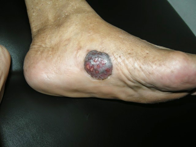
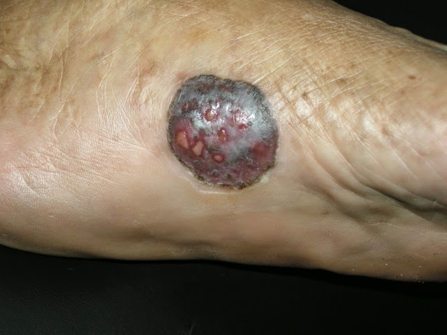
O/E:  shows a localised pigmented tumor 3 x 3 cm with superficial ulcerations on the medial aspect of the sole of left foot.  It has an irregular margin but was well circumscribed.  The nodule is firm on deep palpation.

Clinical Images:


Skin Biopsy
Nests of melanoma cells are seen invading the dermis. The tumour cells are pleomorphic, have vesicular nuclei and eosinophilic cytoplasm. There is increased mitotic activity. Many of the cells contain melanin pigment.

Diagnosis:  Left foot biopsy Malignant melanoma, acrolentiginous type, nodular

Discussion and Questions: We rarely see melanoma here in Malaysia.  The prevalence rate is reported to be about 0.4 per 100 000 population.  I have not had a single case of melanoma the whole of last 2 years.  This patient waited for a year before a diagnosis was made.  What has gone wrong? 

The histopath report unfortunately did not indicate the thickness of the tumor neither is there any mitotic rate or Clark’s level of invasion.  In a study in Malaysia most of the cases are located on the sole of the foot as in this patient.  (12/24 cases) Histologically majority are of the nodular type.  I think based on the report, our patient has a nodular type of melanoma.

Plan: 
Dermatologists in Malaysia don't manage malignant melanoma.  Instead, they are referred to surgeons for excision.  Sentinal node biopsy and CT scan abdomen and chest would be useful for staging of the tumor.  Would PET scan give more useful information for this patient?  Immunotherapy and BRAF inhibitors are probably too expensive  for her.

References:
1. Malaysian J Pathol 2012; 34(2) : 97 – 101
Cutaneous malignant melanoma: clinical and histopathological
review of cases in a Malaysian tertiary referral centre
Jayalakshmi PAILOOR, Kein-Seong MUN and Margaret LEOW*
Departments of Pathology and *Surgery, Faculty of Medicine, University of Malaya
Abstract
Melanoma is a lethal skin cancer that occurs predominantly among Caucasians. In Malaysia, the incidence of melanoma is low. This is a retrospective study of clinical and histopathological features of patients with cutaneous melanoma who were seen at the University Malaya Medical Centre from 1998 to 2008. Thirty-two patients with cutaneous melanoma were recorded during that period. Of these, 24 had sought treatment at the onset of disease at our centre. Chinese patients constituted the largest group (19 cases). The median age of these 24 patients at the time of presentation was 62 years. 16 patients had melanoma involving the lower limb with 12 affecting the sole of the foot. None had melanoma arising from the face. Histopathology showed nodular melanoma in 22 cases (91.6%), with superficial spreading and acral lentiginous melanoma diagnosed in 1 case each. The majority of patients (62.5%) were found to be in Stage III of the disease at the time of diagnosis.

2.  Whiteman DC1, Pavan WJ, Bastian BC. The melanomas: a synthesis of epidemiological, clinical, histopathological, genetic, and biological aspects, supporting distinct subtypes, causal pathways, and cells of origin. Pigment Cell Melanoma Res. 2011 Oct;24(5):879-97.  Free Full Text
Abstract:  Converging lines of evidence from varied scientific disciplines suggest that cutaneous melanomas comprise biologically distinct subtypes that arise through multiple causal pathways. Understanding the respective relationships of each subtype with etiologic factors such as UV radiation and constitutional factors is the first necessary step toward developing refined prevention strategies for the specific forms of melanoma. Furthermore, classifying this disease precisely into biologically distinct subtypes is the key to developing mechanism-based treatments, as highlighted by recent discoveries. In this review, we outline the historical developments that underpin our understanding of melanoma heterogeneity, and we do this from the perspectives of clinical presentation, histopathology, epidemiology, molecular genetics, and developmental biology. We integrate the evidence from these separate trajectories to catalog the emerging major categories of melanomas and conclude with important unanswered questions relating to the development of melanoma and its cells of origin.

Friday, May 23, 2014

A case of severe pemphigus vulgaris

Recently I saw this 40 year old woman who presented with severe blisters and erosions.  It started about 3 weeks ago and spread quite rapidly from the chest to the face and extremities.  Now her mouth is also involved.

Exam:  generalised bulla, majority are flaccid type.  Erosions extensive with crusts esp anterior chest wall.  Superficial erosions on the lower lips.  Nikolysky sign positive.


Skin biopsy:  Section shows skin composed of epidermis and dermis. A suprabasal bulla
containing acantholytic cells is seen. In areas, a tombstone appearance
is seen. Infiltrates of eosinophils and lymphocytes are seen in the
dermis.
Pemphigus vulgaris 
Blood counts and biochemistry are normal.   
She was treated as outpatient.  I have started her on oral prednisolone 40mg daily.  Added oral antibiotics cefuroxime 250mg bd.  Skin dressing with KMNO4 wet compress followed by fusidin/hydrocort cream bd.  Keep skin roof intact.

Questions:

  1. I do not have IF facilities.  Will this lack of facilities affect her prognosis?
  2. what kind of skin dressing would you use?
  3. would you use steroid sparing agent like mycophenolate, MTX , azathioprine or ritazulimab at this juncture?
Thanks for your help.  Your comments are greatly valued.

 References:

Cochrane reviews: Interventions for pemphigus vulgaris and pemphigus foliaceus


Martin LK, Agero AL, Werth V, Villanueva E, Segall J, Murrell DF
Published Online: 
15 April 2009
This review of clinical trials aimed to find out which is the most effective and safest treatment option for pemphigus vulgaris and pemphigus foliaceus.
Pemphigus vulgaris and pemphigus foliaceus are rare diseases characterised by fragile blisters and sores on the skin and mucosa. They are auto-immune diseases which are caused by the body making an antibody against the person's own skin. These diseases are chronic and are not currently curable. Pemphigus vulgaris and foliaceus are managed with drugs which suppress the immune system. The aim of treatment is to suppress blister formation. Systemic glucocorticoids are the cornerstone of management in pemphigus, however adjuvant immunosuppressive and anti-inflammatory agents are commonly used. There are many treatments available, however it is not known which is the most effective or safest treatment option, or which is the best combination.
This review included data from 11 clinical trials involving 404 participants. The studies had very small numbers of participants, so can provide only limited information. Ten different active treatments were studied, including prednisolone, pulsed oral dexamethasone, azathioprine, cyclophosphamide, cyclosporine, dapsone, mycophenolate, plasma exchange, topical epidermal growth factor and traditional Chinese medicine.
This review found insufficient information to conclude which is the most effective and safest treatment plan. We found that mycophenolate mofetil appears to be more effective than azathioprine in controlling disease, although no difference was seen in remission. We found that taking azathioprine and cyclophosphamide decreased the amount of glucocorticoids required. Topical epidermal growth factor decreased time required for lesions to heal by 6 days (median). We found no difference in withdrawal due to adverse events in any study, although differing adverse event profiles were observed for each intervention. We were not able to conclude which treatments are superior overall.
Multiple treatments are available for pemphigus vulgaris and pemphigus foliaceus and there is a variation in dosage plan and combination of drugs used, which makes choice of treatment schedule complex. In addition, response to treatment can vary between individuals. Treatments need to be chosen after careful consideration of the potential benefits and side effects, in the context of the individual's other medical conditions. This review found insufficient information to conclude which is the most effective and safest treatment regimen. Further studies are required to determine the optimal treatment regimen, especially to assess the optimal glucocorticoid dose, the role of adjuvant immunosuppressive medications, and long-term adverse events to improve harm:benefit analyses.