To: Potential Participants
Fr: Henry and David
Re: Format
In order to make things clearer, we suggest you submit your cases in the following way. Don't worry if you don't have all the infomation. If you have clinical pictures and have trouble uploading them, you can send them to one of us and we will do that.
Format for Case Presentation:Title
History
Physical Exam
Pertinent Labs
Path Description
Diagnosis
Questions
Your name and email address.
Best regards
Henry Foong FRCP and David Elpern MD
VGRD Editors
Friday, December 31, 2004
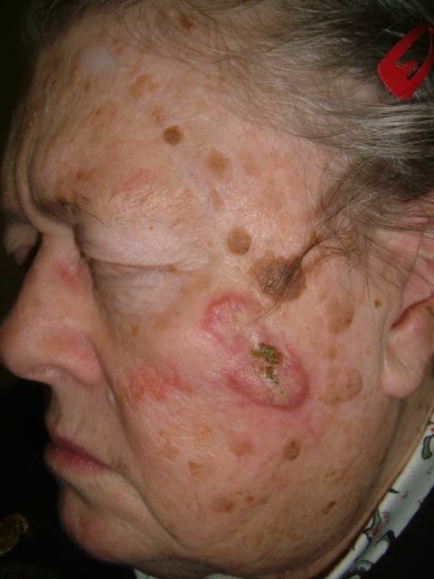

75 yo woman with lesion of face
History: This 75 yo woman was first seen on 12 October 2004 with a lesion on cheek. It had a crust and some purulent material. The lesion was surrounded by large seborrheic keratoses and I thought it was an inflammatory seb ker. Ulcer care with cleaning and Bactroban cream was recommended. I gave her a follow-up appointment and saw her back yesterday.
Physical Exam: There is a 4 cm flesh-colored plaque on the left cheek. The borders are somewhat rolled. There is a crust centrally which when removed shows an ulcer and creamy pus. (see photos)
Lab: Culture obtained. Grew out normal skin organisms
Path: 3 mm punch biopsy obtained. Jan 5, 2005 - Report indicates well-differentiated squamous cell carcinoma
Diagnosis: I suspect SCC or KA. I think the purulent drainage may relate to the squamous cells - I saw a similar case years ago on the back that I thought was a ruptured epidermal inclusion cyst. This lesion has changed in the past two months. It looked quite benign at her last visit.
Jan 5, 2005. I discussed results with patient and she will be referred for micrographic surgery. Exam today reveals a possible submandibular lymph node. I wonder if this is related to chronic inflammation in the tumor rather than mets.
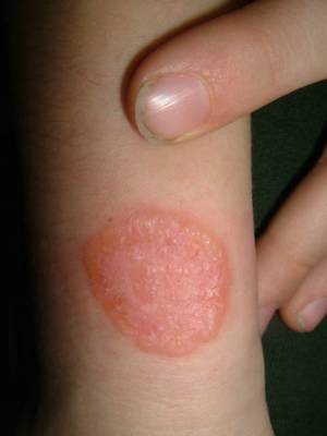
8 yo boy with lesion on wrist
History: The patient is an 8 yo boy with a one week history of an expanding lesion on the right wrist. He plays in a sandbox that is frequented by stray cats. He's been using clotrimazole cream for three days.
Physical Exam: There is a 2 cm diameter plaque on the right wrist. The periphery appears to be vesicular and possibly a bit scaly. No other lesions.
Lab: KOH equivocal - hard to interpret because of cream artifact.
Diagnosis: ? Kerion of glabrfous skin.
I plan to stop the cream, repeat KOH in a few days and have obtained a fungal culture. Will biopsy if still uncertain and process is progressing.
Follow-up of Jan 3, 2005. The patient was seen again and the KOH prep repeated. Today, this was markedly positive (no cream artifact) Dx: Kerion of Glabrous Skin. Usually caused by M. canis. See below for references.
2.5 cm lesion right wrist
References:
1: Alteras I, Feuerman EJ, David M, Segal R.
The increasing role of Microsporum canis in the variety of dermatophytic
manifestations reported from Israel.
Mycopathologia. 1986 Aug;95(2):105-7.
PMID: 3762660 [PubMed - indexed for MEDLINE]
2: Powell FC, Muller SA.
Kerion of the glabrous skin.
J Am Acad Dermatol. 1982 Oct;7(4):490-4.
PMID: 6216270 [PubMed - indexed for MEDLINE]
Physical Exam: There is a 2 cm diameter plaque on the right wrist. The periphery appears to be vesicular and possibly a bit scaly. No other lesions.
Lab: KOH equivocal - hard to interpret because of cream artifact.
Diagnosis: ? Kerion of glabrfous skin.
I plan to stop the cream, repeat KOH in a few days and have obtained a fungal culture. Will biopsy if still uncertain and process is progressing.
Follow-up of Jan 3, 2005. The patient was seen again and the KOH prep repeated. Today, this was markedly positive (no cream artifact) Dx: Kerion of Glabrous Skin. Usually caused by M. canis. See below for references.

2.5 cm lesion right wrist
References:
1: Alteras I, Feuerman EJ, David M, Segal R.
The increasing role of Microsporum canis in the variety of dermatophytic
manifestations reported from Israel.
Mycopathologia. 1986 Aug;95(2):105-7.
PMID: 3762660 [PubMed - indexed for MEDLINE]
2: Powell FC, Muller SA.
Kerion of the glabrous skin.
J Am Acad Dermatol. 1982 Oct;7(4):490-4.
PMID: 6216270 [PubMed - indexed for MEDLINE]
Subscribe to:
Posts (Atom)