Presented by Nai-Chien Yeat
Williams College, Williamstown, Massachusetts
Abstract: 20 year-old Malaysian college student with one day history of an itchy line on right arm.
Yeat's History:
I developed itchy welts all over my body shortly after moving into my new dorm room. A bite on my right wrist caused extensive swelling and intense itching within 24 hours of first discovery. Within 36 hours, a swollen, pruritic red streak extended from my wrist to my upper arm.
A bite on my left ring finger caused extensive swelling and intense itching within 24 hours. Within 36 hours, the swelling and itching had spread to the back of my hand.
After bumping into Dr. Elpern on the street, I started a course of antibiotics (Augmentin) and took antihistamines (Clarityne and Benadryl) to relieve the pruritus.
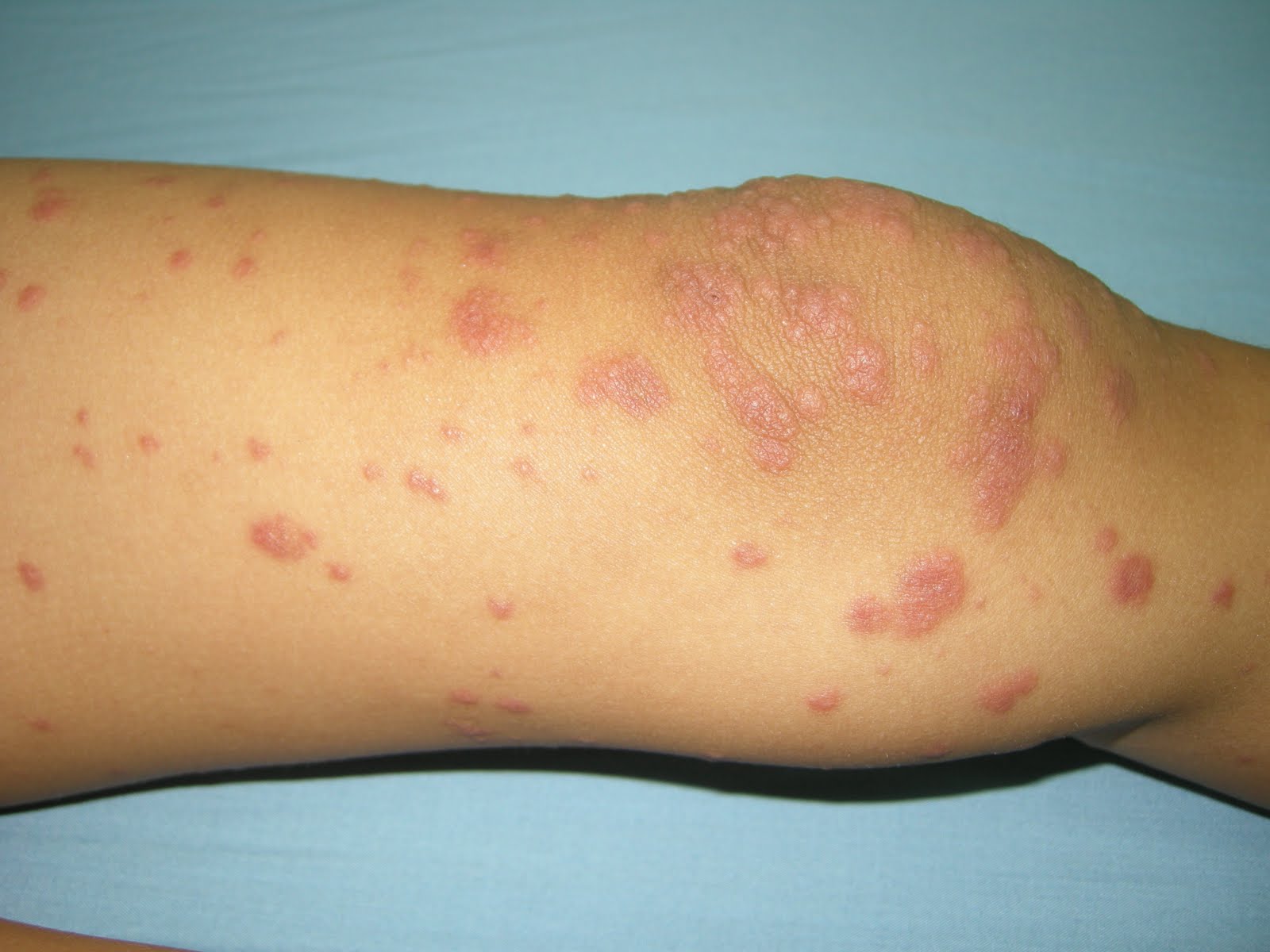
O/E: (DJE) I bumped into Mr. Yeat on Sunday morning, September 4th on the Williams College campus and he showed me his hands and arms. There were erythematous papules with some superficial crusts on the hands and a lymphangitic streak on the volar right arm extending towards the elbow. Other than pruritus, he felt well and had no fever.
 Clinical Photos taken by Mr. Yeat
 Diagnosis:
 Although initially I was concerned about a bacterial lymphangitis, I now think this is most consistent with lymphangitis secondary to insect bite rather than a sign of a bacterial etiology. Yeat knows the initial lesions are bites and he feels well otherwise. I suppose a bite could have been superinfected with strep, so the Augmentin makes sense; but it could also be based on another mechanism. There are a few pertinent references including one from the BMJ which Mr. Yeat found (# 2). I am not convinced this is from bedbugs as many types of arthropod bites apparently can cause lymphangitis. It's curious that so few cases have been reported. This may be because the patients appear to have a bacterial process, are treated with antibiotics and get better as they would over a few days even without the medications. It would be important to know if bed bugs have been found in his dormitory.
(Note from Yeat one week after onset: "The swelling has completely subsided, and you can barely see the red streak that the lymphangitis left behind."
Questions: Mr. Yeat and I will appreciate your thoughts. Do you feel the Augmentin was necessary? Have any of you seen similar cases?
Reference: Superficial lymphangitis after arthropod bite: a distinctive but underrecognized entity?1. Marque M, Girard C, Guillot B, Bessis D. myriammarque@yahoo.fr
Dermatology. 2008;217(3):262-7. Epub 2008 Aug 6.
Abstract
BACKGROUND: Acute bacterial lymphangitis is a common occurrence after skin damage. This diagnosis is often made in case of red linear streaks after arthropod bites, leading to the prescription of oral antibiotics. In this setting, noninfectious superficial lymphangitis after arthropod bites, an eruption rarely mentioned in the medical literature, appears as a diagnostic challenge.
OBJECTIVE: Our purpose was to study the clinical and histopathological features of this underrecognized condition.
METHODS: We collected the observations of six consecutive patients seen between the years 2003 and 2006, who developed an acute linear erythematous eruption along lymphatic vessels, mimicking common bacterial lymphangitis. Standard histological examinations were completed by immunopathological staining using the monoclonal antibody D2-40, a highly selective marker of lymphatic endothelium. Extensive review of the literature about acute noninfectious superficial lymphangitis was performed. Results: The clinical presentation and histological findings excluded an infectious etiology and suggested superficial lymphangitis after an arthropod bite in all the observations.
CONCLUSIONS: This article analyzes the clinical and histological features of noninfectious superficial lymphangitis after arthropod bite, a benign underrecognized condition mimicking common bacterial lymphangitis. Physicians should be aware of this benign reaction to avoid the useless prescription of antibiotics.
2. BMJ Case Reports 2010; doi:10.1136/bcr.09.2010.3310
Acute superficial lymphangitis following pigeon mite biteParvaiz A Koul, Syed Mudassir Qadri
Full Text Online.