Monday, December 30, 2013

Nail Dystrophy x 3 Years

Abstract:  52 yo woman with 3 year history of a nail dystrophy

HPI:  The patient has had this problem for three years.  She works out a lot and recalls trauma to the toe,

O/E:  The left great toe nail is dystrophic,  The nail is quite short and there is brownish green discoloration under the abnormal nail.  There is some hemorrhage under the proximal nail fold.  

Clinical Photos:

Dermatoscopiic Image


Lab:  KOH was positive for hyphae, but fungal culture is negative at 14 days.

Impression:  Nail dystrophy in a 52 year-old woman.  While this is probably traumatic, the long history is of concern and I feel that biopsy should be considered to rule out malignancy.

Questions:  What is your diagnosis?  Would you obtain a biopsy to rule out malignancy given the long history.


Saturday, December 14, 2013

Vellus Hair Cysts


Abstract: 17 yo boy with scores of papules on abdomen, chest, neck

HPI: 
This 17 yo boy has had 2 - 3 mm papules on chest, abdomen and neck slowly progressive since age 10.  Occasionally the lesions become iflamed.

OE:
Scores of 2 - 3 mm somewhat cystic lesions in above areas.  Some are slightly greyish

Clinical Photo:
 Pathology: (Photomicrographs taken by Dr. Deon Wolpowitz, Department of Dermatopathology, Boston University School of Medicine.
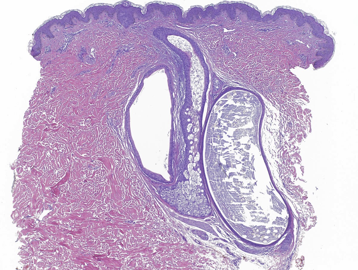
Within the dermis there is a cyst lined by squamous epithelium with a granular layer and containing laminated keratin and numerous vellus hairs.
4x
20x
Diagnosis:  Vellus Hair Cysts

Questions: Has anyone had success treating VHC?  Do they just disappear over time?  Who has seen an adult with with VHC?

Learning Points: (from reference 2)
1. Eruptive vellus hair cysts are a benign, asymptomatic condition, occurring predominantly in children and young adults.
2. Differential diagnoses include milia, comedones, keratosis pilaris, molluscum contagiosum and steatocystoma multiplex.
3.Risk of scarring should always be carefully considered before embarking on surgical or laser treatment.

References:
1.  Vellus Hair Cysts  - eMedicine

2.  Papules on the trunk. Eruptive vellus hair cysts.
Roberts CM, Birnie AJ, Kaye P, Murphy R.
Clin Exp Dermatol. 2010 Apr;3:74-5. doi: 10.1111/j.1365-2230.2009.03494

Sunday, December 01, 2013

Arcuate and Circinate Facial Eruption


Presented by   Dr. Arnulfo Macadangdang
Cebu City, Philippines


Abstract:  18 year-old student with one week history of facial eruption

HPI:  The patient is an 18 yo man with a one-week history of an eruption on face and neck.  It is mostly asymptomatic.  He is an athlete, takes no medications and has no risky behaviors.  He had an upper respiratory infection around a month before onset. 

O/E:  There are arcuate and circinate lesions on cheeks, neck, and forehead.  Two similar lesions on u upper back. The scale is greasy.  No lymphadenopathy.


Clinical Photos:


Lab:  KOH negative


Diagnosis:  This has some features of seborrheic dermatitis, but it is not typical.  Sebosporiasis was another thought.  Evolving psoriasis?  Relationship to "URI" in October?

Questions:  Does this clinical picture suggest any specific diagnosis?

Follow-up:
5 Day F/U photos

 
The patient was seen 5 days after starting desonide 0.05% cream.  His skin lesions have esolved completely with mild post-inflammatory hypopigmentation.  The three lesions on back (not treated) have persisted. No new lesions.  Our tentative diagnosis is atypical seborrheic dermatitis.  Atypical Pityriasis rosea is also considered.  We do not feel serology for syphilis and HIV are indicated at this point, based on a discussion of risky behaviors.   Will taper desonide over the next two weeks and see patient back in a month as necessary.