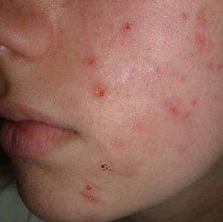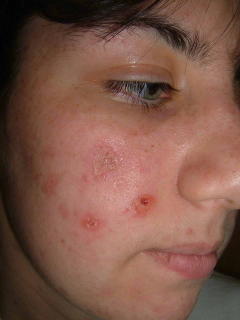

A repeat biopsy on June 27. 2005 showed:
DIAGNOSIS: Skin - Left Temple:
Epidermal necrosis with s cale crust containing neutrophils , sub-epidermal abundant neutrophils and fibrin deposition, superficial and deep perivascular lymphohistiocytic infiltrate with focal neutrophil ic microabscesses, septal and lobular panniculitis with mixed inflammatory cell infiltrate of abundant neutrophils , lymphocytes , histiocytes, and eosinophils and numerous activated endothelial cells, surrounding a medium-sized vessel with marked mixed inflammatory cell infiltrate of neutrophils , histiocytes and occasional eosinophils .
NOTE : These changes are suggestive of a medium-sized vasculitis with overlying necrosis. Elastic tissue stain (EVG) does not reveal the vessel in the deeper sections, therefore, arterial or venular distinction cannot be made. The differential diagnosis includes a large vessel vasculitis such as periarteritis nodosa or early Wegener's granulomatosis. P.A.S. stain is negative for fungal organisms. Fite stain is negative for mycobacteria . However, an infectious vasculitis cannot be entirely excluded . If the clinical suspicion persists, culture studies may be of help . The differential diagnosis also includes , in the appropriate clinical setting , factitial panniculitis with secondary vascular involvement. These are not the changes of lupus erythematosus , pityriasis lichenoides et varioliformis acuta or prurigo nodularis . Serologic studies may be helpful. Clinico-pathologic correlation is suggested.
If dermatitis artefacta is exculded,then we should think about two possibilities:either bed bug urticaria which might simulate prurigo nodularis or papulonecrotic tuberculide and investigate accordingly.Regarding the histopathalogy is abit srange.
ReplyDeleteI'm curious if there was a diagnosis. I have very similar sores on my face, neck, arms, and legs. I've had an inconclusive biopsy, and tested negative for infectious, toxic, inflammatory, and various others. Negative for staph. Doctors are unsure of what's wrong with me as my cbc was inconclusive as well. I began with only two of these sores and now have about 20. They rarely heal and this has been ongoing for over 2 years.
ReplyDelete