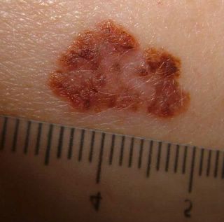
Path Report
DIAGNOSIS: Skin - Right Mid Back:
Malignant melanoma.
Type: Superficial spreading
Greatest thickness: 0.90 mm.
Anatomic level: II
Margins: Complete excised
Radial growth phase: Present
Vertical growth phase: Absent
Mitoses: None
Tumor infiltrating lymphocytes: Present, non-brisk
Ulceration: Absent
Regression: Present
Microsatellites: Absent
Vascular invasion: Absent
Precursor lesion: Not identified
NOTE: The lesion represents a severely atypical compound melanocytic neoplasm characterized by a predominantly intra-epidermal component with marked confluent lentiginous and nested melanocytic hyperplasia , pagetoid spread, extension into adnexae, and by a severely atypical dermal component with papillary dermal regression.
The picture is suggestive of variegated malignant melanoma
ReplyDelete