Wednesday, June 28, 2006

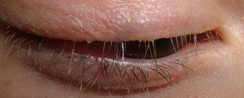
Eye Lash Alopecia

The patient is an eight year old girl who has had a dermatitis of her eyelid margins off an on for around a year. She gets crusted areas which itch and her mother feels she plucks her eyelashes. On the day of the visit the lids looked quiet, but there was definite loss of some eyelashes. There is some evidence of broken lashes.

Question:
Is this eyelash trichotillomania or isolated alopecia areata of the eyelashes? I favor trichotillomania
What is the prognosis for spontaneous recovery?

My plan is to see her if and when the dermatitis is more acute and obtain a culture to see if she has a bacterial blepharitis.


Saturday, June 24, 2006

Bed Bugs

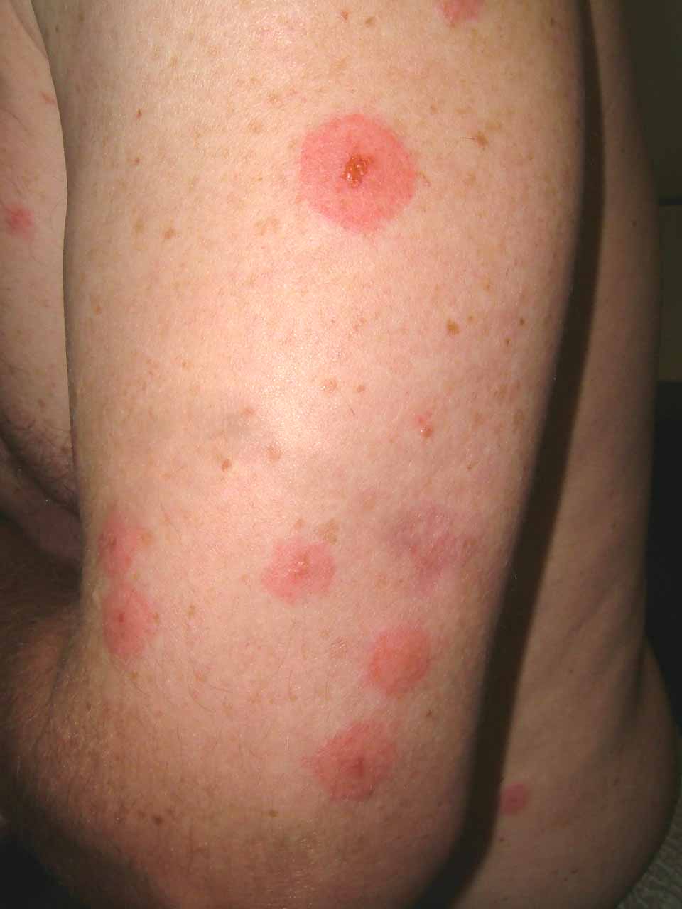
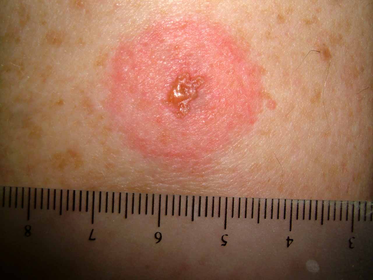
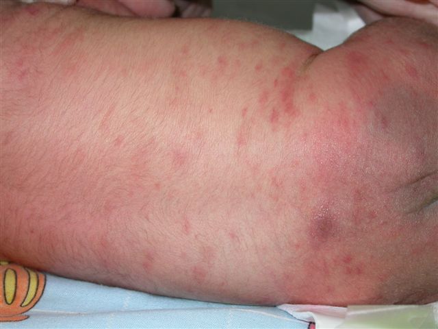
This 35 year-old man went to Mamaronek, New York to watch the New York Open Golf Championship last weekend. The closest motel room he could find was in Bridgeport, Connecticut. The latter is a city with a fairly poor reputation. Two days after returning home to Massachusetts he developed the lesions pictured here on arms and to a lesser extend on torso and legs.

They are consistent with bedbug bites (Cimex lenticularis). For a brief description go to: http://www.emedicine.com/derm/topic600.htm

In November of 2005 a bedbug epidemic was reported in New York in the New York Times. I saw a couple of cases from there here in Williamstown, MA, 150 miles away. I suppose there's a similar problem in Bridgeport now.


Wednesday, June 21, 2006

Lyme Disease

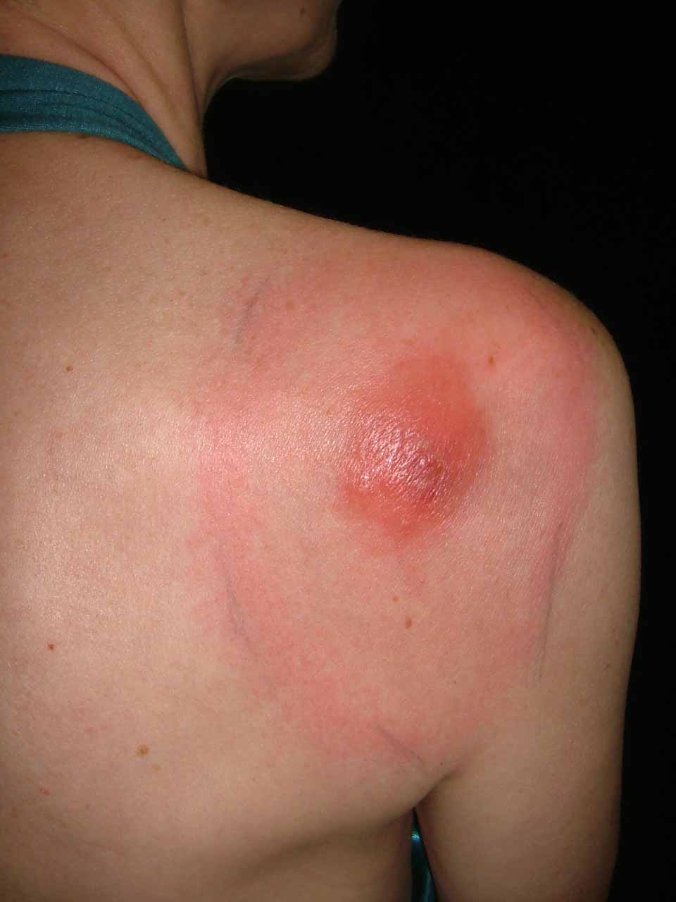
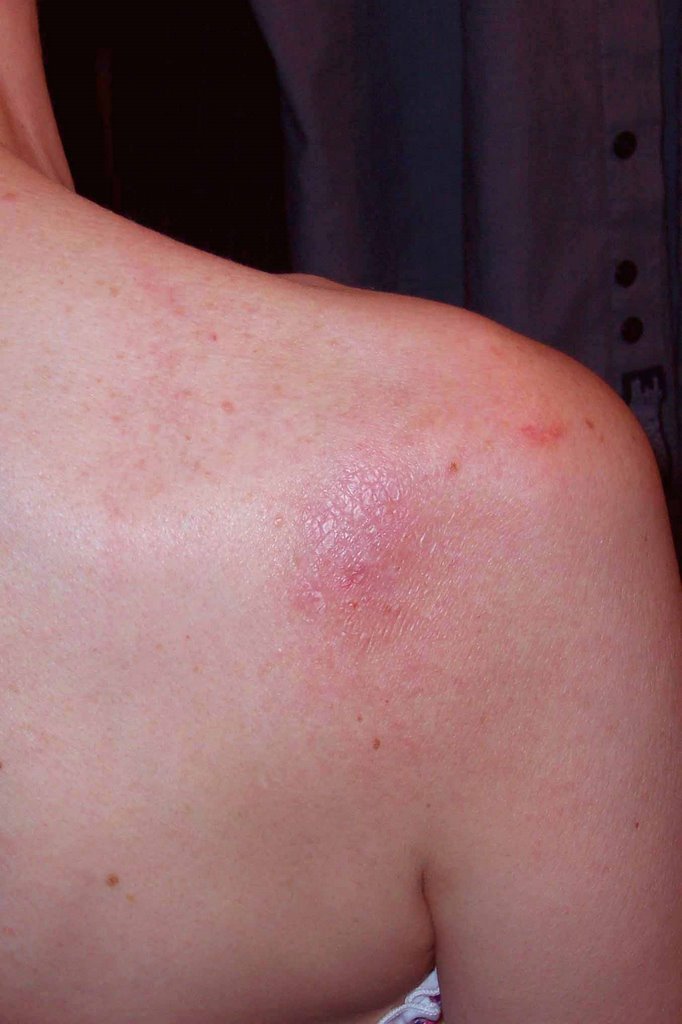
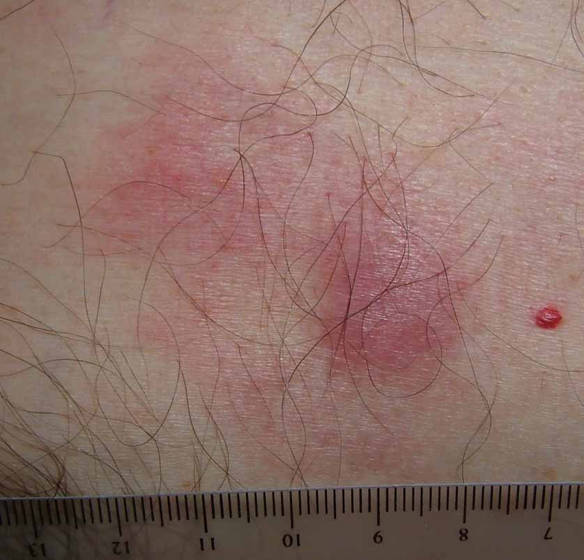
This 52 year-old woman presented as a walk-in patient today. She has a five day history of an erythematous nodule on the right shoulder which has developed a ring around it. The ring measures 9 by 11 centimeters in diameter. Initially, the nodule was painful for a few days; but the pain has subsided. She has had no fever or constutional symptoms and has continued to work as a school teacher.

The patient lives in rural New Hampshire and gardens a fair bit. She was seen two times at a primary care clinic where a diagnosis of spider bite was made. First she was given Keflex, then amoxicillin.

Diagnosis: I am uncertain. I favor Lyme Disease but this has atypical features. The central nodule is unusual, although the peripheral ring looks like Lyme. I also considered atypical Sweet's Disease and a pyogenic process. Lastly, I thought of eosinophilic cellulitis (Well's Syndrome) which I have never seen.

Plan: Since she feels well, I prescribed Doxycycline 100 mg bid, and ordered a cbc, Lyme titers and a G6PD (if the lesion becomes necrotic may want to try Dapsone. I considered biopsy but was strapped for time and thought Lyme most likely. Now, I am uncertain. Biopsy will be done when she is seen in follow-up unless she is significantly better.

Your thoughts will be welcome.


Addendum:

Path Report:
DIAGNOSIS: Skin - (A) Right Shoulder-Periphery-Ring Surrounding Nodule:
Tight, mild superficial and deep perivascular lymphocytic infiltrate .
NOTE : These changes are those of a dermal hypersensitivity reaction to an exogenous antigen and, given the clinical presentation, are consistent with erythema chronicum migrans .

DIAGNOSIS: Skin - (B) Right Shoulder-Nodule:
Broad scale crust containing neutrophils , epidermal hyperplasia , marked papillary dermal edema and a superficial and deep perivascular and interstitial mixed inflammatory cell infiltrate composed of lymphocytes , neutrophils , histiocytes and plasma cells with rare eosinophils consistent with arthropod bite reactio n.
NOTE : These changes may be seen in a tick bite and erythema chronicum migrans . Clinico-pathologic correlation is suggested.

The Lyme Titer is positive.

In my opinion, this is not definitive, but the onus is on me at this time to continue to treat for Lyme.
The patient is on doxycycline and will continue on this at a dose of 100 mg bid for three weeks.
The patient reports that after three days of doxycycline the lesion is smaller. This, too, is not definitive, however, one can not ignore that either.


One Week Follow-up.
The patient feels better. Has mild insomnia.
Although initial lab titer was positive -- the Western Blot was negative indicating an early infection. Our infectious disease consultant expected them to be negative. Early treatment can prevent conversion. Will probably repeat in 2 weeks.

Picture on June 29, 2006

Wednesday, June 07, 2006

Erosio interdigitalis blastomycetica


Why do dermatologists love these cumbersome terms??

The patient is a 30 yo woman with a two week history of a sore area between the 3rd and 4th toes of the left foot. She is in good general health. Not pregnant and on no oral contraceptives. Both parents have Type II diabetes. At the time of onset she was on doxycycline for rosacea.

The examination shows an erosion in the toe web. The KOH prep was positive for pseudohyphae consistent with the diagnosis of erosio interdigitalis blastomycetica

Given her family history she should be worked up for diabetes. The doxycycline may have also played a role.

The role of maceration was discussed. She will keep toes separated and use ketoconazole cream. If this in not effective, I will ask her to dry area after bathing with a hair dryer and continue an imidazole cream or solution.

This is from emedicine.com:
Intertrigo: Intertrigo typically presents with erythema, cracking, and maceration with soreness and pruritic symptoms. Lesions typically have an irregular margin with surrounding satellite papules and pustules. Web spaces of affected fingers or toes are macerated and have the appearance of soft white skin, which is a condition termed erosio interdigitalis blastomycetica (interdigital candidosis).

A good source for the treatment of intertrigo is at: http://www.emedicine.com/derm/topic198.htm

Wednesday, May 24, 2006

Genital Pain in a 60 yo Man

Dear Pre-VGRD,

Thank you for allowing me to present this difficult case.

The patient is a 60-year-old journalist, who presents for evaluation of genital pain that has been present for about six months. He has been in his usual state of health. No new sports or traumas. His medications include atorvastin, buproprion and the generic omeprazole.

He has an annoying, painful sensation on the ventral shaft of the penis. He has noted some hyperpigmentation there and occasionally the skin here gets red. He thinks this lasts for a few hours but he is not sure. The sensation is like that of an abrasion.  He saw a urologist who prescribed clobetasol cream but did not note anything in the area. He used the clobetasol for a few weeks but found that it only seemed to make the pain worse.

EXAMINATION: The examination shows a healthy, appearing 60-year-old man who is a good historian. He has a dark line on the ventral surface of the shaft of the penis. The skin here is completely normal. There was no evidence of erythema today.

IMPRESSION: This is a genital pain syndrome. It may be related to what is called scrotodynia. I also seem to remember some cases that were reported in which there was dermographic urticaria on the genitalia. I could not find references to those. The best that I can come up with at this point is a local genital pain syndrome similar to scrotodynia or vulvodynia. These are poorly understood and the treatments are not particularly effective. If this were genital dermographic urticaria, probably an antihistamine such as hydroxyzine would work.

Those of us who are dermatologists see such cases occasionally. I would appreciate your thoughts.
Marius Grenoble,  QUÉBEC CITY, QC, Canada

Thursday, May 18, 2006

Photosensitive dermatitis from bleaching creams


My friend, David Elpern MD, a dermatologist from Williamstown, USA sent this note to me.

"NY Times
May 14, 2006

A Vision of Pale Beauty Carries Risks for Asia's Women

By THOMAS FULLER

MAKHAM KHU, Thailand - Neighbors gawk and children yell, "Ghost!" The manager of the restaurant where Panya Boonchun worked simply told her she was fired.
The cream that she applied to her face and neck was supposed to transform her into a white-skinned beauty, the kind she saw in women's magazines and on television.
But the illegally produced lotion she bought in a store near this village in southeastern Thailand turned her skin into a patchwork of albino pink and dark brown. Doctors say her condition may be irreversible.
"I never look in the mirror anymore," she said, sobbing during an interview.
Whiter skin is being aggressively marketed across Asia, with vast selections of skin-whitening creams on supermarket and pharmacy shelves testament to an industry that has flourished over the past decade. In Hong Kong, Malaysia, the Philippines, South Korea and Taiwan, 4 of every 10 women use a whitening cream, a survey by Synovate, a market research company, found."

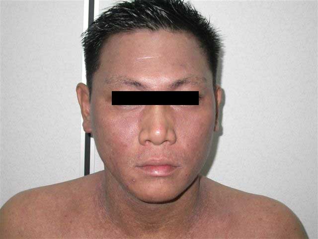
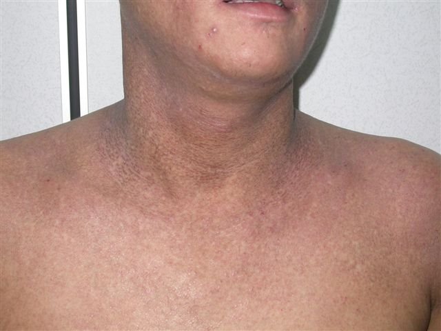
And it so happened the same morning I saw a 33-year-old man who had been applying a bleaching cream from Southern Thailand for his melasma. After several weeks of application, He noticed pigmentary changes on his face which was dusky brownish tinge on the forehead, cheeks and chin. It has now spread to the neck as well. Past one week, he noticed skin eruptions appearing on the forearms as well. He was otherwise well and has no fever or polyarthralgia.
His medication history included OTC garlic pills and fish oil. He is however a heavy drinker.

Examination of the skin showed diffuse dusky erythematous to brownish macules on the patient's ears, face and neck. The eruptions somehow spares the nose. Eczematous eruptions were noted on the forearms bilaterally. There were no hepatosplenomegaly. There were no sclerodactyly changes on the fingers.

My first impression was could this be the "southern thailand bleaching creams ghostly onchronosis due to high concentration hydroquinone cream??

Investigations done showed normal blood counts and blood biochemistry. ANA serology was negative.

I suspect this is some form of photosensitive dermatitis. Porphyria cutanea tarda runs thro my mind as a differential as he is heavy drinker but his LFTs were normal. I have never seen this before.

Wednesday, May 17, 2006

Onycholysis

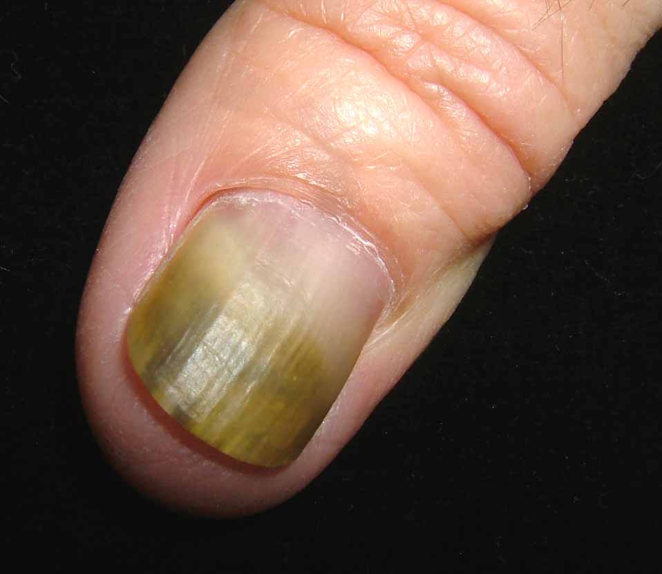
The patient is a healthy 58 year old woman with a 3 year history of onycholysis. Fungal culture has grown out Candida zeylanoides. Bacterial culture negative.

Topicals have not been of help. (imidazoles and thymol in chloroform)
Would fluconazole be of value?
Should the nail be avulsed?
Who has had success treating this kind of problem?

Thursday, May 11, 2006

Pellagra in Alcoholics

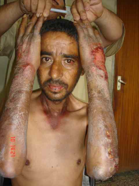
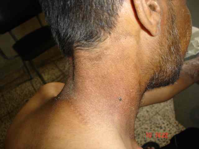
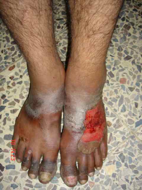
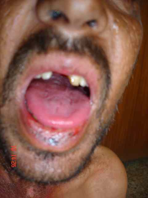
In Iraq, pellagra is only seen in sporadic cases in patients with alcoholism. I describe here a case of pellagra in 30 year old man who used to drink alcohol one litre a day of a local beverage called ARAK. In addition, his nutrition was poor. He developed a typical rash of pellagra in photosensitive area, some lesions are bullous together with beefy tongue. The rash resolved quickly after vitamin replacement but recurred when he resumed alcohol drinking.

I would like to ask my colleagues
1) how often see pellagra
2) what is the cause?


Tuesday, May 09, 2006

Nail Bed Carcinoma

The patient is s 70 yo man who was seen today for a three year follow-up of micrographic surgery for a sqaumous cell carcinoma of the nail bed. There is no evidence of recurrence and the results are cosmetically excellent.

I had another patient with a similar lesion. He was a VA patient and they did not approve him for microgrpahic surgery. He had the distal part of his thumb excised. Bizarre that health insurance trumps the interests of the patient.


Friday, May 05, 2006

Folliculitis and Hair Loss

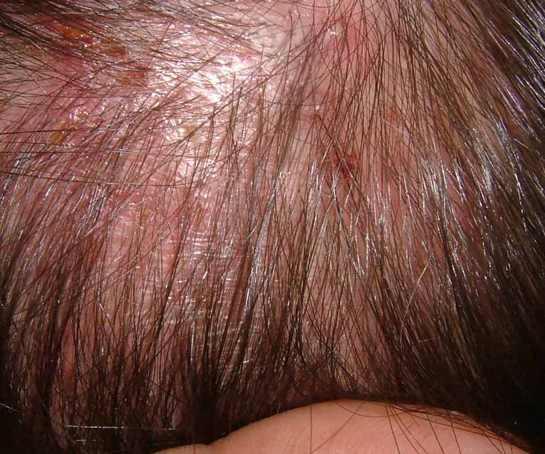
The patient is a 30 year-old man with a 2 year history of folliculitis and mild hair loss. He has erythematous papules and a few pustules scattered over the scalp. Cultures have been negative. Biopsy showed only inflammation c/w folloculitis. Minocycline has been of marginal help, but process recurrs when he stops. He has an occasional papule on his cheeks but no signs of rosacea. His hair is globally thin.

Question: Is this folliculitis decalvans? Would isotretinoin be worth trying? He is also interested in hair transplantation.


Saturday, April 29, 2006

Volcanic Eruption

The patient is an 88 yo woman who came as a "walk-in" today.
She said she had a "volcano erupt" on her leg two weeks ago.

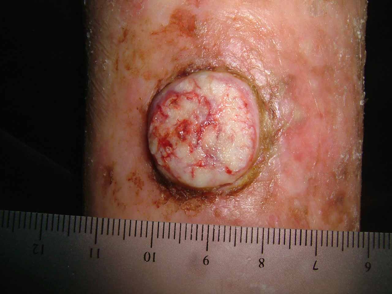
The examination shows an alert actve 88 yo woman with Type II skin. She has a dome-shaped tumor on her right shin which measures 2.1 cm in diameter. It seems to have a collarette surrounding it. She has marked actinic damage.


The history suggests keratoacanthoma. Some dermatologists call this "Squamous Cell Carcinoma -- Keratoacanthoma type." I suspect this is a coding ploy to make them seem malignant.

My question is what to do?
1) It would be hard to close after excision in this site.
2) If it is curetted and dessicated, it make take weeks to months to heal.
3) It could be observed, however, some of these larger lesions can be locally agressive.
4) Intralesional methotrexate is a possibility.

My instinct is to treat with C and E, but I'd like some suggestions. I might follow that with Aldara. I am open to suggestions.

Thank you.

David Elpern

May 4, 2006
At the advice of a few of you, I did a shave biopsy and curetted and dessicated the lesion. The base was mostly gritty (a good sign). I will follow closely. She's 88 year-old. So, I'll observe before any further intervention. May try imiquimod. Will play by ear.

Saturday, April 22, 2006

Cream Complacency

A 60 yo man was seen yesterday for pruritic papules on scalp and extremities. He had seen another dermatologist over the years and had been treated with a host of topicals including liquid nitrogen. Biopsy showed Prurigo nodularis.


[Contents of Bag Brought in by Patient]


After a few years, he developed a febrile illness, was seen in the ER where a CBC showed a WBC of 80,000. He was eventually diagnosed with a rare T-cell lymphoma. Four courses of CHOP have put him in remission.

If his prurigo becomes more active again, one wonders if this will be a harbinger of recurrence.

Sobering lesson. Prurigo may be realated to an underlying malignancy. We can all be lulled into a cream complacency and miss an important clue. I can think of one or two patients over the years with unexplained excoriations who turned out to have a lymphoproliferative malignancy.

Reference:
Seeburger J, Anderson-Wilms N, Jacobs R.
Lennert's lymphoma presenting as prurigo nodularis.
Cutis. 1993 May;51(5):355-8.
Section of Dermatology, Loma Linda University School of Medicine, California.
Abstract:
Lennert's lymphoma is a peripheral T-cell lymphoma that only rarely involves the skin. We present the case of a forty-two-year-old man who experienced severe pruritus for ten months. He was repeatedly diagnosed as having neurodermatitis
and prurigo nodularis before subtle hematologic clues suggested, and subsequent examination of bone and lymph node biopsy specimens confirmed, the diagnosis of Lennert's lymphoma. This report describes the case and presents a brief review
of Lennert's lymphoma.

Friday, April 21, 2006

U.P in a Newborn


I saw a new born today at the neonatal ward. He is the first child, just delivered today by LSCS for fetal distress. Noted to have generalized erythematous wheals and papules. Some of the wheals and papules appear to form blisters. The mother had no history of any infection during her pregnancy. On examination the child appeared comfortable and not in distress. Sleeping and quiet. Afebrile. Generalised erythematous raised papules and wheals were noted on the face, trunk and upper limbs. Some of the wheals blanche with pressure. Tried to elicit darier’s sign – mild erythema but not raised. The wheals were intensely erythematous and inflamed on certain parts of the neck and face.

Clinically he has urticaria pigmentosa

His blood counts : TWBC 3100 Eosinophils 20% 
Platelets 44 000
G6PD - pending

Though the rash appeared intense, the child was comfortable. Would you have started him on oral hydroxizine? Would you investigate further – hematological malignancy, etc the parents were advised about trigger factors (rubbing) and drugs (anesthetics, etc). thanks, Henry Foong

Thursday, March 30, 2006

Unknown

What are your thoughts?
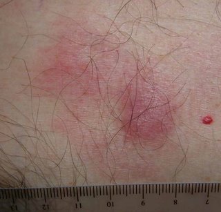


The Pathology Report: ATYPICAL LYMPHOID HYPEPLASIA
"Superficial and deep, nodular and diffuse lymphohistiocytic infiltrate forming follicular germinal centers consistent with atypical lymphoid hyperplasia.

NOTE : Immunostaining reveals a mixture of T-cells (CD3) and B-cells (CD-20). No clonal proliferation is seen on Kappa and Lambda staining. No CD-30 positive cells are noted. These findings are supportive of a lymphoid hyperplasia. If the clinical suspicion persists, follow-up of the patient is suggested"

Given the location, one wonders if this could be secondary to a tick bite (common in our area). See; Pediatr Dermatol. 2001 Nov-Dec;18(6):481-4.

Persistent atypical lymphocytic hyperplasia following tick bite in a child:
report of a case and review of the literature.

Hwong H, Jones D, Prieto VG, Schulz C, Duvic M.

Department of Internal Medicine Specialties, Section of Dermatology, University
of Texas-M.D. Anderson Cancer Center, Houston, Texas 77030, USA.

We report a 6-year-old girl who developed a red papule on the posterior neck at
the site of a previous tick bite. Initial biopsy was performed a year after the
bite and the specimen showed a dense lymphoid infiltrate with admixed CD30+
cells. The patient was referred to our center because of concern about the
development of a CD30+ lymphoproliferative disorder. The lesion was completely
excised. Histology showed no evidence of a clonal lymphoproliferative disorder
or Borrelia infection, but persistence of CD30+ cells. This case demonstrates
that a tick bite reaction can persist for more than 1 year and show
immunophenotypic and morphologic overlap with a CD30+ lymphoproliferative
disorder. Complete history with thorough clinical and histopathologic evaluation
is necessary to arrive at the correct diagnosis.


The patient is a 52 yo engineer who presents with a 2 month history of a 1.5 cm in diameter asymptomatic somewhat "spongy" presternal nodule surrounded on one side with macular non-blanchable erythema.
The clinical appearance is non-diagnostic. This may be an infiltrative process, possibly a malignancy. I have not seen anything like this before with the possible exception of a Merkel Cell carcinoma. Punch biopsies were taken from the nodule and the surrounding erythema.
The results should be back on April 3.


What are your thoughts?

Saturday, March 18, 2006

“NAILING DOWN A DIAGNOSIS”


Presented by
Brian T. Maurer, Enfield, Connecticut


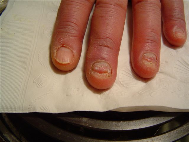
D.S. is a 72-year-old left-handed male with a three-year history of periungual inflammation coupled with periodic cracking, separation and nail loss on the 3rd, 4th and 5th fingers of the left hand. The pads of these fingers also appear erythematous and swollen with dry cracked skin.

Initially, a local dermatologist prescribed topical Lamisil without effect. A subsequent month-long course of oral griseofulvin likewise did nothing. The periungual inflammation seemed to respond to topical erythromycin; the patient reports that all symptoms subsided over the summer months, only to return with the cold autumn weather.

An amateur gardener and golfer, D.S. wears a golf glove on his left hand during summer golfing season (when the condition seems to subside). He reports that his left hand usually feels colder than the right in winter time.

With the exception of one discolored nail of the left great toe, the nails of his remaining fingers and toes appear normal; he exhibits no other form of dermatitis.

Questions: Given the history and clinical course, what is the diagnosis? Suggestions for treatment?

Tuesday, March 14, 2006

Digital Tumor

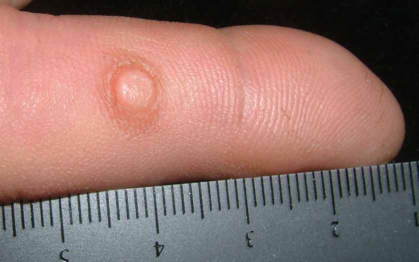
This 61 yo man presented with a 30 year history of an annoying tumor of his right index finger. It interferes with his writing in that it abuts on the area in contact with a pen or pencil. He has pared it down on numerous occasions and it has been unsuccessfully treated with cryotherapy in the past. The clinical picture is that of an acquired digital fifrokeratoma (see reference below). An excisional biopsy is scheduled.


Dupre A, Christol B, Bories M.
[Acquired fibro-keratoma. Report of 8 cases]
Ann Dermatol Venereol. 1977 Oct;104(10):611-5. [Article in French]

The acquired fibro-keratoma is a benign fibro-epithelial tumor of acquired
nature. I was set apart as a distinct entity in 1968 by Bart et al. who named it
"acquired digital fibro-keratoma". It is predominantly, although not
exclusively, located on the fingers and toes, near the phalangeal joints. The
authors report 8 personal cases and make a short general review of the disease.
The lesion is slightly prominent, well defined and surrounded by a
hyperkeratotic collar. It protrudes on the surface of the skin much like a
hernia. Histologically, it is a dermo-epidermal tumor. There is a proliferation
of connective tissue under the papillomatous, hyperacanthotic and orthokeratotic
epidermis. The mature collagen bundles, voluminous fibroblasts and numerous
enlarged capillaries are parallel to the vertical axis of the tumor. The
acquired fibrokeratoma must be differentiated from other cutaneous tumors,
particularly fibromas and Koenen's tumors. The etiology is unknown. However, it
was found in some cases that a trauma had contributed to the development of the
tumor. The only possible treatment is surgical excision.

Friday, March 10, 2006

Interesting Tumor

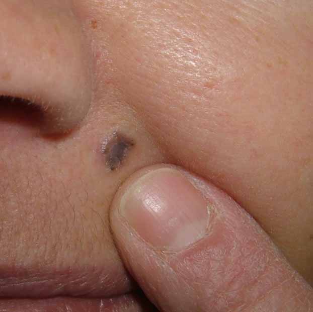
This 48 yo woman presented with a 2 year history of a tumor of the upper lip.
She has type III skin. The lesion in question is 6 mm in diameter.
A biopsy was done. This looks like a pigmented sclerotic basal cell.

She has a sick child and delayed her dermatology visit because she must take son to physicians often.
It's a three hour trip for him. He is 15 years old and has had polycythema rubra vera and essential thrombocythemia since age eight.

Sunday, February 26, 2006

Alopecia with peculiar regrowth pattern

The patient is a 35 year old healthy man who presented with alopecia of the scalp of 1 year duration. Many topical therapies were tried with no response. On examination, there was classical alopecia areata. I gave him IM triamcinolone 40mg injection every 2 weeks with topical Dermovate oinment for 2 months. He came back with hair growth in a wavy pattern in the form of concentric circles (see Figure).. I do not have an explanation. I would appreciate any helpful comments.


Presented by Professor Khalifa Sharquie
University of Baghdad, Iraq

Saturday, February 18, 2006

From the Nail File

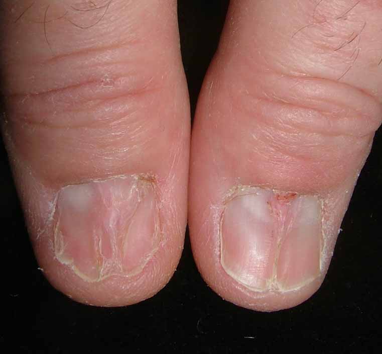
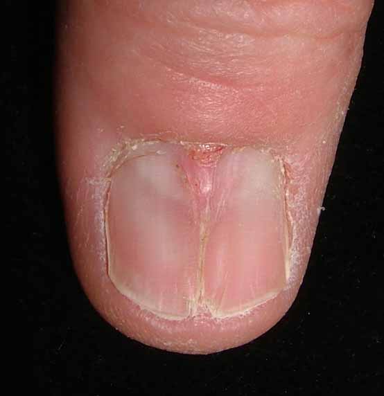
55 year old man with 6 month history of nail distrophy. Only thumbs. He works with his hands cutting film. Trauma likely.

The patient wrote:
"I noticed left thumbnail change in appearance in late August 2005. I scraped the nail down and got fluid near base. The right thumbnail became abnormal after trauma in October of 2005 showing a crack straight up the middle of the nail."


Moderator: I suspect lichen planus. Median nail dystrophy less likely.

Comments??

Saturday, February 11, 2006

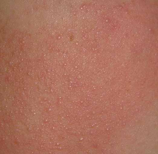
Erythromelanosis Follicularis Facei

The patient is a 15 year old boy with a many year history of ruddy cheeks and a fine follicular eruption in the affected areas. He also has typical keratosis pilaris on his arms and thighs. The diagnosis of erythromelanosis follicularis of the face (also called keratosis pilaris rubra facei) is not difficult. Who has a therapeutic suggestion? I don't think the patient is ready for laser therapy yet. It was difficult to capture the depth of the erythema here which is qute striking. His sister has a similar process but is is milder.


Reference:
Stephan F, Ayoub N, Klein-Tomb L, Tomb R.
[Erythromelanosis follicularis faciei and colli]
Ann Dermatol Venereol. 2002 Jan;129(1 Pt 1):63-5. [Article in French]

BACKGROUND: Erythromelanosis follicularis of the face and neck, originally
described in Japan by Kitamura et al. in 1960, is characterized by a clinical
set of three: well-demarcated erythema, hyperpigmentation and follicular
papules. It affects the face and the neck generally on both sides. Since the
original description, it has seldom been reported in the literature. CASE
REPORTS: This paper reports two patients with unilateral presentation.
DISCUSSION: Having discussed the various differential diagnoses all published
cases were listed and analyzed. The prevalence of this disease appears higher
than is shown by the limited number of cases reported in the literature. It
deserves more recognition. Its nosologic and aetiologic frames still remain to
be clarified.