The Pathology Report: ATYPICAL LYMPHOID HYPEPLASIA
"Superficial and deep, nodular and diffuse lymphohistiocytic infiltrate forming follicular germinal centers consistent with atypical lymphoid hyperplasia.
NOTE : Immunostaining reveals a mixture of T-cells (CD3) and B-cells (CD-20). No clonal proliferation is seen on Kappa and Lambda staining. No CD-30 positive cells are noted. These findings are supportive of a lymphoid hyperplasia. If the clinical suspicion persists, follow-up of the patient is suggested"
Given the location, one wonders if this could be secondary to a tick bite (common in our area). See; Pediatr Dermatol. 2001 Nov-Dec;18(6):481-4.
Persistent atypical lymphocytic hyperplasia following tick bite in a child:
report of a case and review of the literature.
Hwong H, Jones D, Prieto VG, Schulz C, Duvic M.
Department of Internal Medicine Specialties, Section of Dermatology, University
of Texas-M.D. Anderson Cancer Center, Houston, Texas 77030, USA.
We report a 6-year-old girl who developed a red papule on the posterior neck at
the site of a previous tick bite. Initial biopsy was performed a year after the
bite and the specimen showed a dense lymphoid infiltrate with admixed CD30+
cells. The patient was referred to our center because of concern about the
development of a CD30+ lymphoproliferative disorder. The lesion was completely
excised. Histology showed no evidence of a clonal lymphoproliferative disorder
or Borrelia infection, but persistence of CD30+ cells. This case demonstrates
that a tick bite reaction can persist for more than 1 year and show
immunophenotypic and morphologic overlap with a CD30+ lymphoproliferative
disorder. Complete history with thorough clinical and histopathologic evaluation
is necessary to arrive at the correct diagnosis.
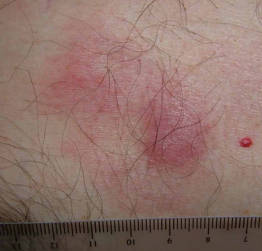
The patient is a 52 yo engineer who presents with a 2 month history of a 1.5 cm in diameter asymptomatic somewhat "spongy" presternal nodule surrounded on one side with macular non-blanchable erythema.
The clinical appearance is non-diagnostic. This may be an infiltrative process, possibly a malignancy. I have not seen anything like this before with the possible exception of a Merkel Cell carcinoma. Punch biopsies were taken from the nodule and the surrounding erythema.
The results should be back on April 3.
What are your thoughts?
Every day we might face such odd cases .This stimulate the fantasy in dermatology.I will consider the possibilty of lymphoma cutis,B-cell lyphoma or condition related to panniculitis
ReplyDeletekhalifa sharquie
I would to prefer more details regarding history and physical examination.In such odd cases these details may shorten a long list of differential diagnoses.I agree ,it may be cutaneous TB or as my teacher's comment a cutaneous lymphoma.The authors described the lesion as spongy with non blanchable erythema .may be one of the vascular tumours??
ReplyDelete