Sunday, March 20, 2005

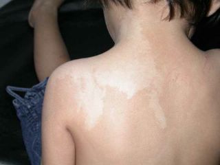
2 yo girl with segmental hypopigmentation

This is a 2 year old girl who has a patch of hypopigmentation on her left shoulder since birth. It extends from back of neck to the left shoulder. It was asymptomatic. Examination showed a hypopigmented macule 5cm by 10cm on the back of left shoulder. It is segmental in distribution and has an irregular border. There was no central hypoaesthesia. She has no ophthalmic or CNS defects.


Clinically she has nevus depigmentosus or some form of pigmentary mosaicism. Biopsy of the lesion was not done.

This is usually a benign skin disorder and is caused by the functional defects of melanocytes and the morphologic abnormalities of melanosomes.

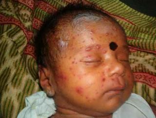
Newborn with Varicella

Posted for Dr. Jayakar Thomas of Royapuram, Chennai, India. Please comment here or reply directly to Dr. Thomas at thomas_j@vsnl.com

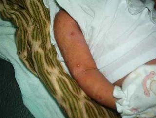
The patient is a 21 day old female infant who presented with multiple generalized vesicles of two days duration. Her mother had developed varicella two days post-partum.

Physical Exam: A normally nourished child with multiple vesicles over erythematous base. Child had low grade fever. There were no oral or genital lesions and no other mucosae were invoved.


Laboratory: Nil relevant


Histopathology: Not done


Diagnosis: Neonatal varicella (NV)


Comments: NV is not seen very commonly. It usually occurs when the mother has an attack of varicella during post partum period as in this case. The child presented here was treated with IV aciclovir with good results.

Varicella occuring in the mother during pregnancy may end up in congenital varicella syndrome in the new born.

Questions: What are the members' experience with neonatal varicella.
Some pediatricians do not use aciclovir and manage with supportive measures and antibiotics. Are they justified?
I personally feel that all children with varicella should be treated with aciclovir or appropraite antivirals. This will definitely bring down the chances of herpes zoster at a later date, given the scarring, pain, and other sequelae of herpes zoster.

Thursday, March 17, 2005

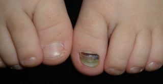
3 yo with nail dystrophy

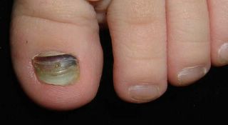
This 3 yo boy has had a malformed left great toe nail since infancy. Recently, he traumatized it and may have had a subungual hematoma. His pediatrician tried to drain it with a hot paperclip, but was unsuccessful. His parents want to know what could be done to improve this nail.


Nail Dystrophy, Child


Nail Dyst - Closeup

Wednesday, March 16, 2005

Sunday, March 13, 2005

Atopic Infant

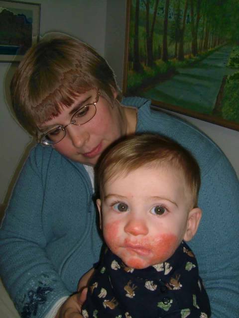
The patient is a one year old boy with an almost life-long history of atopic dermatitis. His father (age 30) has persistent facial eczema. His mother has significant food allergies (nuts and fruit cause angioedema and laryngeal edema). This child's facial eczema has proved difficult to control. Topical steroids have been of value (fluocinalone 0.25% ointment); but topical tacrolimus and pimecrolimus have not been effective. I suspect he may have food allergies. In addition, there is a cat at home. I think he needs to be tested for cat allergy. Food testing is controversial. Would serum IgE measurement be of value? Role of staph superinfection needs to be considered as staph, acting as a superantigen, may be driving this. I'd appreciate your thoughts. DJE


Child S.A.D. Posted by Hello


Child S.A.D. Posted by Hello

Saturday, March 12, 2005

Genital Papules in a 25 yo woman

Presented by:
Dr. Jayakar Thomas
KK CHILDS Trust Hospital, & Apollo Hospitals, Chennai, India
E mail: thomas_j@vsnl.net

The patient is a 25 yo married woman from Chennai, India who presented with itchy papules over the vulval area for the last two years. There was no history suggestive of risk of acquiring any STD.

Physical Exam: Clinical examination revealed a healthy very good-looking young lady with multiple papules over her genital region.

Lab: Nil relevant

Histopathology : Not done

Diagnosis: Verruca plana

Comment: We see verruca vulgaris and condyloma usually in these site.


25 yo woman Posted by Hello
Verruca plana is not common. This lady was in the habit of shaving her pubic hairs which has resulted in spread and persistence of lesions.
Topical 5% imiqumod should help if used with caution.

Question: What do the members feel would be the management strategy?

2o yo man with painful plaque

OFFICE VISIT

MARCH 07, 2005

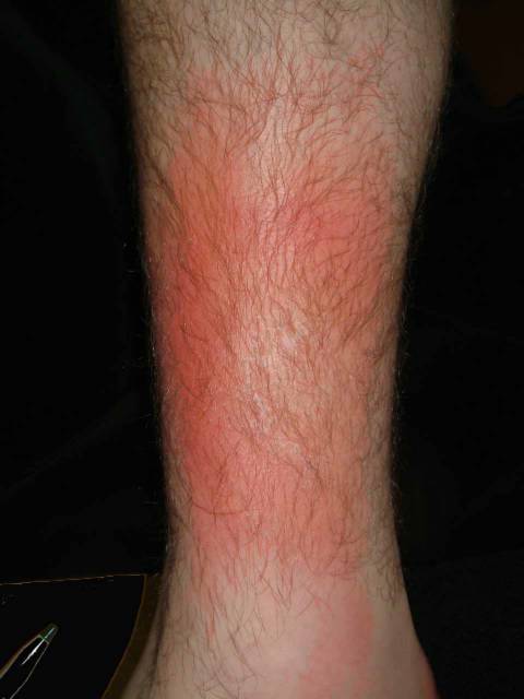
Sam D. was seen today on a same day basis. He has a few day's history of a tender, erythematous area on the right lower leg. This is at the site of a twine ankle bracelet that he had been wearing for two years. He feels a bit run down although he has not had a fever. 

EXAMINATION: The examination shows an area of localized erythema and mild central scaling on the left lower leg just proximal to the ankle medially. It has a sharp margin. KOH prep was negative. There is definite heat at this site.  The regional inguinal lymph nodes are enlarged.

IMPRESSION: Probable cellulitis. Doubt contact. Doubt tinea.  Doubt phlebitis.

PLAN: 

1. Warm compresses. 
2. Dicloxacillin 500 mg q.i.d. 
3. Return for follow-up in four days. 

Follow-up visit March 11
S: Patient feels a bit better energy wise, but leg still red and tender.
He hasn't been able to rest - has classes, lots of responsibilities. Has not been able to do warm compresses more than twice in past 5 days.
No systemic symptoms.
O: No marked change in plaque
A: I still favor a diagnosis of cellulitis.
P: Post on ANAK VGRD for suggestions. How long should this take to resolve?
Clinically, this does not look like erythema nodosum. It began after prolonged microtrauma from a twine ankle bracelet.
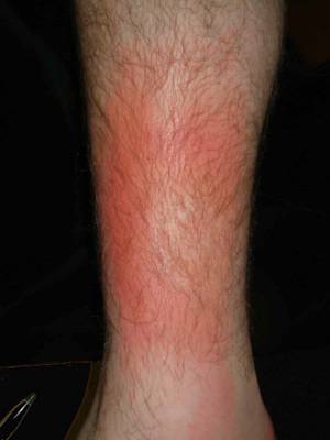


20 yo man with painful plaque Posted by Hello

Friday, March 11, 2005

56 yo woman with erosions for two years

This 56 yo woman has an alomst two year history of intense pruritus. She dates the onset to April 23, 2003 when she was exposed to toxic fumes at work. Since then, she wakes at night excoriating her skin. Other than a mild anemia and eosinophilia, all studies have been unremarkable. She has not worked since April 30, 2003. The is an intelligent person. Divorced, mother of three adult children (one with Turner's Syndrome) she worked in retail and latterly as a security guard. Skin biopsy was not helpful.


3/10/05 Posted by Hello


Back Posted by Hello


CLOSEUP Posted by Hello

Sunday, March 06, 2005

56 year-old woman with metastatic melanoma

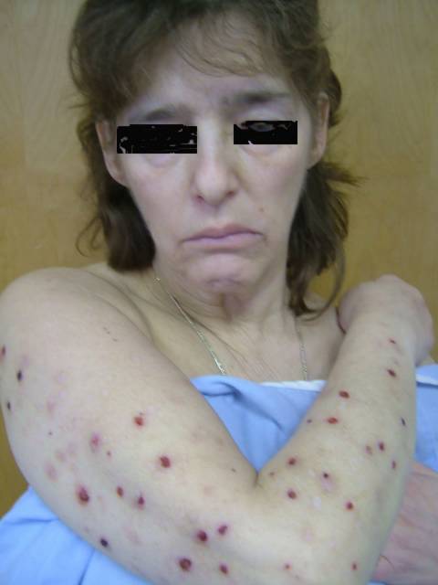
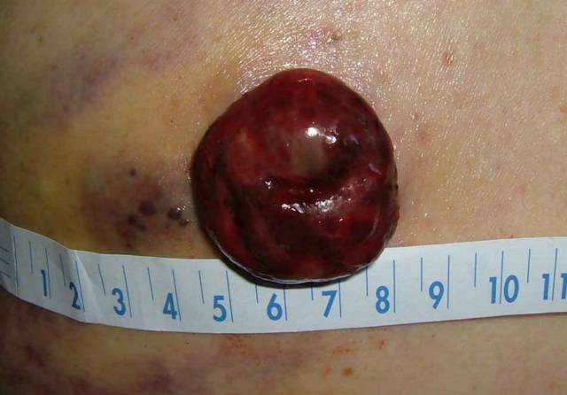
This 56 yo woman presented on February 10, 2005 with multiple subcutaneous nodules, present for around a month. Some were hemorrhagic. Biopsy showed metastatic melanoma. Work-up revealed pancreatic, brain, lung and possibly adrenal mets. The biopsy site has not healed. A four centimeter exophytic tumor has appeared at the site of incision. The patient is lucid and has refused chemotherapy or radiotherapy. The tumor pictured below is a management problem. The part we see is only the visible portion of a larger deeper mass. The site of her primary melanoma has not been identified.


February 10, 2005 Posted by Hello


March 5, 2005 Posted by Hello

72 yo woman with Lupus Pernio

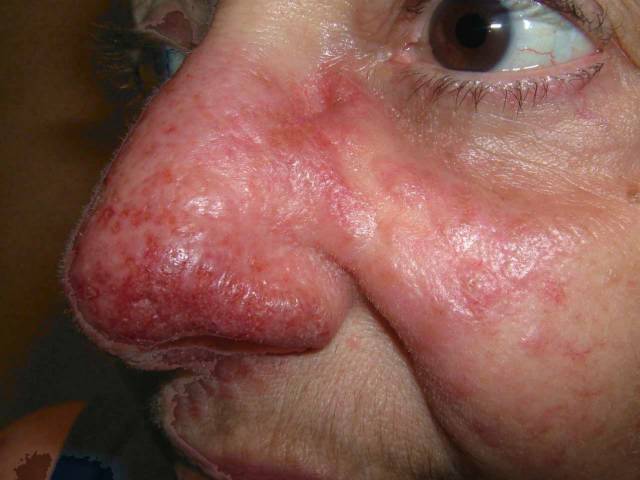
This 72 yo woman has a five year history of biopsy proven lupus pernio. She also has a 30 year history of stable chronic leukemia (I don't know the form). Her rheumatologist treated the sarcoid with hydroxyplaquenil and Methotrexate (without any benefit). The questions: 1) topical tacrolimus 2) Infliximab and possible contraindications b/c of chronic leukemia. Strangely, her insurance carrier has denied tacrolimus ointment, but this is bein contested.


March 5, 2005 Posted by Hello


March 5, 2005 Posted by Hello

Friday, March 04, 2005

53 year old man with 4 month history of lesion back

This 53 year-old man noted a lesion on right upper back around 4 months ago. It has tripled in size since then. On first glance it looks like a BCC, but BCCs rarely grow like this. The patient has Type I - II skin and is covered with freckles. He has a strong family history of skin cancer. I excised this lesion immediately because I was worried that it might be an amenanotic melanoma. If the latter, this is scary since it will be a thick lesion. Say a prayer for this patient that this is BCC.


Nodule Right Upper Back Posted by Hello

Wednesday, March 02, 2005

51 year old woman with incidental lesion

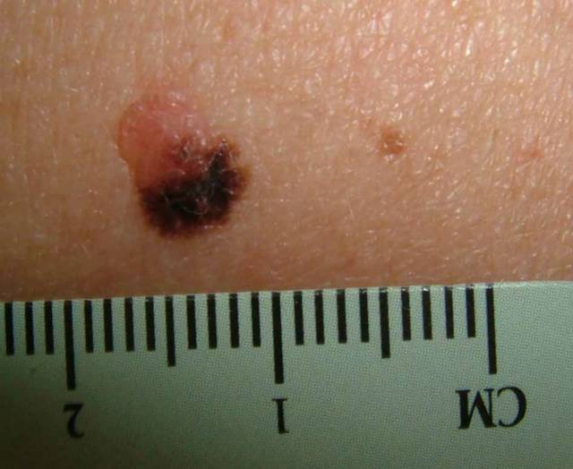
This 7 mm in diameter lesion was noted on the left upper back of a 51 yo woman with type II skin. She was concerned about another lesion on the lower back. The latter lesion was benign in appearance. The lesion pictured below was biopsied and I will post the results in a week. The patient was unaware of this lesion, it being on a part of the back that is difficult to visualize.


incidental lesion Posted by Hello

Friday, February 04, 2005

F/U Case of December 31, 2004

This woman had micrographic surgery on February 2, 2005.
Her tumor was removed in one stage with a simple closure.
Here is a picture taken on February 3, 2005.


1 Day Post Op Posted by Hello

Thursday, January 06, 2005

Wednesday, January 05, 2005

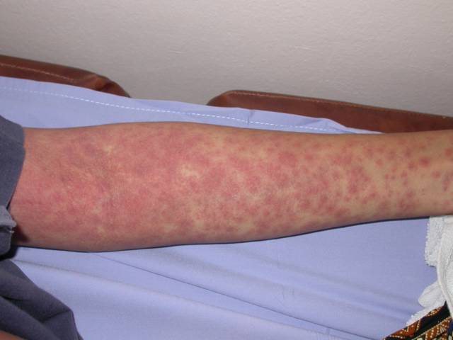
39-yr-old woman with generalised maculopapular eruptions.

The patient is a 39 yr old teacher who presented with generalised skin eruption about 10 days ago. A day prior to that, she developed fever and had seen a GP and was prescribed paracetamol and oral bacampicillin. The rashes had spread to the trunk, lower and upper limbs. She was otherwise well

Pertinent fingings were temp 39C, generalised maculopapular eruptions on the lower limbs and trunk. Some of the lesions have a vasculitic centre. There were no blisters.

Serology and blood chemistry did not show any significant abnormalities. It was the skin biopsy which gave us the final analysis. The biopsy was taken from a lesion on the right leg. Though the lesions looked active, the histology was "mild" no vasculitis was noted; instead there was some interface dermatitis changes. This makes drug eruption as the most likely cause of the rash. Our final diagnosis: Drug rash from bacampicillin.