Wednesday, November 02, 2011

Tumor in Vaccination Site

Abstract: 59 yo woman with six month history of tumor l. arm
HPI: The patient, a kindergarten teacher, was bitten on the hand by a child on March 20, 2011. School policy did not allow the child to be tested for hepatitis or HIV. Therefore, it was recommended that she receive hepatitis B vaccination. She had three shots ( March, June and December 2010) in the left deltoid area. In late January or early February 2011 she developed a tumor at the site of the vaccination.
O/E: There is a 1.2 cm. slightly friable tumor in the above-mentioned area. Dermoscopic exam shows some arborizing blood vessels.

Clinical Photograph:

Pathology: Basal Cell Carcinoma: Nodular and Infiltrating. No epidermal connection is apparent in submitted specimens.


Diagnosis
: Basal Cell Carcinoma in Vaccination site.

Discussion: There have been sporadic reports of skin cancer developing at the sites of vaccination, but never one in a hepatitis B site. The latent period here is short. It's unclear what the initiating factor is. Our patient is a light-complected Caucasian, so has another risk factor, too. We plan to investigate this area further and present a case report with a review of the literature. Your thoughts will be helpful.

Tuesday, October 18, 2011

Temple Bracelet Dermatitis

Abstract: 20 yo woman with allergic contact dermatitis to a bracelet purchased at a temple in Beijing

HPI: This young woman purchased some prayer beads at a Beijing temple in early September, 2011. Within two - three weeks she developed a rash under the bracelet. She treated this with a number of topicals including a neomycin containing cream. She was seen in an ER a few days before she presented to my office and started on prednisone and Keflex.

O/E: A well-defined area of resolving dermatitis on left wrist. It appears to have been bullous.

Clinical Photos:
The clinical picture did not come out well. Second picture is of bracelet on unaffected wrist to show how she wore it.


Diagnosis and Discussion: This contact dermatitis is most likely secondary to wooden beads. There are a few pertinent references (see below). The patient also applied neosporin so we can't rule out that this may have played a role. She does not live near my office and is in college far away. My approach would be to treat with a topical corticosteroid and warn her about neomycin. If this recurs she can be patch tested. At this point, I do not know what kind of wood the bracelet is made of. The references I found were mostly about cocobolo wood. This may prove difficult to determine. Your comments will be welcome. Note: A number of our readers favor rosewood as the culprit (see reference # 3)

References:
1. Hausen BM. Allergic contact dermatitis from a wooden necklace. Am J Contact Dermat. 1997 Sep;8(3):185-7.
Abstract
A 36-year-old female kitchenworker twice developed eczematous lesions corresponding exactly to the area around her neck where she had worn a wooden necklace. Contact dermatitis lasted longer than 1 week. The necklace consisted of 42 brown wooden beads and 63 other wooden parts, 0.5 to 3 cm diameter. Most parts could be identified as Cocobolo wood, Brazilian and East Indian rosewood, and teak. Patch tests with the pure constituents gave +3-reactions to three dalbergions and obtusaquinone, which are known to be the sensitizers of Cocobolo and the above-mentioned rosewoods. Because of these test results, the identification of the species by eye examination could be corroborated. Further detailed questioning revealed that the patient had played a recorder, probably made from Cocobolo (Dalbergia retusa), when a child, to which she unknowingly became allergic.


2. Moratinos MM, Tevar E, Conde-Salazar L. Contact allergy to a cocobolo bracelet. Dermatitis. 2005 Sep;16(3):139-41.
Abstract
Tropical woods are highly valued because of their strength, hardness, and resistance to moisture. These characteristics make them easy to work with and extremely durable, and that is why they have been used in the manufacture of wooden jewelry, musical instruments, furniture, and handles of many different objects. We present a case of a 44-year-old man who developed pruritus, erythema, and blistering around his right wrist, corresponding exactly to the area where he had worn a wooden bracelet. Thin-layer chromatography performed with the extract of the shavings revealed (R)-4-methoxydalbergione and obtusaquinone (the main components of cocobolo wood) and (S)-4'-hydroxy-4-methoxydalbergione (in lower amounts). Patch-testing with sawdust from the bracelet resulted in a very strong reaction. Patch tests with the pure constituents yielded +++ reactions to the main sensitizers of cocobolo, including obtusaquinone, but also to sensitizers present in other rosewoods. This last fact can be explained by cross-reactivity between different dalbergiones. Contact dermatitis from tropical woods is more frequent than thought, owing to their high sensitizing properties. An exhaustive search can identify the allergen responsible in many cases.

3. Hausen BM. [Rosewood allergy due to an arm bracelet and a recorder]. Derm Beruf Umwelt. 1982;30(6):189-92. [Article in German]
Abstract
A 40-year-old woman developed dermatitis of the left forearm after wearing a bracelet manufactured from Brazilian rosewood (Dalbergia nigra All.). Swelling of the lips, itching and vesicles recurred when she played a recorder made from the same timber some years later. Epicutaneous tests were strongly positive after 120 h with 2 of the wood constituents: R-4-methoxydalbergione and S-4,4'-dimethoxydalbergione. The third quinone (S-4'-hydroxy-4-methoxydalbergione) only elicited a weak reaction. Shavings of the wooden bracelet extracted with benzene and ethanol and separation of the residues by thin layer chromatography yielded all 3 dalbergiones in remarkable amounts (congruent to 0,8%). Cross-reactions to the chemically near related R-3,4-dimethoxydalbergione, known as the strongest sensitiser of the dalbergione group, were not obtained, although guinea pig experiments had revealed cross-reactivities. Of the racemic

Thursday, October 13, 2011

Lupus Erythematosus?

presented by Henry Foong:

A case in progress... 14 year-old Malaysian boy with photosensitive eruption


HPI:The patient is a 14 year old student with one year history of erythematous patches on the face, made worse with sun exposure. He is otherwise healthy with no systemic complaints. He has been on no medications byh mouth.

Examination showed few discrete 2-4 cm erythematous plaques on the cheeks, nose, upper and lower eyelids. No alopecia. No scarring.

I Suspect this is lupus erythematous. Acute LE or Subacute LE ? ANA serology and biopsy done. Results pending. Differentials - Jessner's lymphocytic infiltrates


Questions: What are your thoughts pending the biopsy findings? Would you do anything different from what I have done so far?

Friday, September 23, 2011

Painful Brusing in a 29 yo Woman

Presented by Hamish Dunwoodie, MBBS
Moncton, New Brunswick, Canada

Abstract: 29 yo woman with one week history of painful bruising on thighs

HPI: The patient is an otherwise healthy 29 yo woman with a one week history of painful bruises on her thighs. Five years ago she had leucocytoclastic vasculitis of her lower legs and very mild proteinuria. A renal consult felt she probably had mild IgA nephropathy. This has cleared. Her only medication is paroxetine, which she has been on for three months. She denies any trauma. The patient is a single mother of two children (11 and 3 years old) and lives alone with her kids. She was in school recently but is now on disability for "seizures" (although she is on no antiepileptic medications at present). She has been assaulted by a boyfriend in the past, but denies trauma this time.

O/E: There is purpura of the lateral thighs bilaterally. No evidence of LCV any longer. The remainder of the cutaneous examination is unremarkable.

Clinical Photo:


Lab: CBC, Chemistries, Urine Analysis all normal save for trace + rbcs. No proteinuria any longer.

Diagnosis: This is most likely traumatic purpura in a young woman who is reluctant todivulge an accurate history. Gardner Diamond Syndrome (autoerythrocyte sensitization syndrome, psychogenic purpura) was considered as well.

Questions: What are your thoughts?

References: (Full Text Online)
1. Gardner-Diamond syndrome: Difficulties in the management of patients with unexplained medical symptoms. Meeder R, Bannister S. Paediatr Child Health. 2006 Sep;11(7):416-9.

2. Gardner-Diamond Syndrome: bruising feeling. Bostwick JM, Imig MW. Mayo Clin Proc. 2008 May;83(5):572. (This is a short article)

Wednesday, September 21, 2011

Traumatic Ulcer

Abstract: 40 year-old man with non-healing wound

HPI: The patient is a 40 yo man who sustained traumatic abrasions of his leg and arm from a motorcycle accident on May 31, 2011. He has a history of chronic vesicular dermatitis of hands and feet complicated by recurrent staphyloccal cellulitis of legs. The wound on his right knee became infected and he was hospitalized over the summer on two occasions for parenteral antibiotics and debridement. As a result of this wound he has lot his job and his family is living marginally.

O/E: September 14, 2011. There is a nine cm relatively clean ulcer over the right knee. It has shown no tendancy to heal over the past month.

Clinical Photograph:

Diagnosis: Ulcer right knee.

Questions: How would you approach this lesion so that the patient can heal and get back to work? At present, he is getting dressing changes a few times a week and there are no plans for further surgical interventions. It looks like this will take months to heal by secondary intention.

Follow-Up: 10/19/2011 I have seen the patient on two occasions since this posting. The ulcer is ~ 75% better with just daily dressing changes with Vaseline impregnated gauze. He has not needed any further antibiotics. I expect it will be completely re-epitheliazed in two to three weeks.

Sunday, September 11, 2011

Insect Bite Lymphangitis

Presented by Nai-Chien Yeat
Williams College, Williamstown, Massachusetts

Abstract: 20 year-old Malaysian college student with one day history of an itchy line on right arm.

Yeat's History:
I developed itchy welts all over my body shortly after moving into my new dorm room. A bite on my right wrist caused extensive swelling and intense itching within 24 hours of first discovery. Within 36 hours, a swollen, pruritic red streak extended from my wrist to my upper arm.
A bite on my left ring finger caused extensive swelling and intense itching within 24 hours. Within 36 hours, the swelling and itching had spread to the back of my hand.
After bumping into Dr. Elpern on the street, I started a course of antibiotics (Augmentin) and took antihistamines (Clarityne and Benadryl) to relieve the pruritus.

O/E: (DJE) I bumped into Mr. Yeat on Sunday morning, September 4th on the Williams College campus and he showed me his hands and arms. There were erythematous papules with some superficial crusts on the hands and a lymphangitic streak on the volar right arm extending towards the elbow. Other than pruritus, he felt well and had no fever.

Clinical Photos taken by Mr. Yeat


Diagnosis: Although initially I was concerned about a bacterial lymphangitis, I now think this is most consistent with lymphangitis secondary to insect bite rather than a sign of a bacterial etiology. Yeat knows the initial lesions are bites and he feels well otherwise. I suppose a bite could have been superinfected with strep, so the Augmentin makes sense; but it could also be based on another mechanism. There are a few pertinent references including one from the BMJ which Mr. Yeat found (# 2). I am not convinced this is from bedbugs as many types of arthropod bites apparently can cause lymphangitis. It's curious that so few cases have been reported. This may be because the patients appear to have a bacterial process, are treated with antibiotics and get better as they would over a few days even without the medications. It would be important to know if bed bugs have been found in his dormitory.

(Note from Yeat one week after onset: "The swelling has completely subsided, and you can barely see the red streak that the lymphangitis left behind."

Questions: Mr. Yeat and I will appreciate your thoughts. Do you feel the Augmentin was necessary? Have any of you seen similar cases?

Reference: Superficial lymphangitis after arthropod bite: a distinctive but underrecognized entity?
1. Marque M, Girard C, Guillot B, Bessis D. myriammarque@yahoo.fr
Dermatology. 2008;217(3):262-7. Epub 2008 Aug 6.
Abstract
BACKGROUND: Acute bacterial lymphangitis is a common occurrence after skin damage. This diagnosis is often made in case of red linear streaks after arthropod bites, leading to the prescription of oral antibiotics. In this setting, noninfectious superficial lymphangitis after arthropod bites, an eruption rarely mentioned in the medical literature, appears as a diagnostic challenge.
OBJECTIVE: Our purpose was to study the clinical and histopathological features of this underrecognized condition.
METHODS: We collected the observations of six consecutive patients seen between the years 2003 and 2006, who developed an acute linear erythematous eruption along lymphatic vessels, mimicking common bacterial lymphangitis. Standard histological examinations were completed by immunopathological staining using the monoclonal antibody D2-40, a highly selective marker of lymphatic endothelium. Extensive review of the literature about acute noninfectious superficial lymphangitis was performed. Results: The clinical presentation and histological findings excluded an infectious etiology and suggested superficial lymphangitis after an arthropod bite in all the observations.
CONCLUSIONS: This article analyzes the clinical and histological features of noninfectious superficial lymphangitis after arthropod bite, a benign underrecognized condition mimicking common bacterial lymphangitis. Physicians should be aware of this benign reaction to avoid the useless prescription of antibiotics.

2. BMJ Case Reports 2010; doi:10.1136/bcr.09.2010.3310
Acute superficial lymphangitis following pigeon mite bite
Parvaiz A Koul, Syed Mudassir Qadri Full Text Online.

Tuesday, September 06, 2011

Erythema multiforme major

Presented by: Dr. Henry Foong
Ipoh, Malaysia

Abstract: Five Year-old boy with E. multiforme


The patient is a 5 year old boy presented with 3 day history of fever and generalised skin eruptions. Apparently it started with superficial lower lip erosion and the next day he had high fever and generalised skin eruptions on trunk, the upper and lower extremities. There was no family history of similar skin problems.

O/E he was afebrile. Generalised erythematous macules and plaques were noted on the face, trunk and extremities. The lesions were distributed acrally. Some of the macules had sharp margin round shape with concentric rings within it. A vesicle was noted on the centre of the macules. Few typical round macules were noted on the palms and soles. Clinically he has erythema multiforme major

TWBC 14, 900 (N11.4% L75.4% E2%) ESR 19. Mycoplasma antibody is negative. He is now empirically on oral acyclovir and oral clarithromycin.

The most likely cause of the EM is HSV infection in this patient. Wonder if you would use systemic corticosteroids in this patient?

Wednesday, August 31, 2011

Raccoon Purpura

I received this email from an otherwise healthy 23 yo woman who I saw a month ago for an unrelated problem: "I was wondering if you might have any insight to another skin problem I am having. After receiving some terrible news, I have popped a number of blood vessels around my eyes and face to the the point of having dark purple bruises around and on my eyes. I do not know what to do. I look like a victim of abuse and would like to heal my face as soon as possible."
Discussion: One can see eyelid purpura and petechiae with a number of pathologic processes (amyloidosis, coagulopathy) but also after valsalva maneuver, violent vomiting, coughing. I suspect the latter and need more information from the patient. Any thoughts?

Reference: Anesth Analg. 2007 Dec;105(6):1561-3, table of contents.
Periorbital ecchymoses during general anesthesia in a patient with primary amyloidosis: a harbinger for bleeding? Available Free Full Text Online
Weingarten TN, Hall BA, Richardson BF, Hofer RE, Sprung J.
Source
Department of Anesthesiology, Mayo Clinic College of Medicine, Rochester, Minnesota 55905, USA.
Abstract: Primary amyloidosis is a result of proliferation of a population of plasma cells that leads to an increased secretion of monoclonal immunoglobulins (amyloid). Amyloid protein infiltrates increase capillary fragility. Such capillaries can burst, even after minor stress, resulting in periorbital hemorrhage. We describe a 64-yr-old man with primary amyloidosis who underwent general anesthesia. His eyes were gently closed with tape. Upon removal of the tape bilateral periorbital purpura was noted. All coagulation studies were normal. The periorbital hemorrhage was attributed to amyloidosis-induced capillary fragility.

Wednesday, August 24, 2011

Post-Operative Contact Dermatitis

Abstract: 63 yo woman with 5 day history of a dermatitis

HPI: A 63 yo woman developed a dermatitis 2 d post surgery. An arterial line had been placed in the L. radial artery pre-op. The area was first prepped with chlorhexidine, the line was placed, and the area covered with 6 x 7 cm Tegaderm Film. A venous line was placed in the R. external jugular vein and covered with Tegaderm w/o dermatitis.

O/E: An 8 x 8 cm erythematous vesicular and hemorrhagic plaque is seen in the area under the Tegaderm. Island of sparing in center of patch is where angiocath resided. This plaque is cool to touch. Neck completely clear.

Clinical Photos:

Lab and Pathology: Not deemed necessary at this time.

Diagnosis: Irritant vs. Allergic Contact Dermatitis. Not likely Tegaderm since area under patch on neck is clear. I am considering a toxic burn from chlorhexidine under wrist patch. (see Addendum)

Questions: What are your thoughts?

Addendum: The anesthesiologist reviewed his notes and found that he applied Tincture of Benzoin to the area around the arterial line to help keep the Tegaderm in place, but not on the neck for the venous line. Allergic Contact Dermatitis to Benzoin is well-reported. This seems to be the culprit here. Hopefully, wet compresses followed by clobetasol 0.05% ointment will be helpful. We are indebted to the anesthesiologist for reviewing the operative record and educating us! We will patch test her once her eruption has quieted down.

References: (Free Full Text)
1. Indian J Dermatol Venereol Leprol. 2006 Jan-Feb;72(1):62-3.
Contact dermatitis to compound tincture of benzoin applied under occlusion.
Lakshmi C, Srinivas CR.

2. BMC Dermatol. 2004 Mar 31;4:1.
Severe facial dermatitis as a late complication of aesthetic rhinoplasty; a case report.
Rajabian MH, Sodaify M, Aghaei S.
Department of Plastic Surgery Shiraz University of Medical Sciences, Shiraz

Wednesday, August 10, 2011

Two Patients with Longitudinal Nail Dystrophy

This past month, I saw two patients with median nail dystrophies. This is an area that has only rarely been written about. These patients are presented for your interest and thoughts. If you experience difficult with the comment function, you can email DJ Elpern with your thoughts.

Case 1.

55 yo man with a 1-2 year history of a linear striation of the left thumbnail. This is painful with pressure and occasionally spontaneously painful. There is a three mm in diameter pink striation in the left thumbnail beginning at the proximal nail fold. The distal portion of the nail is somewhat deformed and there is the suggestion of an erythematous subungual papule.
Diagnosis: Possible Subungual tumor. I am considering glomangioma. (See below for follow-up)
Questions: Would the best approach be to avulse the entire nail and then do a small elipse? What else would you do here?
Follow-up: The patient first saw a hand surgeon who recommended amputation of the distal portion of the digit. Scared, he saw a second hand surgeon who said he thought this was a glomus tumor and excised it. Pathology confirmed the diagnosis of Glomus Tumor. This photo was taken approximately two months post-surgery.


18 mo post surgery
24 mo post surgery


Case 2.
60 yo woman with 3 month history of an asymptomatic longitudinal split on the left thumbnail. No history of trauma.

Diagnosis: I favor median nail canal (dystrophia unguis mediana canaliformis) here, although at first was concerned about a subungual tumor.
Question: Would you observe or explore and biopsy? Has anyone had success treating this entity?
Follow-up: This lesion was excised by an orthopedic surgeon in November of 2011. It was a Glomus tumor.

Comment: Until I prepared these cases for presentation the diagnoses were less clear to me (perhaps I am wrong anyways). Getting them ready for VGRD-Blog was a good educational exercise. Joubert wrote: "To teach is to learn twice."

Nail References:
Verma SB. Glomus tumor-induced longitudinal splitting of nail mimicking median canaliform dystrophy. Indian J Dermatol Venereol Leprol. 2008 May-Jun;74(3):257-9. (Free Full Text)
Abstract:
Median canaliform deformity of the nail is an uncommon entity, where there is longitudinal splitting of the nail. Longitudinal splitting of the nail is a rare phenomenon and can also occur following number of growths arising in the nail matrix. On examination there was a longitudinal split in the nail plate, beginning in the distal nail fold and extending proximally all the way to the proximal nail fold. There was a small, almost indiscernible, swelling in that area, which was exquisitely tender. The split part of the nail showed a little discoloration. There was no discharge, bleeding, or subungual mass visible. 'Love test' was positive in this case. After nail avulsion, a small 2 mm x 4 mm nodule was exposed and excised. Histopathological examination of the tumor showed a mantle of glomus cells surrounding the blood vessels.

Saturday, August 06, 2011

Congenital Hypopigmented Macules

Presented by Henry Foong, Ipoh, Malaysia
A healthy 16 year old girl complains of asymptomatic 1-2 mm in diameter hypopigmented macules on both shins since birth. There are similar, but to a lesser extent, macules on the arms. Her elder sister has similar lesions. In an older individual with later onset I would have thought of idiopathic guttate hypomelanosis. However, these lesions were congenital and her sister is similarly affected. It does not look like a form of dyschromia but plain hypopigmentation. so unlikely to be dyschromatosis symmetrical hereditaria? or dyschromia cutis amyloidosis? Does not look like pigmentary mosaicism either. Any suggestions? Click images to enlarge.


Impression: Congenital Hypopigmented Macules. Has anyone seen a similar case?

References:
1. Fukai K, et.al. Monozygotic twins with congenital guttate leukoderma. Osaka City Med J. 2005 Jun;51(1):33-6. fukai@msic.med.osaka-cu.ac.jp
Abstract: We report here two cases of congenital guttate hypomelanotic macules observed in monozygotic twins. They both have had discrete leukoderma regions in the axillae, inguinal region and lower abdomen since birth. The size and the shape did not change until at least the age of nine. Development of both patients was otherwise normal. The split-DOPA reaction revealed no DOPA-positive melanocytes in the hypomelanotic skin, but electron microscopy revealed melanocytes that were regular but decreased in number. Cytogenetic analysis of the peripheral leukocytes revealed normal female karyotype in both cases. Considering the unique pattern of the leukoderma lesions which occurred in both monozygotic twins, this might be a new clinical entity.

2. Grosshans E, Sengel D, Heid E. White lentiginosis [ in French] Ann Dermatol Venereol. 1994;121(1):7-10.
Abstract
INTRODUCTION: A congenital guttate hypomelanosis is an unusual feature not yet mentioned in the dermatologic literature.
CASE REPORT: We observed 1982 in a 28 y. female patient numerous guttate lesions, which were flat and pigmented on the light-exposed areas of her limbs, flat or papulokeratotic and depigmented on her trunk. These lesions disclosed a particular histological aspect characterized by a lentiginous hyperplasia of the epidermis, with elongated club-shaped rete ridges, and an unusual loss of pigmentation without disturbance of the keratinization. Further electronmicroscopical and immunohistochemical data were not available. The patient emphasized the congenital occurrence of these lesions, whose fixity could be assessed during a 4 year-follow up time.
COMMENTS: The unusual histological aspect allows the differentiation of these depigmented spots and other known similar conditions: macular leucoderma as sequellae of previous inflammatory diseases, hypomelanotic macules associated with genodermatoses, idiopathic guttate hypomelanoses.
CONCLUSION: This seems to be a not yet described entity which we propose to denominate "white lentiginosis".

Sunday, July 31, 2011

KS in Renal Transplant Patient

Omid Zargari, a dermatologist from Rasht, Iran, is asking for your help regarding a 74 year old man with extensive Kaposi's sarcoma after renal transplantation. The disease began about two years ago, when he was on Cyclosporine (plus prednisolone). At that time, I asked the nephrologist to change CsA with Sirolimus. Now, he's on Pred+cellcept+sirolimus.
I've seen several cases of post-transplant KS. All of them regressed after discontinuing CsA and haven't seen a case with such extent. HHV8 screening is not available here. I referred him to an oncologist, but he refused to start any chemotherapy because he believed this is not a life-threatening condition....considering the amount of impact the disease has put on the QOL of this gentleman, he is seeking for any help...at least a palliation.
What do you suggest?


Friday, July 01, 2011

Smart Phone Fingers?

The patient is a 15 yo girl with a 2-3 year history of painful thumbs. The palmar surface of her thumbs were glazed with decreased fingerprint markings. She has mild hyperhidrosis palmaris. One great toe has mild plantar hyperkeratosis but is not glazed like the thumbs. I noticed a cell phone in her back pocket and asked her to show me how she uses it (see below). She's had this for three years. Her father said she's on the smart phone for hours a day.
Is this a new entity? Contact? Irritant? Repetitive Trauma? Comments?

Friday, April 29, 2011

BCC Tip Nose

The patient is a 70 yo woman who had a nasal bulb lesion biopsied in September 2010. This was an ill-defined area and two, 2-mm biopsies were taken. One showed a superficial and nodular BCC ang the other a melanocytic nevus. This was probably a collision lesion. The patient elected to wait and see what developed.

Today, April 29, 2010, the exam shows a residual lesion with arborizing blood vessels on dermoscopy. This lesion requires definitive treatment either with micrographic surgery or radiotherapy and the patient is leaning towards the former.

Question: With re: Moh's surgery, what kind of closure you you recommend?

Friday, April 22, 2011

Melanonychia Totalis

Abstract: 70 yo African-American woman with black toe-nails for many years.

HPI: This otherwise healthy 70 yo woman was seen for lichen simplex chronicus of the dorsum of the feet. An incidental finding was that of black toe nails. Anamnesis reveals that this has been present for greater than ten years. She is was on no meds by mouth when this developed.

O/E: Most of her toe-nails are black. One or two have longitudinal melanocytic striae. Her finger nails are normal. The toe nails are thickened with subungual hyperkeratosis.

Clinical Photos:


Lab: The KOH was negative and a fungal culture was obtained on April 21, 2011

Diagnosis: Melanonychia. Is this a dermatophyte, a yeast or a saprophyte? We will wait to see what culture shows. What are your thoughts?

Reference:
A case of melanonychia due to Candida albicans
Lee SW, et. al. Clin Exp Dermatol. 2006 May;31(3):398-400.
Abstract: Melanonychia is characterized by tan, brown, or black pigmentation within the nail plate. Fungal melanonychia is rare and may simulate longitudinal melanonychia caused by melanocytic lesions. We report six cases of fungal melanonychia which were confirmed histopathologically or mycologically. On culture, Candida and/or Aspergillus species were isolated in four patients. The nail pigmentation improved after treatment with antifungal agents in all cases, but one patient experienced a new lesion on another nail after cessation of treatment. Fungal infection should be considered as a cause of melanonychia, and fungal melanonychia should be differentiated from the melanonychia caused by melanocytic lesions, particularly by subungual melanoma.

Wednesday, April 13, 2011

Amelanotic Acral Lentiginous Melanoma

Abstract: 61 yo man with 4 - 5 year hx of a tumor on foot.

HPI: The patient is a healthy 61 year old man with a 4 - 5 year history of a slowly growing lesion on the plantar aspect of his right foot. On a recent trip to Jamaica it bled, leading him to consult a podiatrist who astutely did a biopsy. The patient has sarcoidosis which has been treated with weekly i.m. methotrexate for the past two years. (I do not know the dose byt presume it is around 15 mg).

O/E: 2 x 1 cm flesh-colored nodule. Crust in photo is from punch biopsy. Remainder of cutaneous exam unremarkable. No palpable regional lymph nodes. Dermatoscopic exam was not rewarding.

Photo:

Dermoscopic Images:

Lab: Mild leucopenia 3700. Otherwise all chemistries and LDH normal

Pathology: ALM 3.68 (at least) mm thick, (at least) Level IV.
Tumor thickness may be deeper tumor is present at the base of the specimen.
Regression: Not Present
Vascular/lymphatic invasion: Not identified
Mitotic Activity: 7/10 HPF
Tumor Infiltrating Lymphocytes: Non-brisk
Vertical Growth Phase: Present

Discussion: Although this tumor is called "acral lentiginous melanoma" it clearly is a nodular lesion. Might it better be called "acral nodular melanoma?" The patient will need staging and the, depending on findings of staging studies, a wide-local excision with lymph node mapping . He is being referred to the melanoma clinic at Dartmouth Mary Hitchcock Medical Center.

This is an amelanotic acral melanoma that has been present 4 - 5 years by history. Amelanotic acral melanoma are scary lesions as clinically and dermoscopically they do not appear to be worrisome.
It is well-recognized that these can fool practitioners, as they are only rarely seen even by dermatologists and a high index of suspicion is needed. The podiatrist who saw the patient was astute to biopsy the lesion on his first visit.

References:
1. Acral lentiginous melanoma: a clinicoprognostic study of 126 cases.
Phan A, Touzet S, Dalle S, Ronger-Savlé S, Balme B, Thomas L.
Br J Dermatol. 2006 Sep;155(3):561-9.
Department of Dermatology, Hôtel Dieu, Claude Bernard University, 69288 Lyon cedex 02, France.
Abstract:
BACKGROUND: Although the histopathological subtype of melanoma has not been clearly proven to carry independent prognostic significance, acral lentiginous melanoma (ALM) seems to confer a poorer prognosis mainly because disease is often more advanced at the time of diagnosis.
OBJECTIVES: To investigate the distinctive epidemiological and clinical characteristics of ALM, a peculiar histological entity, and to identify prognostic factors.
METHODS: We performed a register-based review of cases from a single large referral centre, the University Hospital Department of Dermatology, Lyons, France. We reviewed patient demographics, the initial presentation of the lesion, and clinical outcome. ALM-specific and disease-free survival were estimated using the KaplanMeier method and compared using the log-rank test. A Cox model was used to identify prognostic factors.
RESULTS: One hundred and twenty-six patients were identified as having histopathology-proven ALM in our melanoma patient register from 1996 to 2004. There were 46 (37%) subungual ALM and 80 (63%) ALM on soles, palms and nonvolar sites. The mean age at diagnosis was 63 years. There were 44 (35%) men and 82 (65%) women, sex ratio M/F 1 : 1.86. The mean Breslow thickness was 2.51 mm (range: in situ to 20 mm). There was no evidence of overexposure to ultraviolet radiation, nor was there found a predisposing genetic trait. Only 16 (13%) patients recalled a history of trauma. Thirty-four ALM (28%) were unpigmented. The median ALM-specific and disease-free survival were 13.5 and 10.1 years, respectively. The 5-year survival rate was 76%. Multivariate analysis identified tumour thickness, male gender and amelanosis as independent clinical prognostic factors for both ALM-specific and disease-free survival.
CONCLUSIONS: Our study provides specific information on the clinical characteristics and outcome of this uncommon histological subtype of melanoma. However, the pathogenesis remains unknown. Breslow thickness, male gender and amelanosis were significantly associated with a poorer prognosis.

2. Acral lentiginous melanoma mimicking benign disease: the Emory experience.
Soon SL, Solomon AR Jr, Papadopoulos D, Murray DR, McAlpine B, Washington CV.
J Am Acad Dermatol. 2003 Feb;48(2):183-8.
Abstract
BACKGROUND: Plantar and subungual melanoma exhibits a higher misdiagnosis rate relative to other anatomic sites. Misdiagnosis and delay in diagnosis are statistically associated with poorer patient outcome. Awareness of atypical presentations of acral melanoma may, thus, be important to decrease misdiagnosis rates and improve patient outcome.
METHODS: We conducted a retrospective case review of plantar or lower-extremity subungual melanoma performed at Winship Cancer Center, a tertiary care, referral center affiliated with Emory University, between 1985 and 2001.
RESULTS: A total of 53 cases of plantar or lower-extremity subungual melanoma were identified. Of 53 cases with a final diagnosis of melanoma, 18 were initially misdiagnosed. Misdiagnoses included wart, callous, fungal disorder, foreign body, crusty lesion, sweat gland condition, blister, nonhealing wound, mole, keratoacanthoma, subungual hematoma, onychomycosis, ingrown toenail, and defective/infected toenail. Of the 18 misdiagnosed cases, 9 were clinically amelanotic.
CONCLUSION: Awareness that amelanotic variants of acral melanoma may assume the morphology of benign hyperkeratotic dermatoses may increase the rate of correct diagnosis and improve patient outcome.

Friday, April 08, 2011

A Complex Patient

The patient is a 61 year-old woman with long-standing insulin-dependent diabetes, rheumatoid arthritis and insulin-dependent diabetes. Her rheumatologist has treated her with methotrexate which she stopped b/c of side-effects. She has also had side-effects (mostly urticaria) with Humira and Remicaide. She was referred for her psoriasis by another dermatologist. Her meds include insulin and prednisone 10 mg per day.

O/E: The patient appears older than her stated age. She appears to have mild facial lipoatrophy. The stigmata of RA is seen in her hands. Her psoriasis is limited to plaques on her back.


Discussion: Given her infirmities and reaction to standard RA and psoriasis meds, I elected to start her on narrow band UVB and clobetasol ointment 0.05% applied after a bath (Soak and Smear protocol).

Questions: Is this real facial lipoatrophy? Is it related to the DM or RA. The patient has not risk factors for HIV or history of abnormal hemograms to suggest immunodeficiency.

Tuesday, April 05, 2011

Dermatomyositis?

Abstract: 84 yo man with three week history of erythema dorsa hands

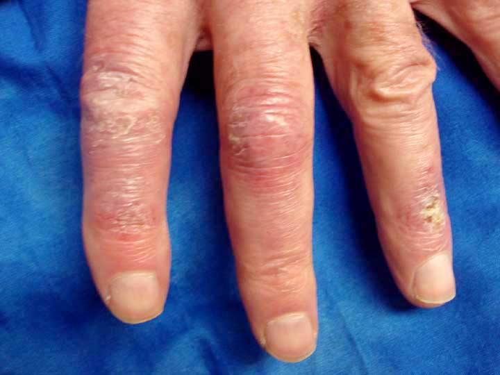
HPI: The patient is an 84 yo man who presents with a three week history of a mostly painless eruption on the dorsal hands (left more than right). I have taken care of his skin for over a decade as he's had a thin melanoma and a number of non-melanoma skin cancers. In addition, he has Parkinson's disease and his only medication is carbodopa. Fourteen years ago, he had prostate Ca successfully treated with seeds. Although mentally alert, the patient has been somewhat frail for years and muscle weakness is difficult to evaluate. There are no other skin findings, no heliotrope, no poikioldermatous changes.

O/E: Dusky erythema on dorsum of left hand with a predilection for the MCP joints. Some crusting. Periungual erythema of proximal nail folds, two fingers, right hand.

Clinical Photos (April 4, 2011)


Lab: The patient saw a rheumatologist who did a thorough w/u for collagen vascular disease with a focus on dermatomyositis. All serologies, chemistries and the hemogram were completely normal. CPK was not done.

Impression: The dermatitis is suggestive of dermatomyositis (DM). It's early spring here and he may have been more exposed to light. At this point, I am considering an early and evolving DM. Amyopathic or hypomyopathic DM takes six months to confirm. Considering his age, a thorough evaluation for malignancy might be considered. This appears to be photo-located, so I considered PCT but will hold off on urinary porphyrins for the time being.

Questions: Your thoughts will be appreciated.

Addendum: Two colleagues (Amanda Oakley from NZ and Fran Storrs from Portland, OR, USA) suggested chilblains. I called the patient and asked him if he might have been out more recently without gloves. He told me his hands are usually cold and he has a 200 yard (~ 200 mtr) walk to his mailbox. He hasn't been wearing gloves recently. It's been a cold spring here -- I think chilblains is a more likely diagnosis and I asked him to wear gloves outside when it's < 50 F (10 C) and get back to me in a week. The extend of his involvement is more than we usually see with chilblains, but the dx makes good sense. I'll affix a f/u in a couple of weeks.

Wednesday, March 30, 2011

Alopecia Universalis


HPI: The patient is a 77-year-old woman who was seen for alopecia, which has been present for about eight months now. This followed chemotherapy for nonhodgkin’s lymphoma. She has a history of alopecia areata decades ago which resolved on its own.

O/E: The examination shows that this patient has alopecia universalis. She has a few eyelashes but no eyebrows, no body hair, no scalp hair.


DX: Alopecia universalis following chemotherapy. This is unusual. There is one report of alopecia universalis following treatment for hepatitis C with ribavirin and interferon.


PLAN: I am going to get a list from her of the medications she was treated with for NHL and see if there are any reports on this. I will also run this be some colleagues.


Question: Has anyone seen a similar patient?


References:

Wednesday, February 02, 2011

Unusual Nail Bed Tumor

Abstract: 67 yo man with 1 - 2 month hx of a nail bed tumor.

HPI:
67 yo man with 1 - 2 month hx of a nail bed tumor. The overlying nail has been destroyed. Lesion is asymptomatic.

O/E: 8 mm diameter flesh-colored tumor left thumb nail. No pigment. Lesion is solid, not friable.

Clinical Photos


Pathology: Hyperkeratosis, parakeratosis, epidermal hyperplasis. There are ectatic blood vessels in an edematous and fibrotic stroma. Iin the DDx is an unusual traumatized hemangioma or subungual fibroma.

Microscopic Photos
courtesy of Deon Wolpowitz, Boston University SkinPath


Diagnosis: Benign nail bed tumor, not otherwise specified. Presented for ideas.

Questions: What is your diagnosis, and how would you approach this lesion?

Plan: The patient has been referred for defintive surgery to a Mohs surgeon.