Presented by Will Shepard, M.D.
Gillette, WY
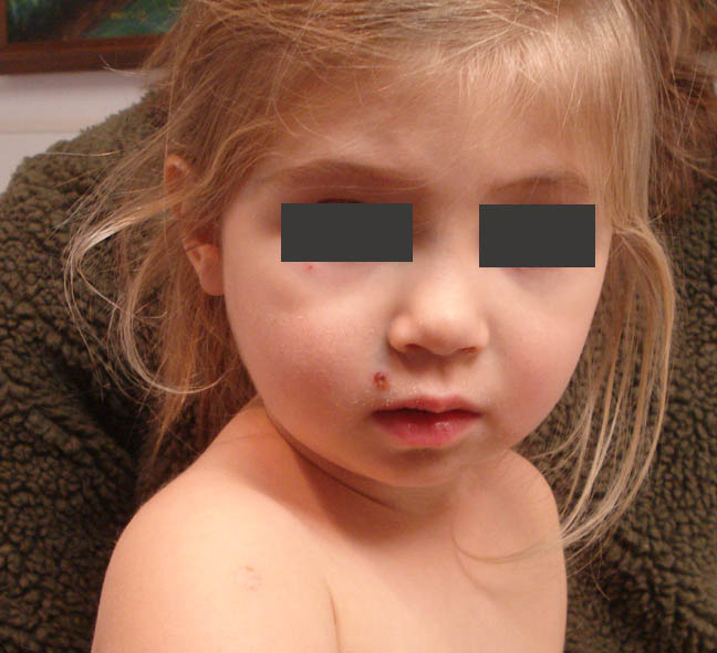
The patient is a two year-old girl with a two week history of
oral and skin lesions. She has been well
and healthy otherwise and all of her milestones have been normal. The present
illness began with two ulcers on her tongue.
A few days later she started to develop skin lesions, first on the
arms. The new lesions start with
erythematous macules and became crusted after 12 – 18 hrs. She has continued to develop new lesions on
the torso, face and extremities. Throughout
this period she has been healthy, no fevers, appetite normal and in no discomfort.
O/E: The tongue
lesions have disappeared. The skin
lesions are few in number and measure 0.5 to 1 cm in diameter. They are scaly annular macules on an
erythematous base.
Clinical Images:


 |
| Rough area from Bandaid |

Impression: The onset of an acute problem with first oral and then skin lesions in an otherwise healthy toddler suggests a viral process.
References:
1
[Paraviral exanthems]. [Article in German]
Fölster-Holst R, Zawar V, Chuh A. Hautarzt. 2017
Mar;68(3):211-216.
Abstract: Paraviral exanthems are distinct skin diseases due
to infections with different viruses. Although no virus has been identified so
far in some exanthems, the main age of manifestation, the clinical course of
the exanthem, and the extracutaneous symptoms are suggestive for a viral
genesis. While many viral infections are a direct result of the infection,
paraviral exanthems reflect the response of the immune system to the infectious
pathogens. Viruses cannot be identified in the skin. Typical paraviral
exanthems include Gianotti-Crosti syndrome, pityriasis rosea, pityriasis
lichenoides, papular-purpuric gloves and sock syndrome, and asymmetrical
periflexural exanthema. Unilateral mediothoracic exanthem, eruptive
pseudoangiomatosis are rare and eruptive hypomelanosis has been described
recently.
We have seen any number of infants and toddlers in our practice with similar outbreaks of oral and cutaneous lesions in late autumn of 2017. Most of them have presented with fever and many more oral and cutaneous lesions in the same distribution, including palms and soles. We have attributed these outbreaks to Coxsackie viral infections.
ReplyDeleteAlthough this patient could certainly fall into a similar diagnostic category, the lack of a febrile prodrome and a paucity of lesions give one pause to make that diagnosis.
The lesion on the border of the tongue is reminiscent of a bite mark. Several of the cutaneous lesions (perioral and thigh) are suggestive of impetigo. The prominent cafe-au-lait spot on the left abdomen is obviously an incidental finding, but begs the question: are there other hyperpigmented macules or axillary freckling?
Interesting presentation, which includes a number of differential diagnoses to consider.
Comment of Dr. Yoon Cohen, Pediatric Dermatologist: As the flu season and winter, I would consider infectious etiologies whether bacterial or viral, especially if there is a history of eczema. With oral lesion as well as well torso and extremities involvement, one would consider viral etiologies such as hand foot mouth disease, eczema coxsackium or even eczema herpeticum, however, these rashes usually tend to accompany with the flu-like prodromal symptoms. The patient does not appear to be ill other than her skin lesions. Since there are only several discrete lesions, impetigo is a reasonable consideration. If there is a new lesion, bacterial and viral cultures could be considered.A diluted bleach bath 2-3 times weekly can be helpful
ReplyDeleteWas there any other members of the family having similar lesions? Though the history suggest it is a viral illness, but the deep crusted discrete lesions, some of which appeared weepy suggest that this could be a streptococcal/staphylococcal infection. Suggest do a swab for culture and treat with oral antibiotics for at least 5 days to cover both strep and staph. Wet compress with dil. KMNO4 and topical fucidin ointment would be useful.
ReplyDelete