People's College of Medical Sciences
Bhopal, India
Abstract: 12 year old boy with shortness of breath, intermittent chest pains and skin lesions.
History: This 12 year-old boy was admitted to the pediatric service with a three month history of shortness of breath. He has been having sleepless nights and we witnessed his distress in the echo room when he developed severe chest pain ( no sweating etc) and it remarkably subsided after 5 minutes of standing up after the echo examination! He has had skin lesions since the age of four.
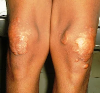
O/E: We saw him in the echocardiography room. On examination he had these remarkable cutaneous lesions in the elbows, legs and perianal region and over the Achilles tendons. There were reddish-yellow nodules over the extensor aspects of the knees and elbows and discrete subcutaneous nodules over the Achilles tendons.
Clinical Photos:


Lab: Serum cholesterol 641 mg%. His echo showed a global hyopkinesia with dilated left atrium and ventricles.
Diagnosis: Familial Hypercholesterolemia with Tuberous and Tendon Xanthomas.
Questions:
1. What further diagnostic studies are needed?
2. Do you think this is the homozygous variant?
3. We have yet to find a suitable explanation for his variable chest pain that aggravates only on lying down and subsides on standing. Could it be due to a myxomatous tissue near the coronary ostia?
3. What is the evidence surrounding the efficacy of drugs and even LDL apheresis for familial hypercholesterolemia?
5. What are the chances of failure to respond to therapy and what is the long term prognosis?
References:
1. Christopher Sibley and Neil J Stone . Familial hypercholesterolemia: a challenge of diagnosis and therapy. Cleve Clin J Med. 2006 Jan;73(1):57-64
Abstract
People with familial hypercholesterolemia (FH) have dramatically high levels of low-density lipoprotein cholesterol (LDL-C), which can lead to accelerated atherosclerosis and, if untreated, early cardiovascular death. Although the heterozygous form of FH is often unrecognized, detecting it early can enable risk reduction before premature coronary heart disease occurs. Available Free Full Text on PubMed
2. Beigel R, Beigel Y. Homozygous familial hypercholesterolemia: long term clinical course and plasma exchange therapy for two individual patients and review of the literature. J Clin Apher. 2009;24(6):219-24
Heart Institute, Chaim Sheba Medical Center, Tel-Hashomer, Israel. beigelr@yahoo.com
Abstract
Familial hypercholesterolemia (FH) is an autosomal dominant disease. Homozygous FH (HFH) manifests with severe hypercholesterolemia since birth (cholesterol levels >5-6 the upper normal limit), which, if untreated, leads to early onset accelerated atherosclerosis and premature coronary death, usually before the 2nd or 3rd decades of life. Various invasive procedures (iliocecal bypass, porto-caval shunt, liver transplant, and gene therapy) have been introduced for lowering low density lipoprotein (LDL) aiming at reducing atherosclerosis and improving survival of HFH patients. Of all the various methods, LDL apheresis has become the most attractive. Although its impressive effect on LDL-C reduction is well established, its long-term (of more than 10 year) effect on the atherosclerotic process and specifically cardiac end-points in HFH is hardly documented. We herewith report on the longest term lipophoresis so far reported in two HFH patients, each treated with plasma-exchange and LDL-apheresis for more than 20 years. The observations provide an opportunity to focus on various aspects regarding not only the procedure itself but also its effect on various clinical endpoints. By this description together with reviewing the literature, we discuss several issues, some of them are generalized while others are individualized, dealing with the approach of long term LDL apheresis in HFH.


A classsical case of tuberous xanthomata. Check family history, Do a complete lipid profile and a cardiology consult. Would use oral statins to reduce the cholesterol and triglycerides levels and hopefully dimimish size of xanthoma. Would not treat the xanthoma surgically - they tend to recur.
ReplyDeleteRemarkable case. Difficult to find in the western world where advanced primary health care system guarentees early detection of such cases and less complications.I agree with doctor henry foong, though lesions are quite large but serum lipid control leads to reversal of xanthoma mass.
ReplyDeleteYes, tuberous xanthomata from homozygous fam. hypercholesterolemia. These patients often have significant pericardial effusions which on pericardiocentesis look like liquid gold. The positional chest pain is likely pericarditis from the effusion, and no, not secondary to xanthomata near the coronary ostia.
ReplyDeleteMichael LaCombe
This is from Roy and Yitzhak Beigel whose reference is on the blog:
ReplyDeleteThe case itself is interesting yet raises some questions before a management decision can be made:
There is data missing from the physical examination:
What are findings upon cardiac and lung auscultation? are the lungs clear? what are his vital signs, especially when he complains of chest pain and shortness of breath.
What is the lab workup, especially regarding his CBC and cardiac enzymes?
What does the ECG show during his complaints? and what does the entire Echo report say, does he have aortic stenosis?
Regarding his cholesterol levels this case resembles a case of FHH - what is the family history, are there any other family members involved?
In regard to management and therapy for both short and long term I think you will find the attached manuscript helpful.
The excessively high total cholesterol (>600 mg%) coupled with extensive xanthomas clinch the diagnosis of homozygous FH. Although unsightly, the xanthomas themselves have little bearing on the clinical course of this disease, the endpoint of which is arteriosclerosic heart disease. Even so, it would be prudent to treat the child with statins—perhaps multiple agents—to lower the serum cholesterol.
ReplyDeleteAs Dr. Lacombe notes, chest pain that worsens with lying supine and improves with sitting up is a classic finding in pericarditis. No mention is made of the presence of a pericardial effusion on the echocardiogram. I wonder about the findings of the dilated left atrium and ventricles. This child might have valvular heart disease—perhaps mitral regurgitation or aortic stenosis, the latter of which is a common finding in homozygous FH. Again, we are given little information about the state of his valves on echo or auscultatory findings. He definitely warrants a consultation with a pediatric cardiologist, if one is available.
Given the boy’s age and current status, the prognosis is guarded.
In this case, although the dermatologist can render a sound diagnosis, further workup and treatment rest in the hands of the cardiologist.
Brian T. Maurer, PA-C