Queens Memorial Medical Center
New York, New York
This 68-year-old woman presented for evaluation of painful lesions under the skin that have been present for 3-4 months. She has a history of psoriasis, which is in remission, fibromyalgia, hypertension. There is no personal history of malignancy. No exotic travel. She had smoked over two ppd for decades.
EXAMINATION: The examination shows a woman who
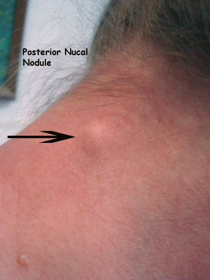
appears slightly older than her stated age. She has 6-8 freely movable
subcutaneous smooth-surfaced lesions on the back, posterior nuchal area, and the upper
chest. The largest is ~ 5 mm in diameter. The remainder of the exam plus breast palpation was unremarkable. No adenopathy appreciated.

INITIAL MPRESSION: Subcutaneous skin lesions, present for only a
short period of time. Etiology is unclear.
PLAN: An excisional biopsy was taken today from the lesion
on the right upper back.
Pathology:
The first two are H&E of nodule in the fat, showing
atypical cells with duct-like vacuoles. The second two are representative
immunoperoxidase stains. GATA 3 is the nuclear one (dot-like pattern) and
mammoglobin is the cytoplasmic staining.
These darkly beautiful photomicrographs were taken by Dr. Lynne Goldberg at the Boston University School of Medicine's Department of Dermatopathology.
These darkly beautiful photomicrographs were taken by Dr. Lynne Goldberg at the Boston University School of Medicine's Department of Dermatopathology.


Plan: Mammography and breast ultrasound. Referral to oncologist.
Mammography shows masses in r. breast. Ultrasound guided biopsy planned and specimen will be sent for Estrogen receptor, progesterone receptor and Her-2 (human epidermal growth factor receptor).
Note: In this age of "Social Distancing" it is unlikely that this disgnosis would have been make expeditiously in a woman with no history of an underlying malignancy. We will add more as her case progresses.
Your thoughts will be appreciated.
References:
1. Mammaglobin, a Valuable Diagnostic Marker for Metastatic
Breast Carcinoma Zhiqiang Wang1, et. al. Int J Clin Exp Pathol (2009) 2, 384-389
Abstract: Identification of
metastasis and occult
micrometastases of breast
cancer demands sensitive
and specific diagnostic
markers. In this
study, we assessed
the utility of
a mouse monoclonal
antibody to human
mammaglobin for one
such purpose. Immunohistochemical stains
were performed on
paraffin-embedded sections from
a total of
284 cases, which
consisted of primary
breast invasive carcinomas
(41 cases) with
matched metastases to ipsilateral axillary lymph nodes, metastatic
breast carcinoma to liver (1 case) and kidney (1 case), non-breast neoplasms
(161 cases), and normal human tissues (39 cases). The results showed 31 of the
41 cases of primary breast cancer with axillary lymph node metastases were
positive for mammaglobin (76%). In the meantime, we documented expression of
mammaglobin in occasional cases of endometrial carcinoma (17%). Our data
further validated that mammaglobin is a valuable diagnostic marker for
metastatic carcinoma of breast origin, although endometrial carcinoma should be
considered as a major differential diagnosis.
2. GATA3 Expression in Common Gynecologic Carcinomas: A
Potential Pitfall. Tatjana Terzic et.
al. Int J Gynecol Pathol, 38, 485-492 2019
Abstract: GATA binding protein 3 (GATA3)
immunohistochemistry is primarily used as a marker of breast and urothelial
differentiation, particularly in metastatic settings. In the gynecologic tract
it also serves a robust marker for mesonephric and trophoblastic tumors. Full
Abstract: pubmed.gov PMID:
30059453
No comments:
Post a Comment
We welcome your comments. We endeavor to serve your patients and you. If you want us to respond, please add your name and email address. Some people have trouble uploading comments. In that case, please send comments directly to djelpern@gmail.com. Thank you.