Saturday, February 28, 2009

Diseases Don't Read Textbooks

Abstact: 5 yo girl with enlarging plaque on back.

HPI: The patient is a 5 year old girl seen on February 27, 2009 with a 10 day history of an enlarging plaque on the left back. She had a similar, but less dramatic lesion in April 2008 which was treated with cefuroxime for two weeks. Her family lives in a wooded area and her mother had Lyme Disease last year. The patient feels well, may have had some mild arthralgias according to her mother. No neurological symptoms. She is allergic to penicillin, amocicillin and sulfonamides.

O/E: 17 x 12 cm plaque left back. 2 x 2 cm plaque right arm. These lesions are somewhat urticarial in appearance. The center of the larger lesion is paler than the periphery.

Photos:


Lab: Lyme titers pending

Diagnosis: Presumptive Lyme Disease. She was started on cefuroxime by her pediatrician.
Questions:
1) What else would you consider in the differential diagnosis
2) Can one have ECM more than once? This child had something similar 10 months ago.
3) Presuming this is Lyme -- how long shoud she be treated?

Reasons Presented: Lyme Disease is unusual in the winter. Can one have "primary lesions" with a reoccurence? In a young patient where tetracyclines are contraindicated with a proven allergy to penicillins, what is the best third line drug and how long to administer.

Saturday, February 21, 2009

Interesting Follow-up: Paronychia in a Child

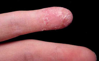
In October 2007, we presented the case of an eight year old girl with chronic paronychial inflammation located on the left index finger (Paronychia in a Child). She had no other dermatoses. The patient is adopted so we have no family history. We assumed this was some kind of localized psoriasis or acrodermatitis continua. Clobetasol ointment was prescribed which she has used since. (Photo above from 10/2007)

The patient was seen in follow-up recently. The paronycial inflammation had subsided but the finger tip was still abnormal, especially on the palmar surface and there is now hypopigmentation and atrophy distal to the area of inflammation. This latter is likely secondary to the clobetasol. Her topical therapy was switched to calcipotriene cream (the ointment is no longer available in the US.)

Photos:
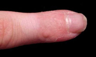


Questions:
1) What do you think the diagnosis is?
2) Side-effects on the fingers from super-potent topical corticosteroids are rarely reported. One suspects that they are not that unusual. When does the treatment get worse than the disease? (I should have been more diligent in follow-up)
3) Who thinks that these preparations can cause bone changes?
Your comments will be appreciated.

References:
1. Deffer TA, Goette DK.. Distal phalangeal atrophy secondary to topical steroid therapy. Arch Dermatol. 1987 May;123(5):571-2.

2. Tosti A, Fanti PA, Morelli R, Bardazzi F. Psoriasiform acral dermatitis. Report of three cases. Acta Derm Venereol. 1992;72(3):206-7.
Department of Dermatology, University of Bologna, Italy.
The authors report 3 patients affected by psoriasiform acral dermatitis, a distinctive clinical entity characterized by a chronic dermatitis of the terminal phalanges, associated with marked shortening of the nail beds of the affected fingers. The skin biopsy showed in all cases the pathological features of a subacute spongiotic dermatitis. X-ray examination of affected fingers showed no bone or soft tissue changes. Differential diagnosis of psoriasiform acral dermatitis included psoriasis, atopic or contact dermatitis and corticosteroid-induced distal phalangeal atrophy.

3. Brill TJ, Elshorst-Schmidt T, Valesky EM, Kaufmann R, Thaçi D. Successful treatment of acrodermatitis continua of Hallopeau with sequential combination of calcipotriol and tacrolimus ointments. Dermatology. 2005;211(4):351-5.
Department of Dermatology, J.W. Goethe University, Frankfurt, Germany.
Acrodermatitis continua of Hallopeau (ACH) is a rare type of pustular psoriasis affecting the digits. We report on a 43-year-old female patient who had been suffering from ACH for more than 20 years. Despite the fact that the disease was localized on one finger during the whole period, several topical and systemic treatments resulted in only temporary or partial improvement of the lesion. Although the monotherapies with calcipotriol and tacrolimus ointments gave no satisfying results in the long-term management of the disease, the combination of both agents led to a continuous improvement of the patient's skin condition. Copyright 2005 S. Karger AG, Basel.

Saturday, February 14, 2009

R/O Subungual Melanoma

70 yo man referred for suspected subungual melanoma.

HPI: The patient is a retired engineer with a one month history of subungual pigmentation. He suffers from Waldenstrom's macroglobulinemia and peripheral neuropathy. If he had injured his toe, he would not know.

O/E: The left middle toenail shows brown-blackish subungual pigmentation. It was difficult to appreciate if this was melanin or blood both clinically or dermoscopically. Hutshinson's sign is negative.


Dermoscopy before 3 mm punch biopsy

Diagnosis: Probably subungual hematoma. Need to r/o melanoma.

Procedure: Modification of Haneke Technique.

1. Patient soaks foot in warm water for 20 - 30 minutes
2. Carefully drive a 3 mm punch through the nail with care not to cut into the nail bed.
3. Lift off the cut disk of nail and observe the nail bed.


Dermoscopy after 3 mm punch biopsy and H2O2 to defect

In this case, what appeared to be dried blood was present. The area was cleaned with hydrogen peroxide and a normal appearing nail bed was see. There was no pigment noted. Dr. Hanecke's technique utilizes a Hemocult stick to test scraping from underside of nail, however, our strips were outdated and not reliable.

Note: Dr. Eckhart Haneke pioneered this technique but is not acknowledged in the literature. Here are his comments to this case: "Thank you very much for your email and the links, which I saw for the first time. Thank you also for giving me the credit.
You are completely right that we do not even need the hemoccult test strip for the correct diagnosis, but it is very convincing and impresses the patient. And of course, it is one more proof.
Also clinically, as this is no streaky lesion a melanoma is improbable - however, a very fast growing melanoma can appear like this.
When you apply hydrogen peroxide and the pigment disappears this is due to the hemodestructive action of H2O2 on erythrocytes: hemoglobin has a pseudocatalase action splitting H202 into H2O and O. That is why hydrogen peroxide is also a very good disinfective agent and I use it to cleanse my dermatosurgery field from blood."