The patient is a 28 yo man referred on a walk-in basis with an eight month history of a bullous eruption on the dorsum of his left hand. He gets one or two painless lesions a month. No history was sent over with the patient and he is a poor historian. His medications include Welbutrin, Clozeril and Soma (carisprodol/aspirin)
Exam:
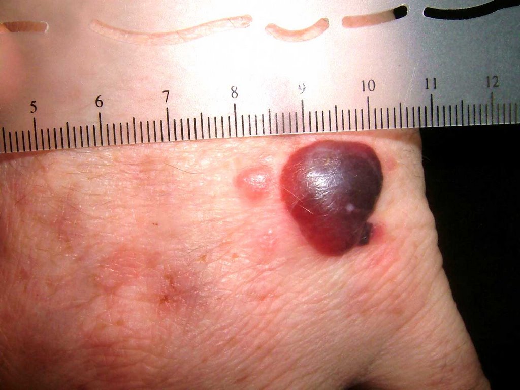
The only findings are on the dorsum of the left hand. Here, there is a hemorrhagic bulla, a vesicle and areas of mild erythema at sites of previous lesions.

Biopsy:
I suspect fixed drug or pseudoporphyria cutanea tarda. I biopsied the small papule and the edge of the bullae and made a follow-up appointment.
Path Report:
A subepidermal separation, individually necrotic keratinocytes , and a sparse superficial perivascular lymphocytic infiltrate with occasional neutrophils .
NOTE : (A and B). Amphophilic globular material is seen deposited in the dermis and around blood vessels in both specimens. This material is P.A.S. stain positive, and stains negative for amyloid and elastic tissue. The differential diagnosis could include porphyria cutanea tarda , although this is usually less inflammatory. A subepidermal autoimmune bullous disorder could also be considered. An additional biopsy for direct immunofluorescence may be of help.
Laboratory Studiess:
CBC normal, LFTs normal, ferritin normal. Hep B and C negative, All urinary porphyrins well within normal levels. Uroporphyrin: 9.6 ug (nl < 30.0) Coproporphyrin 40 ug (normal < 65/24 hr)
Discussion: This is likely pseudoporphyria cutanea tarda. A similar case has been reported. Your comments are welcomed.

Looks like a tense subepidermal blister as opposed to fragile intraepidermal blister (presumably Asboe-Hansen and Nikolsky signs are negative). Any milia in healing sites of prior blisters? Med list is important? Consider urinary uroporphyrins.
ReplyDelete--Rick Sontheimer
This is an interesting case presentation. In view of his age and site of presentation being tense bullae in a sun exposed areas, i would consider pseudoporphyria ( include drug induced) but need to exclude 3 important causes ie Porphyria Cutanea Tarda, Erythropoietic Porphyria (usually in childhood thoguh) and Bullous Lupus. A good detailed drug history and family history of hepatitis, porphyria and photosensitivity would help.
ReplyDeleteHenry
It is interesting case.FDE is a far possibility as the rash should recur in all sites of previous old lesions and the pictur is not that of FDE.Localised bullous pemphigoid is recognised entity and it is on top of differential diagnoses.Regarding porphyria,it unusaul to be seen in a localised pattern.Biopsy and immunoflourescence will make picture more clear.
ReplyDeletekhalifa sharquie