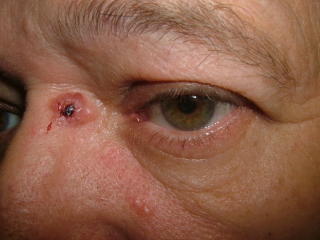
LATE BREAKING!! BIOPSY HERE SHOWED THE LESION TO BE AN INTRADERMAL NEVUS.
I should have paid more attention to the history.
MORE... The patient underwent an excision - and the final report was a Basal Cell + an intradermal nevus. Quite unusual. The initial clinical impression was more accurate that the incisional biopsy. This is sobering.

This 57 yo man presented with an 8 mm in diameter papule that has bled since he started to wear glasses a year or so ago. He has been aware of a slowly growing lesion in this area since age 19 (38 years ago).
The lesion has been biopsied and I await the results. It appears to be a BCC. The long history underscores the benign behavior of many of these lesions. While we have all seen case reports of aggressive BCCs that have caused loss of eyes, ears, nose - these are likely in the very small minority. It is the growth characteristics and behavior of these lesions which is likely key, not their appearance micorscopically. Our therapy for these indolent tumors may be too aggressive based on the slow growth and lack of metastatic potential.
Truly amazing. I will wait for the histology results before contemplating further. It is unusual but if HPE shows BCC I would till go for complete excision - too close the eye to warrant other therapy I think.
ReplyDeleteHenry
Clinically you could never have interpreted that lesion to be anything other than an ulcerated BCC.The problem is I would probably have excised and grafted it without doing a biopsy!
ReplyDelete