The patient is an 11 yo girl with difficult-to-treat warts present for 4 years. Her pediatrician has used cryotherapy, and a vesicant. They have tried salicylic acid under occlusion for a month and the warts have grown.
The child is very self-conscious. What would you do?
This is a rapid publication site that replaces Virtual Grand Rounds in Dermatology (vgrd.org). Please join and feel free to post cases. You can share the URL with friends. Since 2000, VGRD has been a valuable means to share cases in real time from one's home or office. "AND GLADLY WOLDE HE LERNE AND GLADLY TECHE" has served as an enduring and inspirational motto. For more information, see the "About Page."
Tuesday, February 23, 2016
A Thing of Beauty
The patient is a 7 year-old boy with a lesion present since infancy. He was adpopted at age 11 months and prior history is unknown. The lesion has changed only minimumly over the past few years.
O/E: 7 mm lesion with macular dots and two darker pigmented papules on the left abdomen. He has a small area of segmental vitiligo on the left neck.
Ckinical and Dermatoscopic Images:
Workup: None at this time
Diagnosis: Nevus spilus, Speckled Lentiginous Nevus
Note: An association of segmental vitiligo with SLN has not been reported.
Reference:
Speckled lentiginous naevus: which of the two disorders do
you mean?
Happle R1. Clin Exp
Dermatol. 2009 Mar;34(2):133-5
Abstract: Speckled lentiginous naevus (synonym: naevus
spilus) no longer represents one clinical entity, but rather, two different disorders can be
distinguished. Naevus spilus maculosus
is consistently found in phacomatosis spilorosea, whereas naevus spilus
papulosus represents a hallmark of phacomatosis pigmentokeratotica. The macular
type is characterized by dark speckles that are completely flat and rather
evenly distributed on a light brown background, resembling a polka-dot pattern.
In contrast, naevus spilus papulosus
is defined by dark papules that are of different sizes and rather unevenly
distributed, reminiscent of a star map. Histopathologically, the dark spots of
naevus spilus maculosus show a 'jentigo' pattern and several nests of
melanocytes involving the dermoepidermal junction at the tips of the papillae,
whereas most of the dark speckles of naevus spilus papulosus are found to be
dermal or compound melanocytic naevi. The propensity to develop Spitz naevi
appears to be the same in both types of speckled lentiginous naevus, whereas
development of malignant melanoma has been reported far more commonly in naevus
spilus maculosus.
Tuesday, February 16, 2016
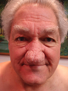
Rhinophyma
The patient is a 73-year-old semi-retired carpenter who presents
for evaluation of lesions on his back and eyelids. Surprisingly, he did not mention his nose.
O/E: He has two epidermal inclusion cysts on the back some
and some small skin tags around the right upper and lower lids (these lesions were snip
excised).
More significantly, the skin of the distal nose it grossly thickened
and patulous.
Clinical Photos:
Diagnosis:
Rhinophyma, Grade 3.
Although he did not initially express concerns regarding his
nose, when I mentioned that there are treatments, the patient was very
interested.
References:
1. Basal cell carcinoma masked in rhinophyma.
2. Basal cell carcinoma and rhinophyma. Leyngold M et. al. Ann
Plast Surg. 2008 Oct;61(4):410-2.
Abstract: Rhinophyma, the end stage in the development of
acne rosacea, is characterized by sebaceous hyperplasia, fibrosis, follicular
plugging, and telangiectasia. Although it is commonly considered a cosmetic
problem, it can result in gross distortion of soft tissue and airway
obstruction. Basal cell carcinoma (BCC) is a rare finding in patients with
rhinophyma. The objective of this study is to review the literature of BCC in
rhinophyma and report on a case. A 70-year-old male presented with
long-standing rosacea that resulted in a gross nasal deformity. The patient
suffered from chronic drainage and recurrent infections that failed
conservative treatment with oral and topical antibiotics. The patient decided
to proceed with surgical intervention and underwent tangential excision and
dermabrasion in the operating room. Since 1955 there have been 11 cases
reported in the literature. In our case, the pathology report noted that the
specimen had an incidental finding of a completely resected BCC. The patient
did well postoperatively and at follow-up remains tumor-free. Despite the
uncommon occurrence of BCC in resection specimens for rhinophyma, we recommend
that all specimens be reviewed by a pathologist. If BCC is detected,
re-excision may be necessary and careful follow-up is mandatory. Larger studies
would be needed to determine the correlation between the 2 conditions.
Monday, February 15, 2016
Cryotherapy Gold Mine
CPT Codes
17000 – Cryotherapy for One Lesion Reimbursed at: $59.88
17003 – Cryotherapy for > 1 Lesion Reimbursed at: $8.05/lesion
Cryotherapy pattern of six dermatologists with practices in same area of New England. All in private practice. This only shows Medicare reimbursements (that probably account for 34 -- 40% of gross income).
Saturday, February 13, 2016
Excoriated Acne with Hyperpigmentation
A 37 yo Haitian woman was seen complaining about hyperpigmented lesions on face and back. By history, these followed acne which she has excoriated. Her health is good otherwise and she takes no medication by mouth.
O/E: Hyperpigmented papules and macules on face and torso in a young woman with Type V skin. Some lesions show mild excoriation.
Clinical Photos:
Diagnosis: Post-inflammatory hyperpigmentation in excoriated acne.
Comment: In my area, we have few patients with Type V and VI skin. This is a common problem, but I do not have much experience treating it. What is your protocol? I started her on tretinoin 0.05% cream as this may be covered by her insurance.
Note: A Pubmed search for hyperpigmented acne scars retrieves only three references and all are to laser surgery which this patient can not afford. This problem must be extraordinarily common; yet the biomedical literature is strangely silent about it.
Friday, February 05, 2016
Active Nevus
17 yo boy seen for another problem. Atypical nevus noted on left shoulder.
O/E: Type III-IV skin. 5 mm papule with slight play of color clinically. Pt. unaware of lesion.
Dermoscopic images shows pigment dots of varying sizes.
Dx: Actively growing nevus. Easy to excise with a 6 mm punch. Best to do this of just observe? Patient will be leaving for college in another city in a few months.
Thursday, February 04, 2016
Unusual Facial Dermatosis
Abstract: 16 yo girl with 3 month history of annular dermatosis mid-forehead.
HPI: This healthy 16 yo has had a 4 cm roughley circular plaque on the mid-forehead. Takes no medications by mouth and is not aware of any OTC meds taken infrequently, She has bued 1% hydrocortisone cream and also a topical imidazole without improvement.
O/E: The lesion has fairly sharp borders. The surface is somewhat gready. KOH prep failed to demonstrate hyphae or spores. The remainder of the cutaneous examination is unremarkable.
Clinical Photo:
Diagnosis: The picture is suggestive of seborrheic dermatitis, but the circular nature is unusual. What are your thoughts?
Plan: Betamethasone valerate 0.1% cream b.i.d. x 2 weeks sand then Elidel cream. If it does not respond or if it recurs a biopsy will be done.
Monday, February 01, 2016
Unilateral/Segmental Vitiligo in a 9-year-old boy receiving Melagenina as treatment
HPI: The patient
is a 9-year-old boy who developed loss of pigmentation on the right side of his
face over a 3-month-period. The depigmentation of the skin progressed rapidly
with no antecedent eruption, redness or trauma. There was no history of
exposure to a chemical or irritant prior to depigmentation of the skin.
No medical history of hypothyroidism or other medical
conditions. No family history of
vitiligo or autoimmune diseases. He often spends 2 to 6 hours in the sun
playing outside only beginning to wear sunscreen recently.
Diagnosis: He appears to have a segmental or
unilateral vitiligo.
He lives in Cozumel, Quintana Roo, Mexico and has been
evaluated by a dermatologist locally who confirmed the diagnosis
clinically. No biopsy was obtained.
Labs: TSH,
complete blood count and chemistry panel were normal.
Prior/Current Treatment:
Melagenina solution
Since the patient is a citizen of Mexico, he and his family
were able to travel freely to Cuba and obtained an appointment in the Vitiligo
clinic evaluation and treatment after a 6-month wait. Their first visit to Cuba
was in August, and they are expected follow-up in 6 months. He was given Melagenina
solution to be applied twice a day to the depigmented areas of skin. Melagenina
solution can only be obtained in Cuba at this time. It is derived from placental extract that is
mixed with an alcohol solution. He will return to Cuba in February 2016 for a follow-up
visit and to obtain more Melagenina.
Images:
His mother has noticed some repigmentation of the treated areas. The pictures shown are after using the
treatment for 4 months.
Second Opinion in USA
and Plan:
We recommended adding tacrolimus (Protopic 0.1%) ointment and a
low-dose steroid such as mometasone furoate cream to his Melagenina treatment
regimen to be applied twice a day. The patient was counseled on the importance
of using a titanium and zinc oxide waterproof sunscreen on the face to prevent
further darkening of the surrounding area and to protect the areas of
depigmentation.
Discussion:
Vitiligo is a common skin disorder
affecting about 1 to 2% of the world population. It commonly affects children
and can be seen in different patterns.
This patient appears to have a unilateral or segmental pattern but not
necessarily dermatomal.
It has been shown that segmental
vitiligo in children is relativley common and less frequently associated with
systemic autoimmune diseases or endocrine disorders.
Treatment in the USA and Mexico
includes using narrow band UVB phototherapy or psoralen with UVA phototherapy
as well as topical low-dose steroids and tacrolimus combinations. Narrow
band UVB phototherapy is considered one of the most efficacious treatments and can
be used alone and in combination with topical steroids and tacrolimus. Some patients are
also treated with the excimer lasers and have undergone melanocyte transplants.
Melagenina or placental extracts are not used currently.
In the General de
Mexico hospital, up to 50 cases of vitiligo are seen per day. Many efforts are being made to increase
awareness about vitiligo. One controversial issue in Mexico has been the
exploration of naturalist physician care and unresearched treatments options. As
we are aware, this is a consideration in the U.S. as well. Although the medical
community wants to be open to new ideas involving topical and oral nutritional
and botanical substances, in Mexico the concern is that patients will use their
limited financial resources on unsubstantiated treatments. Phototherapy clinics
treat vitiligo patients in the larger cities of Mexico, but unfortunately many
patients, including this patient, could not travel regularly to these
established clinics due to financial and transportation limitations.
1)
How
safe, well regulated and efficacious is the Melangenina solution in the
treatment of vitiligo? Should this be something explored more for patients in
the USA?
2)
Should
the patient inform the physicians in Cuba that they will be adding other
topical medications to the regimen?
Out of respect to the Cuban
dermatologists, we encouraged them to inform the clinic that a second opinion
was sought out and new medications were started. The patient and his family were unsure if the
treatment with Melagenina was part of a clinical
trial.
3)
Should
we consider oral minipulse therapy with methylprednisolone?
Although there are relapses and other
considerations with oral steroid use in children, a few case reports and
clinical trials have shown some benefits.
Lo, Yuan-Hsin,
Gwo-Shing Cheng, Chieh-Chen Huang, Wen-Yu Chang, and Chieh-Shan Wu.
"Efficacy and Safety of Topical Tacrolimus for the Treatment of Face and
Neck Vitiligo." The Journal of Dermatology 37.2 (2010): 125-29.
Web.
http://onlinelibrary.wiley.com/doi/10.1111/j.1346-8138.2009.00774.x/full
Majid, Imran et al. “Childhood vitiligo: response to methylprednisolone oral minipulse therapy and topical fluticasone combination.” Indian Journal of Dermatology 54.2 (2009): 124–127. PMC.http://www.ncbi.nlm.nih.gov/pmc/articles/PMC2807150/
Shrestha, S., AK Ha,
and DP Thapa. "An Open Label Study to Compare the Efficacy of Topical
Mometasone Furoate with Topical Placental Extract versus Topical Mometasone
Furoate with Topical Tacrolimus in Patients with Vitiligo Involving Less than
10% Body Surface Area." Nepal Medical College Journal 16.1 (2014):
1-4. Web.
Xu, Aie, Dekuang
Zhao, and Yongwei Li. "Melagenine Modulates Proliferation and
Differentiation of Melanoblasts." International Journal of Molecular
Medicine Int J Mol Med (2008): n. pag. Web.
http://www.spandidos-publications.com/ijmm/22/2/193
Confluent and Reticulated Papillomatosis
The patient is a 21 yo man with a six month history of subtle scaly patches on both axillae. He was treated by is internist with ketoconazole cream without effect. A KOH prep was done and was negative.
Clinical Image:
Patches are slightly yellowish and there are islands of sparing.
Pathology: Hyperkeratosis, papillomatosis (increased compared to specimen B), mild epidermal hyperplasia and a superficial perivascular lymphocytic infiltrate consistent with confluent and reticulated papillomatosis.
NOTE: The differential diagnosis could include acanthosis nigricans. PAS stain is negative for fungal organisms.
Photomicrographs courtesy of Jonathan Ho MD MS, Department of Dermatopathology, Boston University School of Medicine
Diagnosis: confluent and reticulated papillomatosis.
This is a difficult diagnosis clinically and histologically. It's a type of "dermatological non-disease." Without the help of an experienced dermatopathologist I doubt that this diagnosis would have been arrived at.
The patient will be treated with minocycline, 100 mg bid for a month. If this is CARP the process will be resolved.
Clinical Image:
Pathology: Hyperkeratosis, papillomatosis (increased compared to specimen B), mild epidermal hyperplasia and a superficial perivascular lymphocytic infiltrate consistent with confluent and reticulated papillomatosis.
NOTE: The differential diagnosis could include acanthosis nigricans. PAS stain is negative for fungal organisms.
Photomicrographs courtesy of Jonathan Ho MD MS, Department of Dermatopathology, Boston University School of Medicine
Diagnosis: confluent and reticulated papillomatosis.
This is a difficult diagnosis clinically and histologically. It's a type of "dermatological non-disease." Without the help of an experienced dermatopathologist I doubt that this diagnosis would have been arrived at.
The patient will be treated with minocycline, 100 mg bid for a month. If this is CARP the process will be resolved.


