This is a rapid publication site that replaces Virtual Grand Rounds in Dermatology (vgrd.org). Please join and feel free to post cases. You can share the URL with friends. Since 2000, VGRD has been a valuable means to share cases in real time from one's home or office. "AND GLADLY WOLDE HE LERNE AND GLADLY TECHE" has served as an enduring and inspirational motto. For more information, see the "About Page."
Monday, October 31, 2005
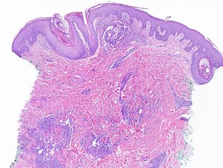
BCC Scalp
The patient is a 75 yo man with a three month hisory of a lesion at the vertex of the scalp.
Exam: Man with Type II - III skin. 2 x 1.6 cm diameter erythematous plaque scalp. There are some small crusted areas.
Biopsy shows superficial BCC.
Question: Is Aldara appropriate?
C + E will take ages to heal.
Excision could n ot be closed without a graft or a flap.
Wednesday, October 26, 2005
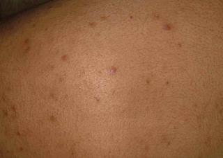
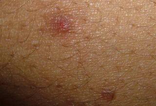
19 year old student with acneiform eruption
History: 19 year-old man with a 5 year history of acneiform eruption predominately on torso and proximal extremitiesl Some facial involvement, but less than on torso. He is in good health, no history of diabetes or steroid use. No antibiotics for months. In the past he has used benzoyl preoxide ceams and washes, topical retinoids, topical antibiotics and tetracycline and its derivatives. Mome has ever helped.
Exam: Healthy young man with Type IV skin. On torso and proximal extremities he has discrete erythematous papules and an occasional pustule. No cysts. Face largely clear.
Lab: nil
Pathology: Initial reading showed marked perifollicular lymphoneutrophilic infiltrate c/w acute folliculitis. PAS negative.
Diagnosis: Probable Pityrosporon folliculitis
Discussion: Are the yeasts incidental or indicative of pityrosporum folliculitis. I asked for more cuts and this showed numerous PAS (+)"fungal spores" in the follicular ostia.
I have started him on itraconazole 200 mg per day and Nizoral 2% shampoo to torso. The literature does not have good guidelins for how to treat this; but I suspect 4 - 6 weeks woth oral meds.
Please suggest diagnostic and therapeutic alternatives.
Thursday, October 20, 2005
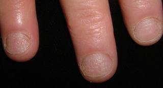
Atypical Nail Pits
Case for Discussion:
This 40 year-old woman presented for evaluation of a nail dystrophy present for 2-3 months.
She has a history of Hashimoto's thyroiditis. About 9 months ago she developed vitiligo.
Her health is otherwise normal
Meds. Synthyroid and iron
Lab: Thyroid Perox AB 248 (Nl. 0 - 34 IU/ML)
ANA < 1:40
Physical Exam:
Vitiligenous patches left neck and left upper back
Around 4 finger nails show a distinctive pitting. The pits are fairly uniform and the affected nails are rough and lusterless. There are no cutansous lesions of psoriasis.
Discussion and Questikon:
The picture is atypical for psoriatic nail pits but that is not excluded.
I favor a relationship to the underlying autoimmunity that has caused the Hashimoto's and vitiligo.
The picture is similar to that seen with alopecia areata, but the patient has not has any alopecic patches.
Nail dystrophy has occasionally been described before the development of A. areata. And patients with Hashimoto's thyroiditis have a higher than expected incidence of alopecia areata.
We welcome your thoughts or suggestions.