Monday, March 28, 2016

Congenital Triangular Alopecia

The patient is an 11 y.o boy seen for an unrelated problem.  His mother noted an alopecic area in her newborn and attributed it to something the obstetrician did,

O/E:  There is a 1.5 xm area of alopecia on the left fronto-temporal area.
Diagnosis: Temporal (Congenital) Triangular Alopecia

We have presented two similar cases in the past.

Here is a free full text article from the Indian Journal of Dermatology

Sunday, March 27, 2016

Labial Macules

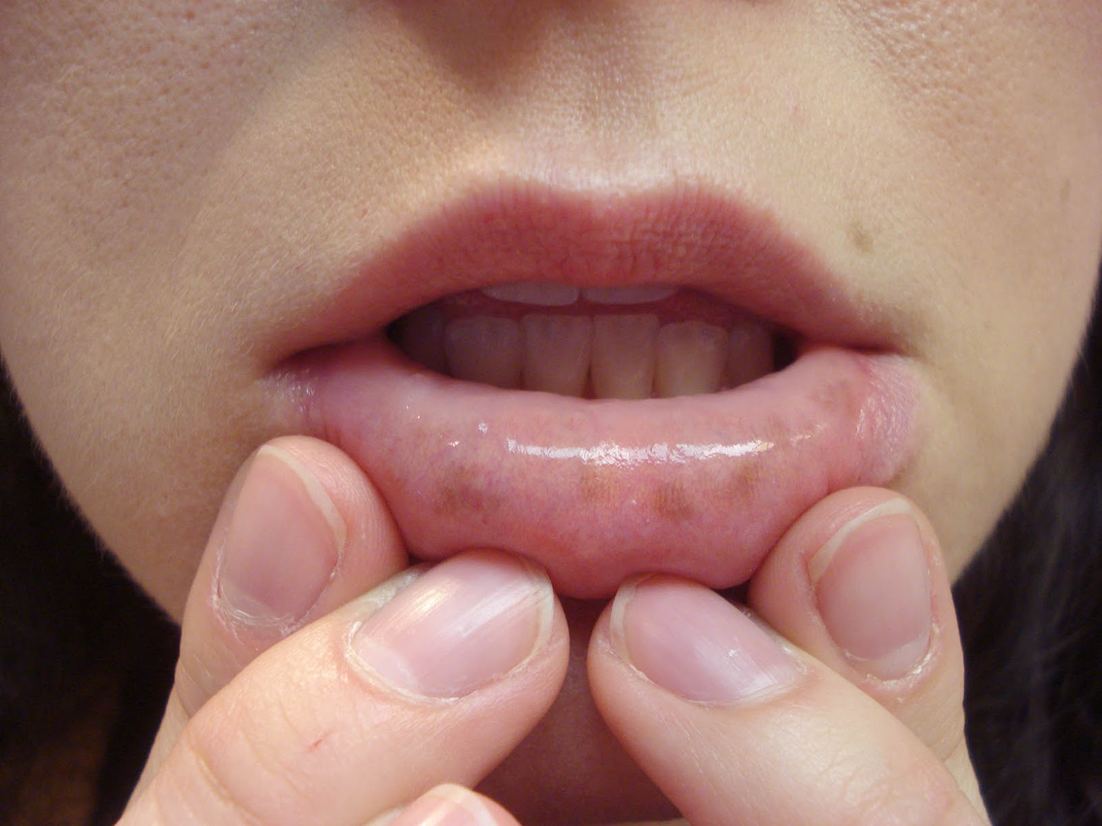
The patient is a 33 year-old woman who recently noted pigmented macules on her lower lips.  She admits to having an anxiety disorder and being preoccupied with her health (which is otherwise excellent).  Her anxiety dates back to the death of her father from colon cancer at age 52 (when she was 10 years old).  She was seen for an unusual cheilitis in 2010 and her case was presented on VGRD then.

O/E:  There are six lightly pigmented macules on her lower lip.  No other pigmentation on her oral mucosae. No other pigmented macules noted.

Discussion: While these macules could be post-inflammatory changes from her allergic cheilitis, it's hard to ignore her family history and the possibility that they could be a forme fruste of Peutz-Jeghers Syndrome.  Endoscopy seems, to me at least, to be indicated.  With classical PJS the pigmented macules may be present even in infancy or childhood.  Visualizing her G.I. tract is important here, if only to allay this woman's anxiety.

Your thoughts will be appreciated.

Saturday, March 19, 2016

Bullae and Erosions in a Newborn


Presented by Dr. Henry Foong
Ipoh, Malaysia

This 20-day-old baby presented with multiple blisters on the hands and feet.  The mother first noticed them on Day 2 after a normal delivery when there were blisters on the hands.  Subsequently, she noticed blisters on the feet as well.  These blisters ruptured easily on friction or minimal rubbing. Recently he has difficulty in swallowing as well. There was no fever.  No family history of similar problems and no history of consanguinity in the family.


He has seen paediatricians and dermatologists but his condition did not improve.

The child looks fretful.  Superficial erosions with crusts were mainly noted on his hands and feet.  A superficial bulla 0.5 x 0.5 cm was present on the left index finger.  Mucosa of the lips appeared eroded.  These was no nail involvement.

Do you see this condition? How do you manage this in your institution? I guess this will be a tough management problem for the child and the family.


 Diagnosis:  This child most likely has epidermolysis bullosa, probably recessive dystrophic EB, although the Hurlitz type needs to be considered as well. 

Our pediatric dermatologists can help with management suggestions. 

Definitive diagnosis can be obtained with biopsy.  In the past, electron microscopy was utilized, but there may be simpler studies.


Note:  At an appropriate time, if the diagnosis is more certain, the parents might want to visit the DEBRA site. There is a resources in Malaysian, in addition: DebraMalaysia.

References:
1. Recessive Dystrophic Epidermolysis Bullosa
A Review of Disease Pathogenesis and Update on Future Therapies
Luis Soro, DO et. al  J Clin Aesthet Dermatol. 2015. 8(5): 41–46.
Abstract: Objective: Review the pathogenesis of recessive dystrophic epidermolysis bullosa and provide an update on research currently underway that is aimed at treating and potentially curing this severe skin disorder. Design: Review article. Setting: Private practice and large teaching hospital. Participants: None. Measurements: N/A. Results: Currently, patients with recessive dystrophic epidermolysis bullosa are managed with only supportive care. However, there are several promising new treatment avenues that may help patients in the future. These include gene therapy, cell therapy, and protein-based therapy. Each approach offers distinct advantages and disadvantages. Conclusions: The advances in understanding the molecular basis for epidermolysis bullosa over the last few decades has led to significant progress in devising new treatment options. Though many of these approaches remain several years away from regular implementation, it is an exciting time for research in the field.  Free Full Text.

2. Recessive Dystrophic Epidermolysis Bullosa: Advances in the Laboratory Leading to New Therapies. Woodley DT, Chen M. J Invest Dermatol. 2015;135(7):1705-7.  Free Full Text.


Tuesday, March 08, 2016

In the Maze of the Technical Exhibit


zombies roam these halls
collecting priceless samples
in overstuffed bags


after exhausting themselves in the cavernous maze, a relaxing dinner sustained the weary tropers and their consorts.