HPI: This 27 yo woman, whose wedding is August 4th, 2012, presented on July 19th with a one day history of a facial eruption. She has a history of rhus allergy and had been at the beach collecting leaves for an architecture project a couple of days before the onset of symptoms. At the time, she was wearing a bathing suit and a wrap-around towel. The rash first appeared on her face and left lower abdomen. She has a history of acne vulgaris which is quiescent now. She was on prednisone 20 and 10 mg a day for a week, ~ 10 days before this episode.
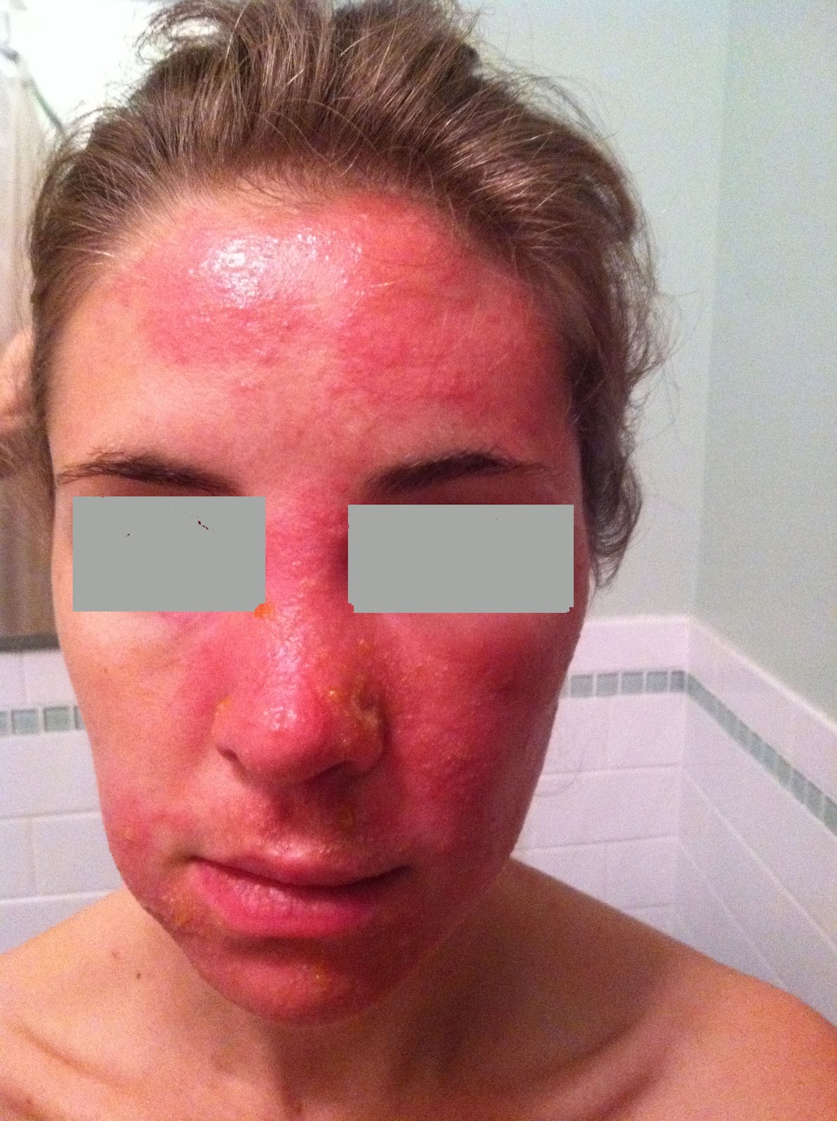
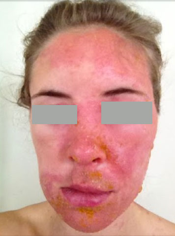
O/E: July 19th: Marked erythema and induration on left forehead, cheek, chin. Vesicles and small bullae scattered in the area; A few erythematous streaky patches lsft hip. Over the next few days, in spite of treatment the process progressed to involve abdomen, neck and fingers. On July 23, there are new erythematous-bullous areas on the left lower abdomen and hip. (see photos).
Clinical Photos: See below
Therapy:
July 19th: Prednisone 20 mg b.i.d. (her weight is 54 kg). Cool tap water compresses. Hydroxizine 20 - 30 mg hs.
July 21: Because of progression of dermatitis, prednisone increased to 50 mg per day in divided doses. Dome Boro compresses 3 x per day, Silvadene cream because of some erosions at site of bullae. Prednisone is causing her to feel anxious and panicky. Lunesta 1 - 2 mg added for sleep.
July 23: I am surprised that new lesions continue to develop in spite of an adequate dose of prednisone (see photos). Have added a "soak and smear" protocol for body lesions and desoximetasone cream bid for dermatitis on body and once daily for face.
Diagnosis: Severe Allergic Phyto-Contact Dermatitis secondary to rhus. We saw this at onset and in spite of a reasonable dose of prednisone and cool compresses it has progressed. Steroid dose was limited because of CNS symptoms and initially I was reluctant to use topical steroids due to her history of acne; but have just started desoximetasone cream on 7/23. The timeline is important as she is getting married in 12 days. It is odd that this has progressed after what is usually an adequate dose of prednisone, and I am worried that increasing the prednisone may cause more anxiety and insomnia. Most likely this is a severe anamnestic response to urushiol and she may indeed need a higher dose of prednisone or she may not be absorbing it.
Questions:
1) How would you handle the facial erythema?
2) Topical corticosteroids as well as oral steroids can exacerbate acne. Should we add a moderate to strong topical corticosteroid for a few days to suppress erythema?
3) She is anxious and has insomnia secondary to prednisone already. I feel prednisoen is the key to improvement, but am reluctant to push the dose. Your thoughts?
4) Is there a role for topical tacrolimus?
Your suggestions re: diagnosis and management will be appreciated.
Photos:
July 19


July 20
July 21
July 22

July 23


 |
| New lesions 4 d p start prednisone July 24 Real Improvement Noted Today  7/30 Pretty Much Better  |


